Anatomical Illustration for Research Papers: Standards and Workflow
Create anatomical illustration for research papers: journal figure rules, heart and organ labeling, AI disclosure, and when to hire a medical illustrator.
Anatomical illustration for a research paper is not a prettier photograph and not a career-guide drawing class. It is a figure that must survive peer review: correct spatial relationships, readable labels at column width, a legend that does not contradict the text, and a file that meets the journal’s DPI and format rules.
This page is for authors who need an organ, approach, or labeled schematic in a manuscript, review, or grant. It is not a guide to becoming a Certified Medical Illustrator. If you need botanical plates, molecular cartoons, or a general history of the craft, start with the broader scientific illustration guide instead.
Medical Illustration Generator
Generate a labeled anatomical figure from a written brief — organ, plane, and structures named up front. Review every label before it goes near a manuscript.
Draft an anatomical figure →What “anatomical illustration” means in a manuscript
In a paper, an anatomical illustration usually does one of four jobs:
| Job | Typical figure | What reviewers punish |
|---|---|---|
| Orientation | “Here is the organ and the plane of section.” | Wrong laterality, missing anterior/posterior |
| Relationship | “Structure A lies deep to B and gives rise to C.” | Crowded labels, arrows that point at the wrong fascia |
| Procedure / approach | Surgical corridor, catheter path, biopsy site | Cosmetic shading that hides the path |
| Comparison | Healthy vs pathologic, pre vs post, species vs species | Inconsistent scale or lighting between panels |
A good anatomical figure answers the caption without the reader opening a textbook. A bad one is a stock heart with six labels that do not match the Results.
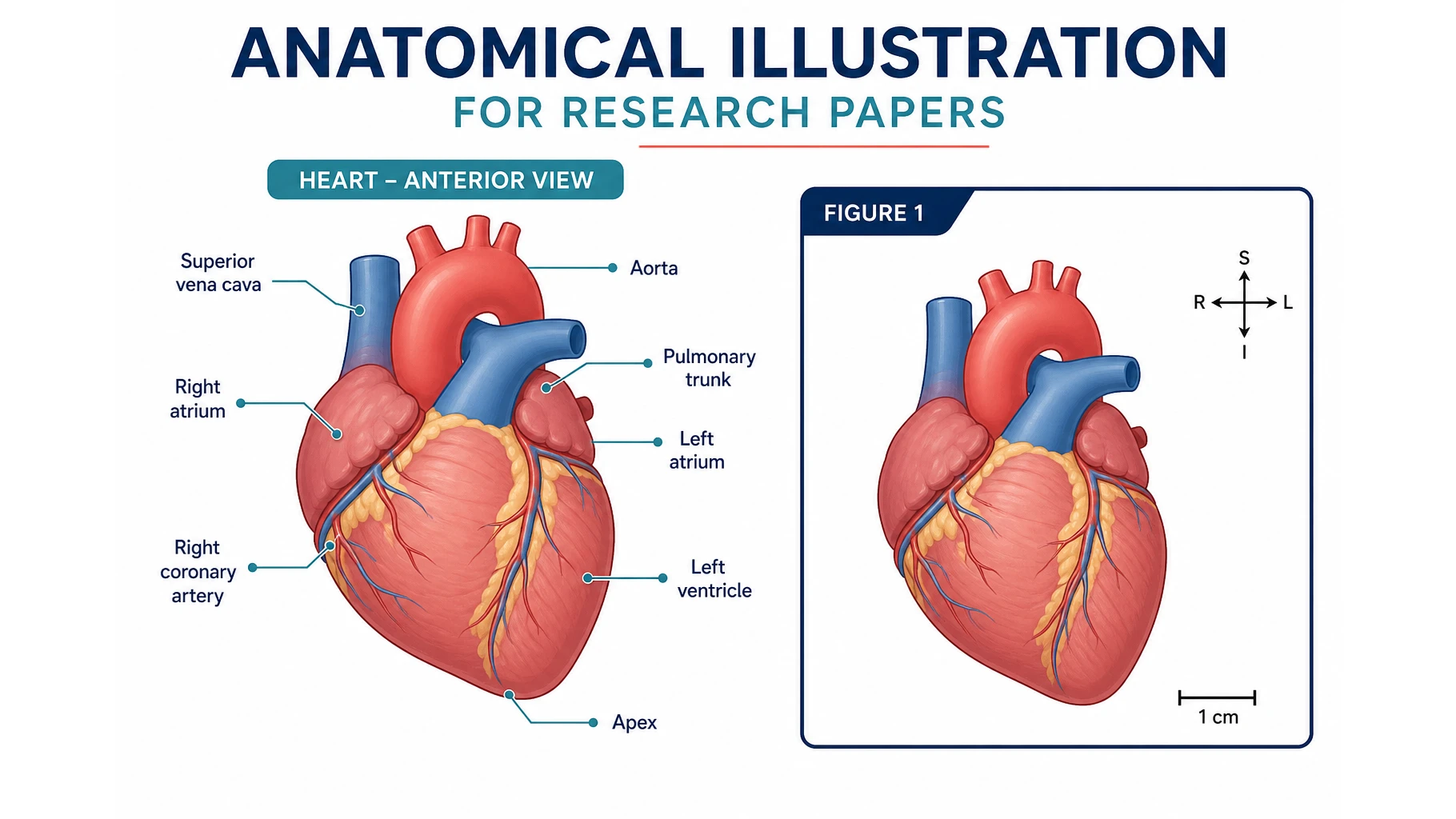
Heart figures are the example this article will keep returning to, because “heart illustration” is the query authors actually type when they mean “labeled cardiac anatomy for Figure 1.” The same rules apply to kidney, brain, joint, and whole-body plates.
A research heart figure should name the chambers, valves, and vessels that the paper discusses — and leave off the ones it does not. Extra labels look complete and read as noise.
Journal rules that decide the file before you pick a tool
Do not generate anything until you have the target journal’s artwork page open. Requirements differ, but the recurring constraints are consistent across Dove Press figure guidelines, Springer’s artwork notes (for example Anatomical Science International), and Elsevier’s author artwork instructions.
Resolution and format
- Photographic / continuous-tone color: 300 dpi at final print width
- Combination (photo + labels): often 600 dpi
- Line art: often 1000–1200 dpi
- Common submission formats: TIFF, EPS, high-quality PDF, sometimes JPEG. Some journals reject PDF figures; read yours.
- RGB unless the journal demands CMYK for print.
Size
- Design at column width (often ~85–90 mm) or full page width (~170–180 mm), not at a 4K desktop canvas you will shrink later.
- Labels must remain legible after the journal reduces the figure. If a 9-pt label dies at column width, it was never a figure.
Type and color
- Sans-serif (Arial, Helvetica) at a consistent size. No drop shadows, no outlined letters.
- One color language for the whole paper. If Figure 2 uses a red artery / blue vein convention, Figure 5 cannot invent a third palette. See scientific color palettes for papers and fonts for figures.
Scale and ethics
- Micrographs need a scale bar and a numeric definition in the legend. Magnification (“400×”) is not a substitute — Experimental Biology and Medicine says this explicitly.
- Cadaveric or patient-derived images need the consent / anonymization path your IRB and journal require. An illustration that still identifies a patient is a compliance failure, not a style issue.
Captions
- The caption carries the title and the explanation. Keep lettering on the image short (
RA,LV,*). Define abbreviations in the caption, not in a hidden sidebar.
Generative AI and anatomical figures: read the policy first
This is the decision that should happen before you spend credits.
Elsevier. Elsevier’s generative AI policies for journals split images into three buckets, not a single ban:
- Explanatory images (flow charts, schematic conceptual illustrations, experimental workflows) — AI is allowed if you check accuracy and disclose the tool, version, and how it was used in the caption and the article’s AI statement.
- Primary research / data images (microscopy, histology, blots, radiology, patient photos, and any figure that is supposed to be the observation) — AI must not create or alter them.
- Graphical abstracts — Elsevier says to use a dedicated scientific-illustration tool (their examples are BioRender and Mind the Graph), not a general-purpose image generator. Cover art is a separate editor decision.
A labeled anatomical schematic (orientation, relationship, approach) is usually bucket 1. A generated plate that pretends to be a specimen, a scan, or a measured morphology is bucket 2 and will get the paper rejected. If you are unsure which bucket your figure is in, treat it as bucket 2 or hire an illustrator.
Other publishers. Policies are not uniform. Some titles allow AI-assisted graphical abstracts with disclosure; some are silent and will still reject a figure that looks generated. Read FEBS Press policies and your target journal, not a blog summary. Graphical-abstract size and layout are a different job — see how to make a graphical abstract.
What you should do in every case
- Read the journal’s AI and artwork pages, not a blog summary (including this one).
- If AI is allowed, disclose the tool, version, and what it did, in the methods or acknowledgments as the journal asks.
- You remain responsible for every label and every spatial relationship. “The model drew it” is not a defense.
ConceptViz does not call generated figures “journal-ready.” Paid exports can include an AI Use & Commercial License Statement (a use record, not a publisher acceptance letter). Acceptance is the editor’s.
A workflow that produces a usable anatomical figure
1. Write the claim the figure must prove
One sentence. Examples:
- “The left anterior descending artery runs in the anterior interventricular groove and is the vessel occluded in this cohort.”
- “The surgical corridor reaches the cerebellopontine angle under the tentorium, lateral to the trigeminal nerve.”
If you cannot write the sentence, you are decorating.
2. Choose the view on purpose
Anatomy has standard views because they reduce argument:
- Heart: anterior, posterior, four-chamber, short-axis, right-left lateral
- Brain: lateral hemisphere, midsagittal, inferior cranial-nerve view
- Joint: standard orthopedic planes, not a random 3/4 glamour angle
Name the view in the prompt and in the caption (Anterior view of the heart). If the paper’s radiology is a four-chamber echo, do not submit an anterior surface drawing and hope the reader maps it.
3. List the structures that earn a label
Cap the list. A paper about mitral annular disjunction does not need every pulmonary vein named. A teaching whole-body plate might; a research figure usually should not.
Write the list before you generate:
View: anterior heart
Must label: RA, RV, LA, LV, aorta, pulmonary trunk, LAD
Must not label: coronary sinus, pectinate muscles, every pulmonary vein
Style: flat didactic color, white background, Arial-style labels, no drop shadow
4. Generate or sketch, then run a structured review
Whether the first draft comes from a medical illustrator, Illustrator, or a generator, review in this order:
- Laterality — patient’s left, not your left. Hearts and brains are where this fails first.
- Connections — does the pulmonary trunk leave the right ventricle? Does the IVC enter the right atrium?
- Layer — are you looking at the surface, a cut, or a see-through? Mixing those without saying so is how “the LAD is inside the chamber” happens.
- Label-to-structure — every leader lands on the structure it names. No leaders to empty space.
- Consistency with the text — if the manuscript says “anomalous origin from the right sinus,” the figure cannot show a normal left origin.
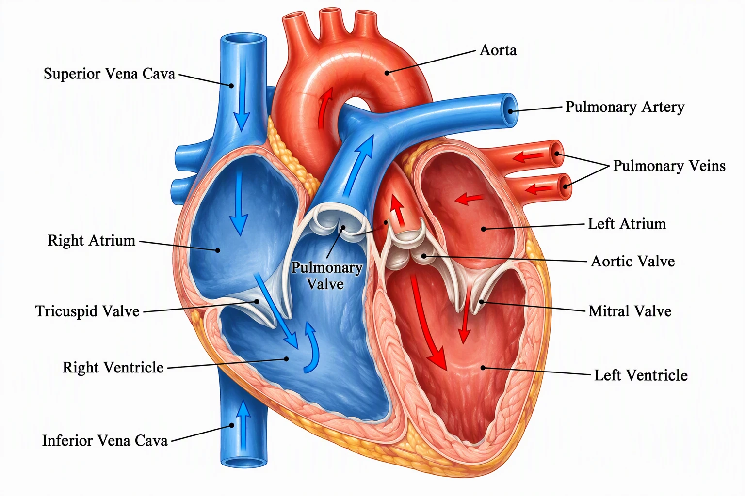
Didactic color helps students. In a paper, the same palette must stay consistent across panels and must not invent vessels the study never measured.
5. Export for the journal, not for the slide deck
- Crop excess margin.
- Flatten or embed fonts if you submit PDF/EPS.
- Name files
Figure1.tif,Figure2.tifas the journal asks. - Keep an editable source. Reviewers will request a label change.
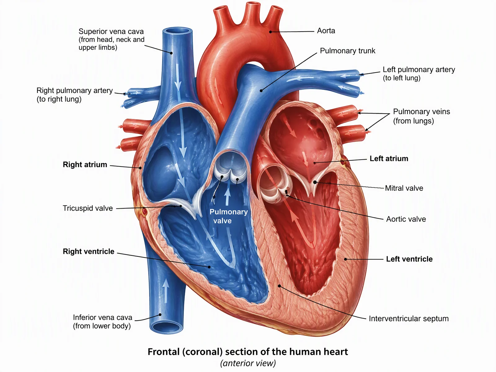
For a heart-only figure, the dedicated heart diagram generator is a narrower brief than a general medical plate. For line-style plates (skeleton, joint), use the anatomical drawing generator.
Heart Diagram Generator
A focused cardiac figure when Figure 1 is a heart and nothing else.
When to hire a medical illustrator (and when not to)
Hire a trained medical illustrator — ideally someone with AMI credentials or a graduate program background — when:
- The figure is a journal cover, a textbook plate, or a grant’s visual centerpiece
- The anatomy is rare, surgical, or disputed, and a wrong furrow would change the claim
- You need vector layers a production editor can restyle
- The publisher bans generated images and you do not have time to redraw
Skip the commission when:
- You need a working sketch for coauthors this afternoon
- The figure is a standard orientation view you will replace after review
- The journal allows a simple labeled schematic and your coauthors can check it
Typical custom medical-illustration quotes for a single complex plate still land in the high hundreds to low thousands of dollars and a one- to three-week calendar. That is rational for a cover. It is how grants die for a generic four-chamber cartoon.
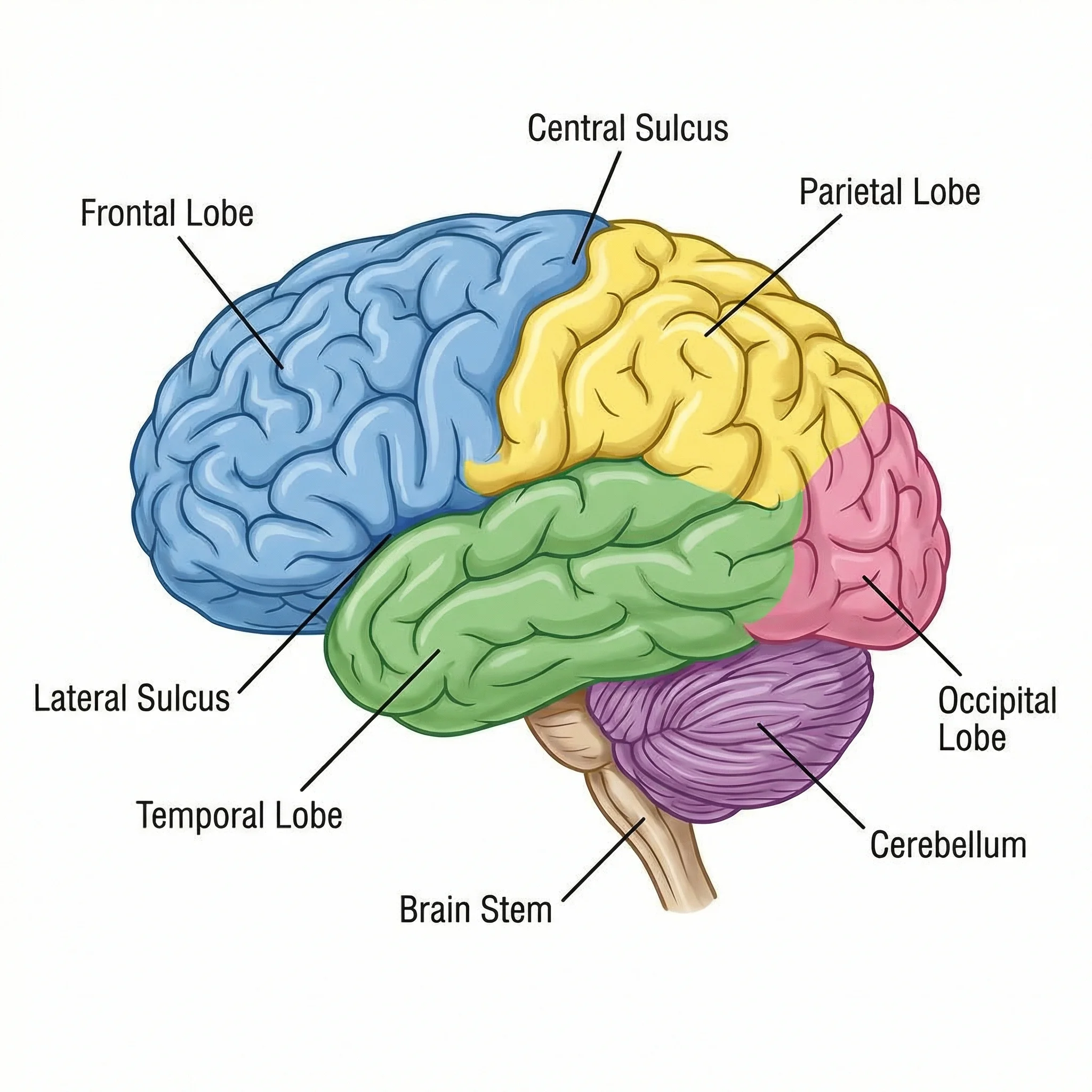
CNS figures fail on laterality and on cranial-nerve order more often than on “art quality.” Check the claim, then the shading.
Color, labels, and the heart-specific traps
Cardiac figures collect a small set of unforced errors:
- Red/blue = oxygenated/deoxygenated, except when you are drawing coronary arteries (arterial blood, drawn red) on the surface of a blue-coded right ventricle. Say what the colors mean in the caption.
- LAD vs posterior descending laterality depends on dominance. If the paper does not determine dominance, do not draw a confident PDA origin.
- Four-chamber vs anterior surface. Authors paste an anterior drawing into a four-chamber legend. The labels then “fit” and are still wrong.
- Valve names vs cusps. “Aortic valve” is not the same label as “right coronary cusp.”
- Scale. A heart drawn next to a schematic catheter must share a scale language or the catheter looks like a garden hose.
The same class of error shows up in joints (left vs right knee), kidneys (hilum orientation), and brains (patient left on coronal slices).
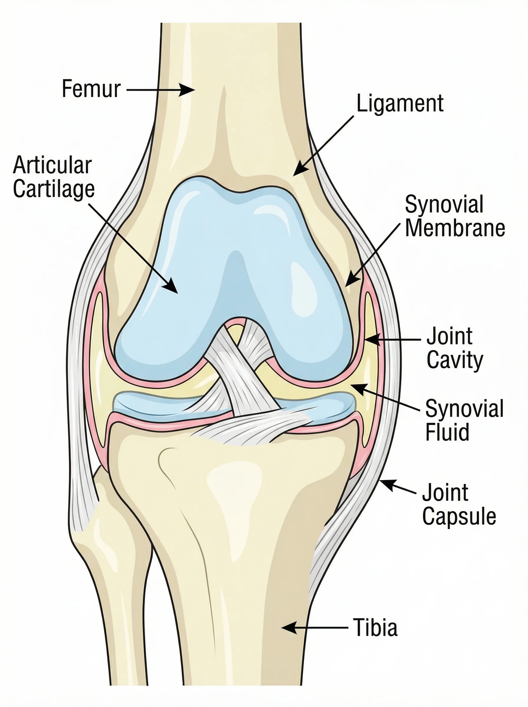
Orthopedic plates need the standard clinical view. A dramatic 3/4 render that hides the joint line is a poster, not a Results figure.
How this sits next to BioRender and Illustrator
BioRender pricing is a seat-and-license problem. BioRender is strong when the figure is an icon-assembled experimental cartoon. It is weaker when you need a specific anatomical plane that is not in the icon set.
Adobe Illustrator (or Inkscape) is still the honest tool for final vector labels on a figure you will submit to a journal that wants EPS and will ask you to move one leader 2 mm.
A generator is the honest tool for a checked first draft: you write the view and the must-label list, you get a plate in minutes, and a human — preferably a coauthor who does this anatomy for a living — rejects or accepts each label.
For priced picks across all four categories (icon libraries, drawing software, 3D atlases, AI generators), see our best medical illustration software comparison.
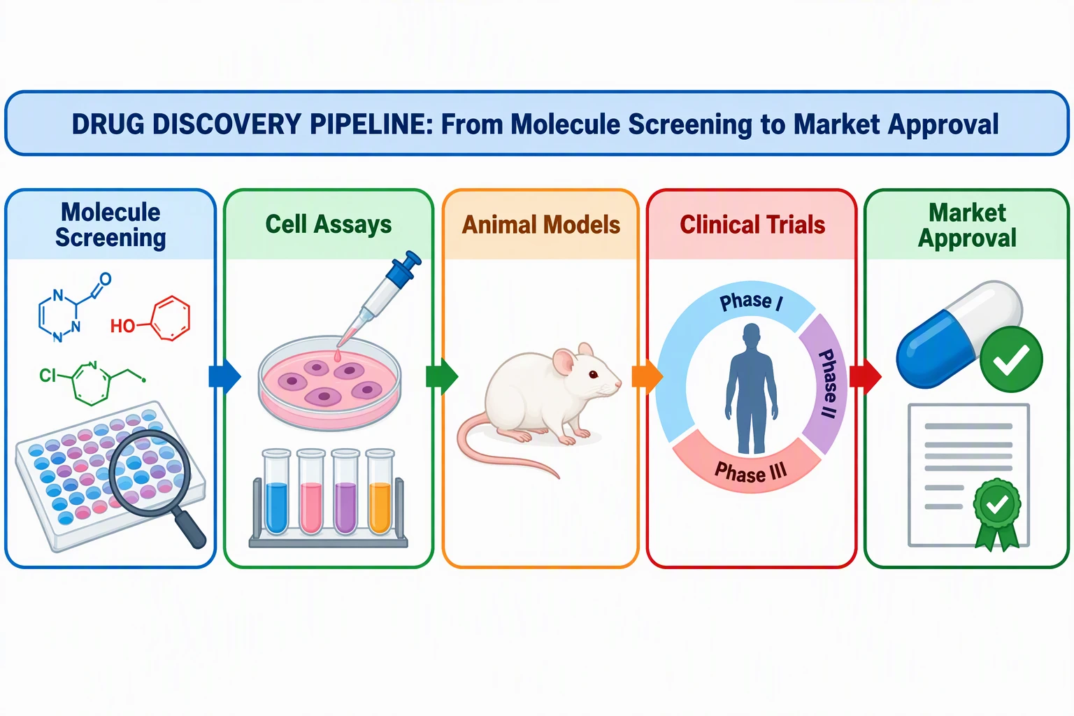
Graphical Abstract Maker
If the journal wants a single-panel visual abstract rather than an anatomical Results figure, that is a different size and a different AI policy.
Draft a graphical abstract →A short pre-submission checklist
Print this next to the figure:
- Journal artwork page read this week (DPI, format, width, AI policy)
- View named in the caption
- Laterality checked by a second author
- Every on-figure abbreviation defined in the caption
- No structure labeled that the paper does not discuss
- Colors consistent with other figures
- File named and sized for the journal, not for Twitter
- Rights / consent / AI disclosure handled
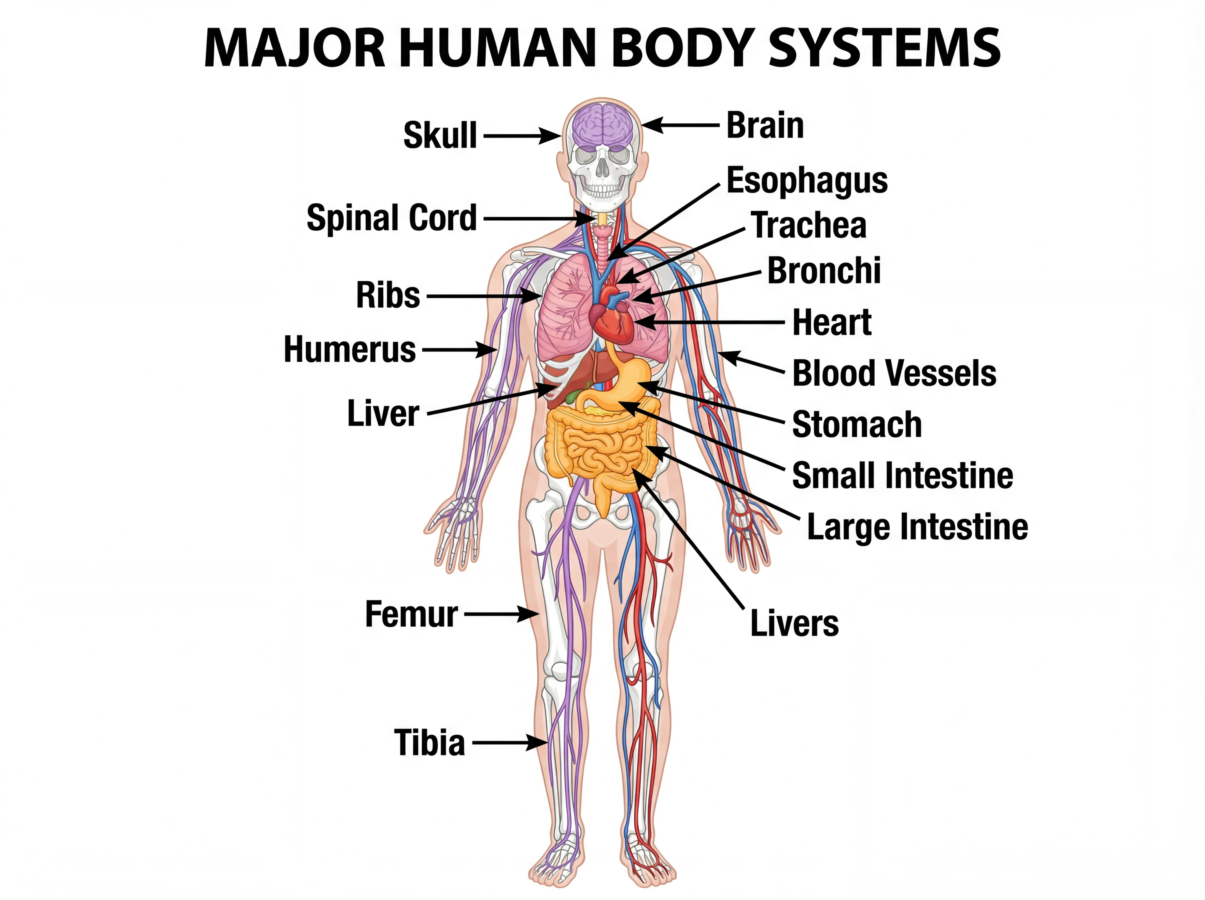
Overview plates belong in reviews and teaching figures. A primary research paper almost always wants one organ, one plane, and fewer labels.
Frequently Asked Questions
What is anatomical illustration in a research paper?
It is a figure that shows spatial anatomy — organs, planes, vessels, nerves, or approaches — so a reader can understand a claim without opening an atlas. It is judged on accuracy, labels, and journal file rules, not on painterly style.
What resolution should anatomical figures be?
Most journals want at least 300 dpi for color continuous-tone figures at the final printed width, 600 dpi for photo-plus-label combinations, and 1000–1200 dpi for line art. Always use the target journal’s artwork page.
Can I use AI to make anatomical illustrations for a journal?
It depends on the figure type and the journal. Elsevier allows AI for some explanatory schematics if you disclose the tool in the caption; it bans AI on primary research images such as microscopy or patient photos; and it wants dedicated illustration tools, not general-purpose generators, for graphical abstracts. Read the current policy before you submit.
When should I hire a medical illustrator?
Hire one for covers, textbook plates, rare or surgical anatomy, or journals that ban generated images. A standard orientation figure that coauthors can check can start as a draft and move to vector labels.
How do I label a heart figure correctly?
Name the view (anterior, four-chamber, short-axis), label only the chambers and vessels the paper discusses, keep left and right in patient coordinates, and define every abbreviation in the caption. Do not mix an anterior drawing with a four-chamber legend.
Should anatomical figures use red and blue for arteries and veins?
Often yes for didactic figures, but the caption must say so. Coronary arteries on a blue-coded right ventricle still carry arterial blood. Never let color contradict the physiology the paper claims.
What file format do journals want?
TIFF, EPS, and high-quality PDF are the usual set. Some journals refuse PDF figures or Office files. Submit one figure per file with the journal’s filename pattern.
Is a generated anatomical figure the same as a graphical abstract?
No. A Results anatomical figure is evidence. A graphical abstract is a single-panel advertisement of the paper and has its own pixel size and, often, a different AI policy. Do not reuse one file for both without checking both rules.
Bottom line
Anatomical illustration in a paper is a claim in picture form. Pick the view, cap the labels, obey the journal’s file and AI rules, and have a second anatomically literate author reject the figure if laterality or connections are wrong.
If you need a first labeled plate today — and the figure is an explanatory schematic your journal allows, or you will redraw before submission — start with the medical illustration generator. If the figure is a cover, a rare surgical plate, or anything that will be read as primary data, commission a medical illustrator.
If you are still choosing a vendor rather than a view, use the AI scientific illustration comparison or the 1-to-1 BioRender vs ConceptViz.
Categories
More Posts
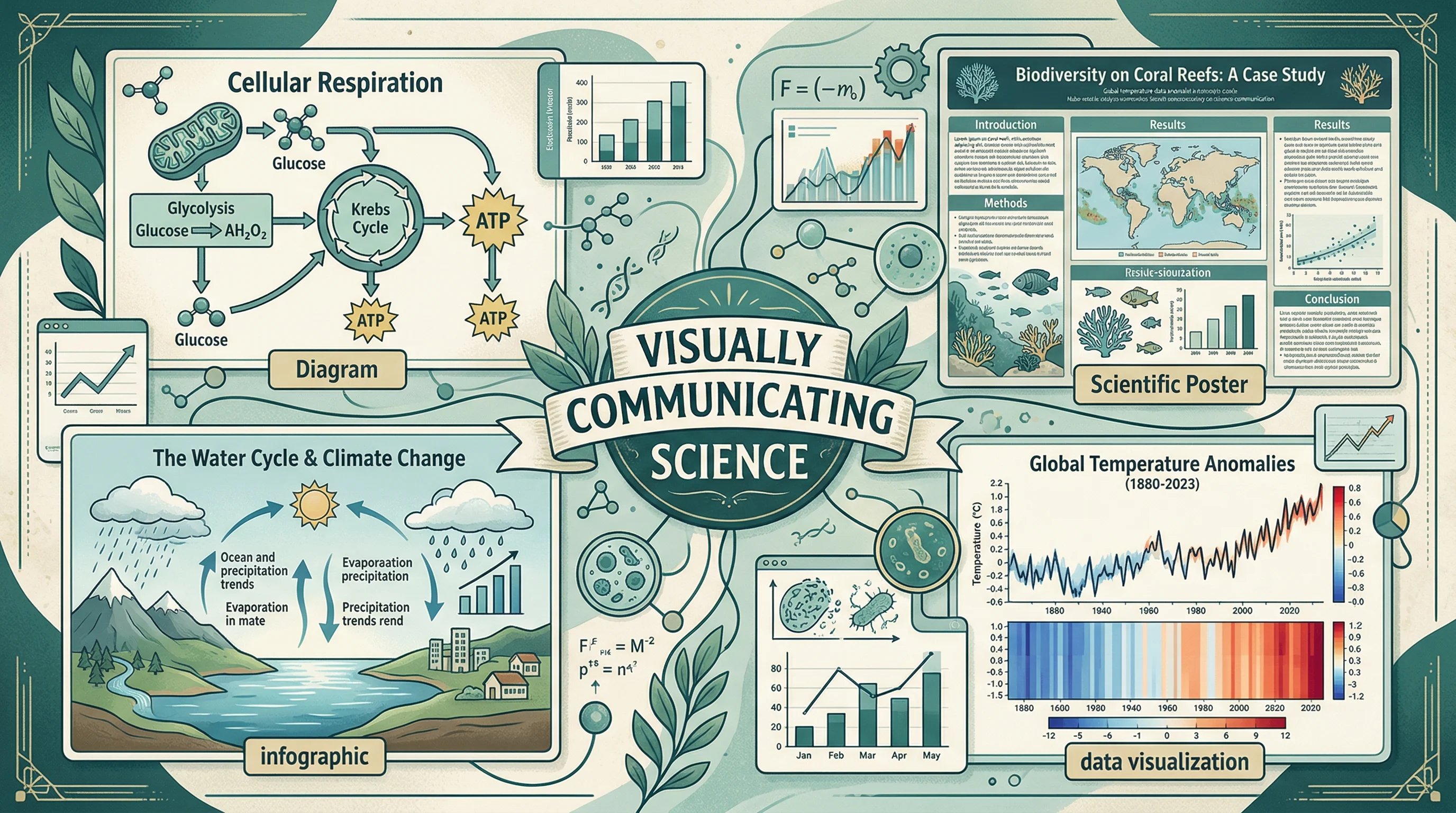
How to Illustrate Science: Methods, Tools & Examples (2026)
Learn 8 proven ways to illustrate science for research papers, posters, and presentations. Covers diagrams, infographics, graphical abstracts, and more.
Scientific Infographic Design: Complete Guide for Researchers and Scientists
Master the art of creating effective scientific infographics. Learn design principles, data visualization techniques, and best practices for research.
Best Scientific Color Palettes: Hex Codes & Examples
Compare Okabe-Ito, Viridis, Paul Tol and other scientific color palettes for research figures. Includes colorblind-safe hex codes, use cases and code snippets.