Heart Diagram Generator for Labeled Heart Anatomy
Generate a labeled human heart diagram from a description. Show the four chambers, the valves, the major vessels, and blood flow — choose labeled for study notes or blank for worksheets and quizzes, then download your image.
Heart Diagram Generator
Free to try ·
Your heart diagram will appear here
Describe the heart structures and click Generate
Heart Diagram Examples
Labeled, blood-flow, valve, and blank versions for class and revision
Labeled Heart Anatomy
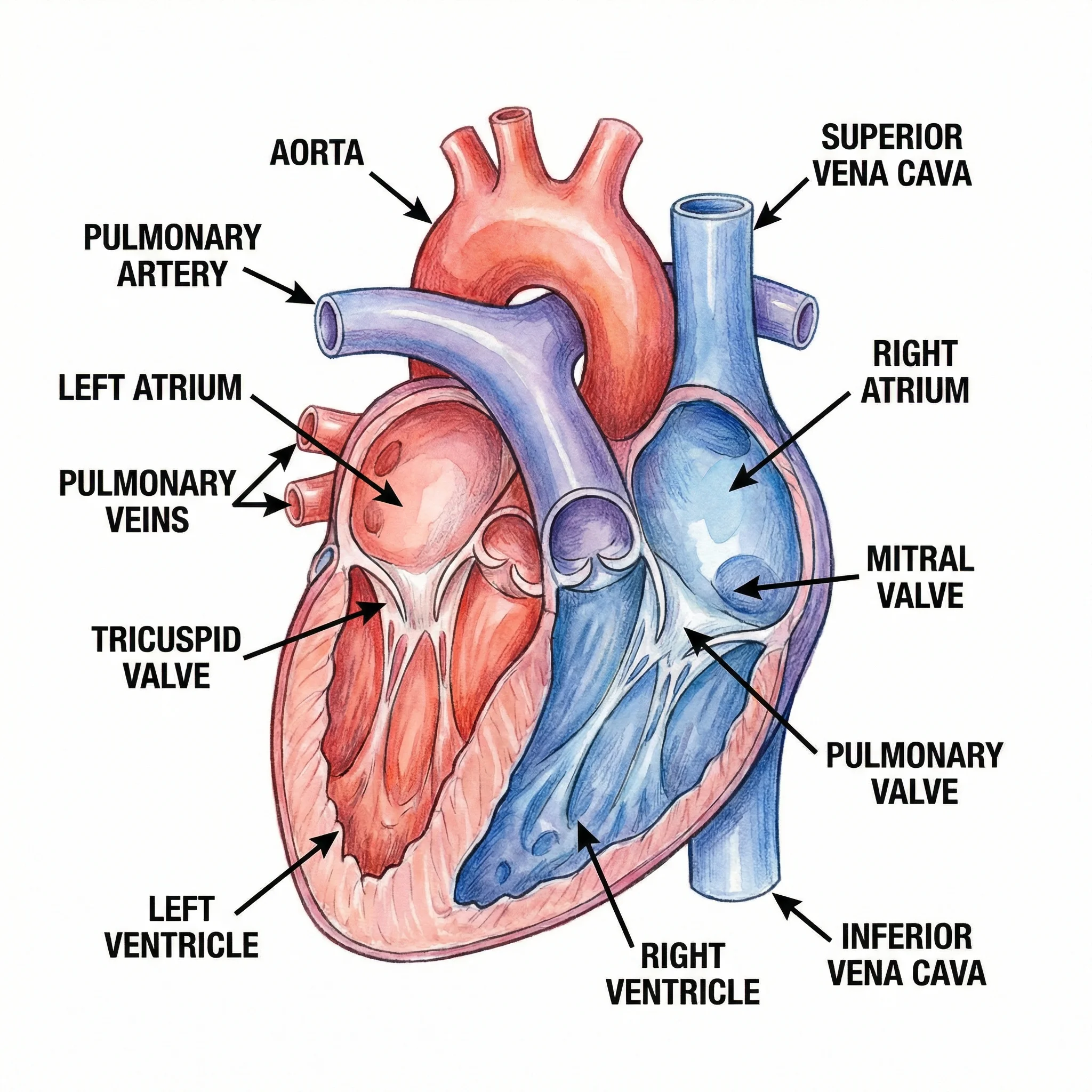
A full anterior-view diagram with every chamber, valve, and major vessel labeled.
Blood Flow Pathway
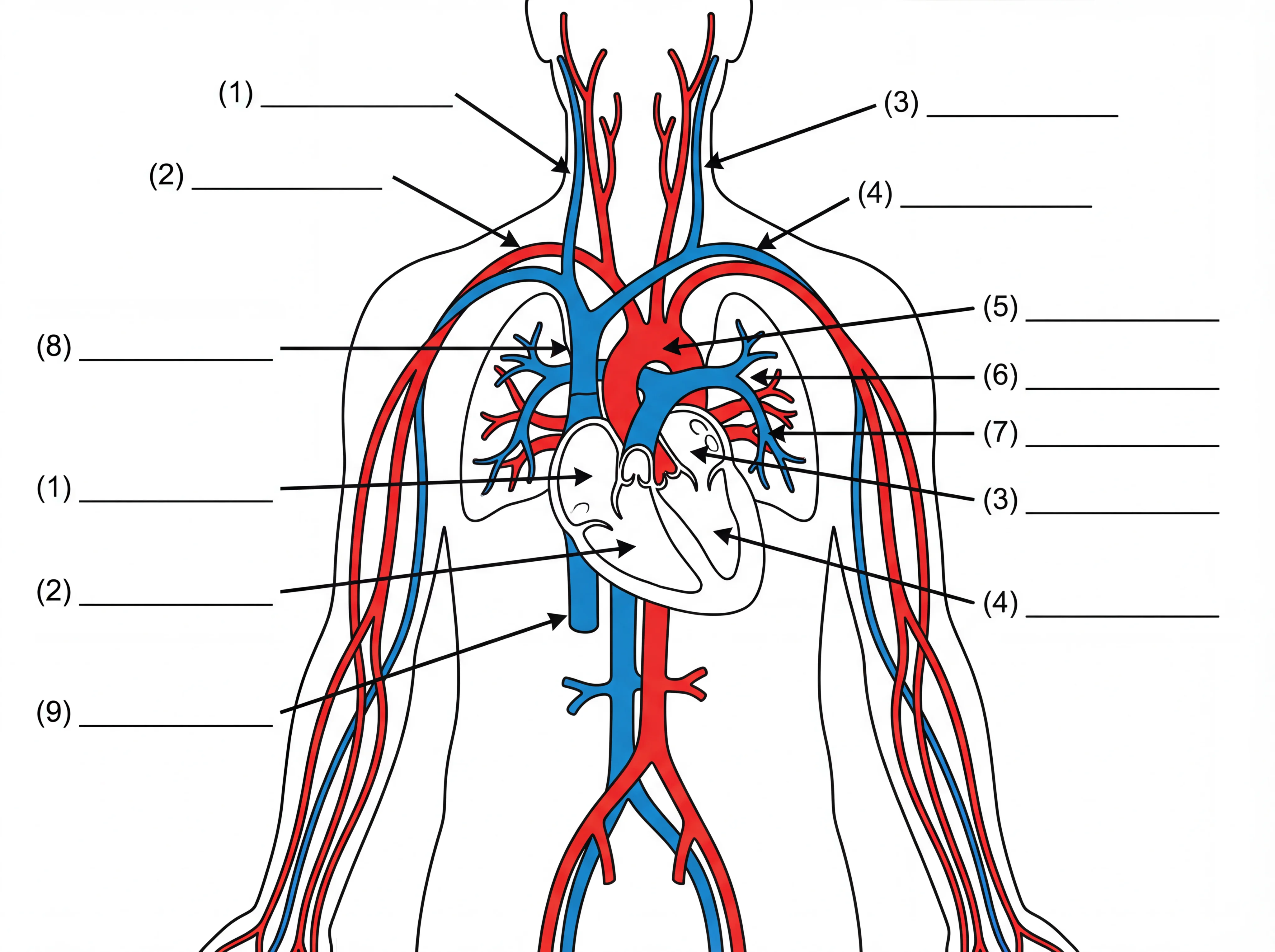
Numbered arrows trace the path of blood from the vena cava through the lungs and out the aorta.
Simple Heart for Kids
A bright, simplified version with big labels for elementary and middle-school classes.
Heart Cross-Section
A frontal section showing the septum, wall thickness, papillary muscles, and chordae tendineae.
Heart Valve Diagram
A superior view of all four valves, with insets showing each one open and closed.
Blank Worksheet Version
An unlabeled outline with numbered blanks — ready to print as a worksheet or quiz.
What is a heart diagram?
A heart diagram is a labeled drawing of the human heart that shows its internal structures and how they fit together. A good diagram identifies the four chambers, the four valves, and the major blood vessels, and usually uses red and blue to show where blood is oxygenated and deoxygenated. It is one of the most common figures in biology and anatomy lessons because it turns an abstract organ into something students can read, label, and revise. This generator builds that diagram from a plain-English description, so you can get the exact view and level of detail you need.
The labeled parts of the heart
- Four chambers: the right atrium and left atrium on top, and the right ventricle and left ventricle below — the right side handles deoxygenated blood, the left side oxygenated blood.
- Four valves: the tricuspid valve and mitral (bicuspid) valve between the atria and ventricles, and the pulmonary valve and aortic valve where blood leaves the ventricles. They keep blood moving in one direction.
- Major vessels: the superior and inferior vena cava bring blood in, the pulmonary arteries carry blood to the lungs, the pulmonary veins return it, and the aorta carries oxygenated blood to the body.
- Septum: the muscular wall that separates the left and right sides of the heart so oxygenated and deoxygenated blood do not mix.
How blood flows through the heart
Blood follows a fixed double loop. Deoxygenated blood returns from the body through the vena cava into the right atrium, drops into the right ventricle, and is pumped through the pulmonary valve and pulmonary arteries to the lungs — this is the pulmonary circulation. In the lungs it picks up oxygen and returns through the pulmonary veins to the left atrium, drops into the left ventricle, and is pumped through the aortic valve into the aorta to supply the whole body — the systemic circulation. A blood-flow diagram uses blue arrows for the deoxygenated path and red for the oxygenated path so the two loops are easy to follow, which is exactly what the blood flow sample prompt produces.
Labeled vs. blank diagrams for worksheets and quizzes
A fully labeled diagram is best for teaching, study notes, and revision because every structure is named with a leader line. A blank or unlabeled version keeps the same outline and leader lines but replaces the names with numbered blanks, so students can fill them in — perfect for worksheets, homework, and quizzes. With this tool you can ask for either: request a labeled diagram for your notes, then generate a matching blank version for an assessment. The blank worksheet sample prompt is set up to produce a quiz-ready figure with numbered structures.
How to generate a labeled heart diagram
- Describe the diagram you want — name the view (anterior, cross-section, or valve plane) and which structures to label.
- Choose the level of detail: a simple version for younger students, or a detailed one with the septum, papillary muscles, and chordae tendineae.
- Say whether you want blood-flow arrows, and whether labels should be filled in or left blank for a worksheet.
- Generate the image, review it for accuracy, then download it to drop into slides, notes, or a printable handout.
Using heart diagrams in the classroom
Teachers use these diagrams to introduce the cardiovascular system, build labeling worksheets, and create quick formative quizzes; students use them to revise the chambers, valves, vessels, and blood flow for exams. Because the diagrams are AI-generated, treat them as teaching illustrations rather than clinical references: always review the output against your textbook before sharing it, as AI can occasionally mislabel or misplace a structure. These figures are for education only and are not medical advice. For broader anatomy, the related tools below cover other body systems and medical illustrations.
Frequently Asked Questions
Related Biology Tools
 Biology
Human Body Systems Diagram Generator
Create labeled diagrams of the circulatory, respiratory, digestive, and other body systems.
 Biology
Anatomical Drawing Generator
Draw labeled anatomy figures for organs, the skeleton, muscles, and more.
 Medical
Medical Illustration Generator
Generate clean medical illustrations of organs and structures for study and presentations.