Plant Cell Diagram Labeled, Blank & Printable
Free plant cell diagrams — labeled, blank, coloring, and exam-review sheets. View, download, or print any version, or customize your own.
Free Plant Cell Diagrams
Every version below is free to view and download — no sign-up.
Labeled Plant Cell Diagram
Every organelle labeled — cell wall, chloroplasts, large central vacuole, nucleus and more. Ready for study, notes, or slides.
Core diagrams
Structure & focus
Comparison
Print & study
Test yourself: plant cell
A quick self-test — questions and answers reshuffle each time, with a review of anything you miss at the end.
Where does photosynthesis take place in a plant cell?
Need a custom version?
Change the grade level, colors, or which organelles are labeled — describe it and generate your own.
Customize Your Plant Cell Diagram
Free to try ·
Your custom plant cell diagram will appear here
Ask for a labeled version to study, or a blank one for a worksheet
What is a plant cell diagram?
A plant cell diagram is a labeled cross-section that shows the parts of a plant cell — the boundary structures and the organelles inside. Because real cells are microscopic and roughly three-dimensional, a diagram simplifies the cell into a clear cutaway so each organelle can be named and located. Use a fully labeled version for studying, or a blank, unlabeled version as a worksheet or quiz.
Labeled vs unlabeled plant cell diagrams
- Labeled diagram: every organelle is named with a clean callout line — ideal for learning the parts, revising before an exam, or illustrating notes and slides.
- Unlabeled (blank) diagram: the same cell with empty leader lines and no names, so students can fill in the organelles themselves — perfect for worksheets, quizzes, and homework.
- Coloring and review sheets: black-and-white line versions and exam-review layouts let students color, label, and self-test from the same diagram.
- Use the labeled version to teach, then hand out the matching blank version to assess — the cell layout stays consistent across both.
The organelles in a plant cell
- Cell wall: the rigid outer layer made of cellulose that gives the plant cell its fixed, box-like shape and support.
- Cell membrane: the thin layer just inside the wall that controls what enters and leaves the cell.
- Large central vacuole: a single big, fluid-filled sac that stores water and keeps the cell firm (turgid); it often takes up most of the cell.
- Chloroplasts: green organelles containing chlorophyll where photosynthesis happens, capturing light to make glucose.
- Nucleus: the control center that holds the cell’s DNA, usually with a visible nucleolus inside.
- Mitochondria: the sites of respiration that release energy; plus the endoplasmic reticulum, Golgi apparatus, and ribosomes that build and process proteins, all suspended in the cytoplasm.
Plant cell vs animal cell: the key differences
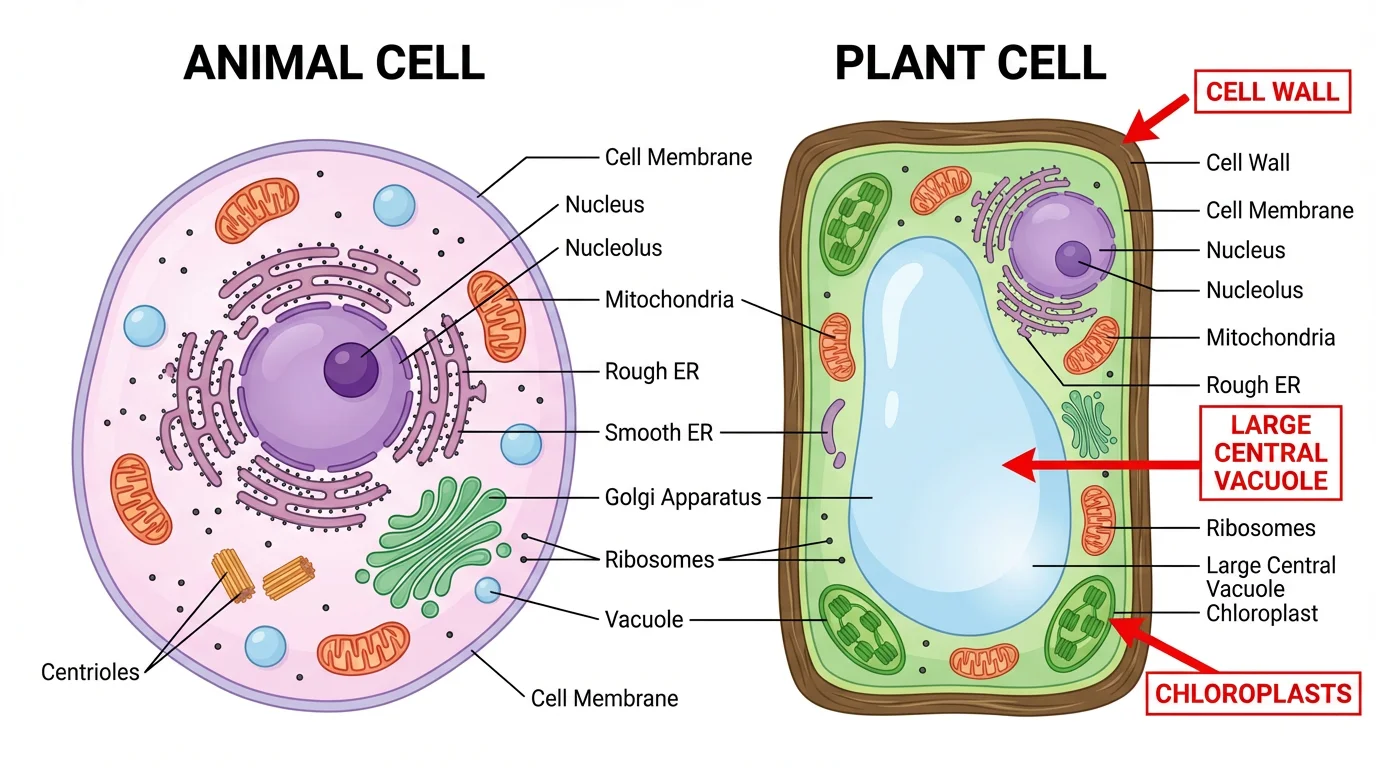
Plant and animal cells share many organelles — both have a nucleus, mitochondria, endoplasmic reticulum, Golgi apparatus, and ribosomes. The differences are what students are usually asked to identify. A plant cell has three structures an animal cell lacks: a rigid cell wall, chloroplasts for photosynthesis, and a single large central vacuole. Animal cells, by contrast, have centrioles and only small, scattered vacuoles, and they take an irregular round shape because they have no wall. The comparison sheet places the two side by side so those distinctions are easy to see.
Using plant cell diagrams in class and study
A consistent diagram is one of the most effective ways to teach cell biology. Teachers can post a labeled version while introducing organelles, then issue a blank version of the same cell for students to label from memory — a quick, low-prep formative check. Students revising on their own can do the reverse: study the labeled diagram, then test themselves on the unlabeled one. The coloring-page and exam-review layouts add a hands-on option for younger learners and a print-ready format for revision, all built around the same plant cell.
Frequently Asked Questions
Related Biology Tools
 Biology
Animal vs Plant Cell Comparison
Compare plant and animal cells side by side, with shared and unique organelles labeled.
 Biology
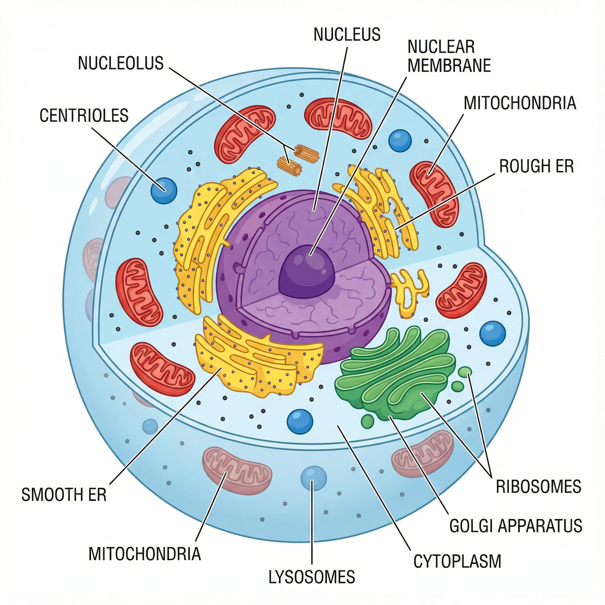
Animal Cell Diagram
Labeled animal cell diagrams with the nucleus, mitochondria, and other organelles.
 Biology
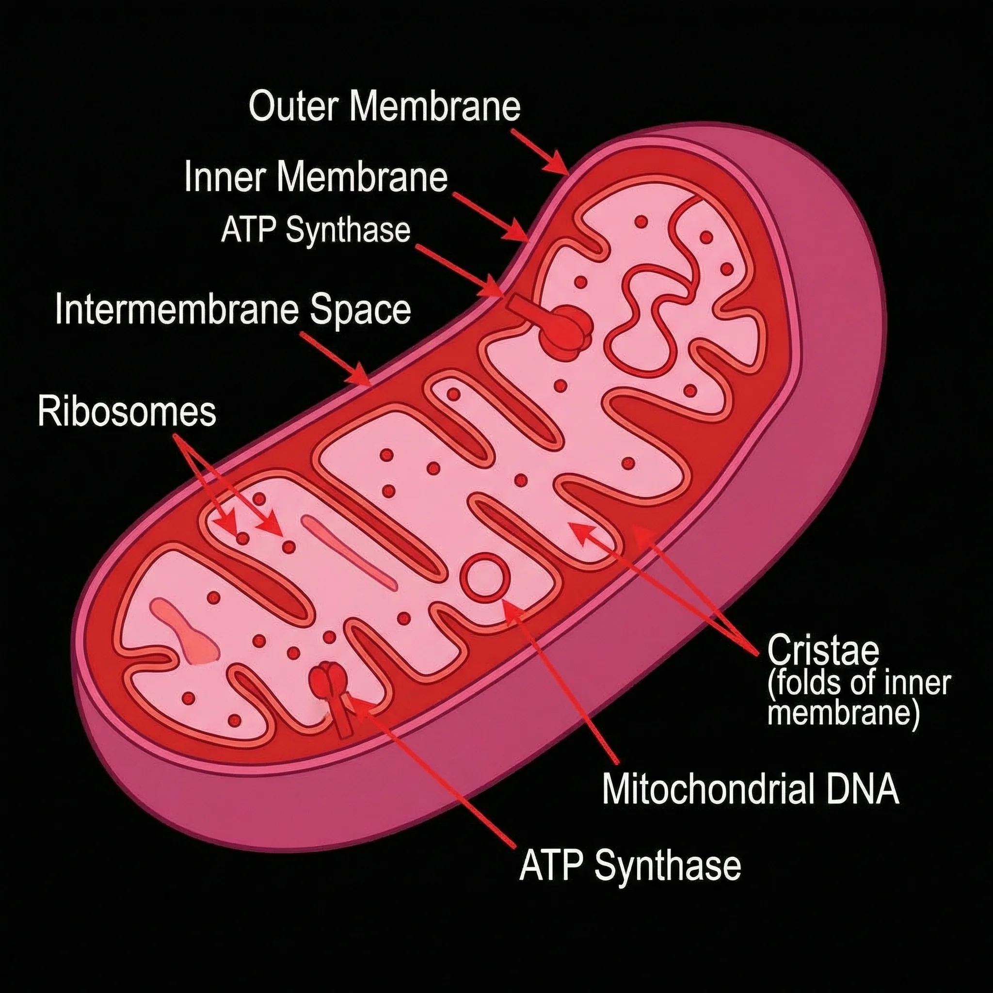
Mitochondria Diagram
Labeled mitochondria diagrams — the site of respiration inside plant and animal cells.
 Biology
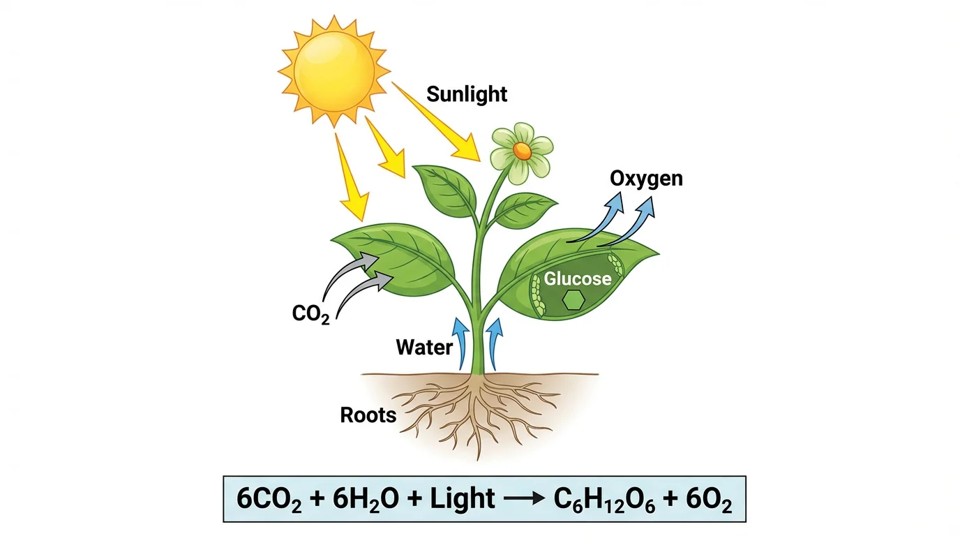
Photosynthesis Diagram
See how chloroplasts turn light, water, and CO₂ into glucose, step by step.
 Biology
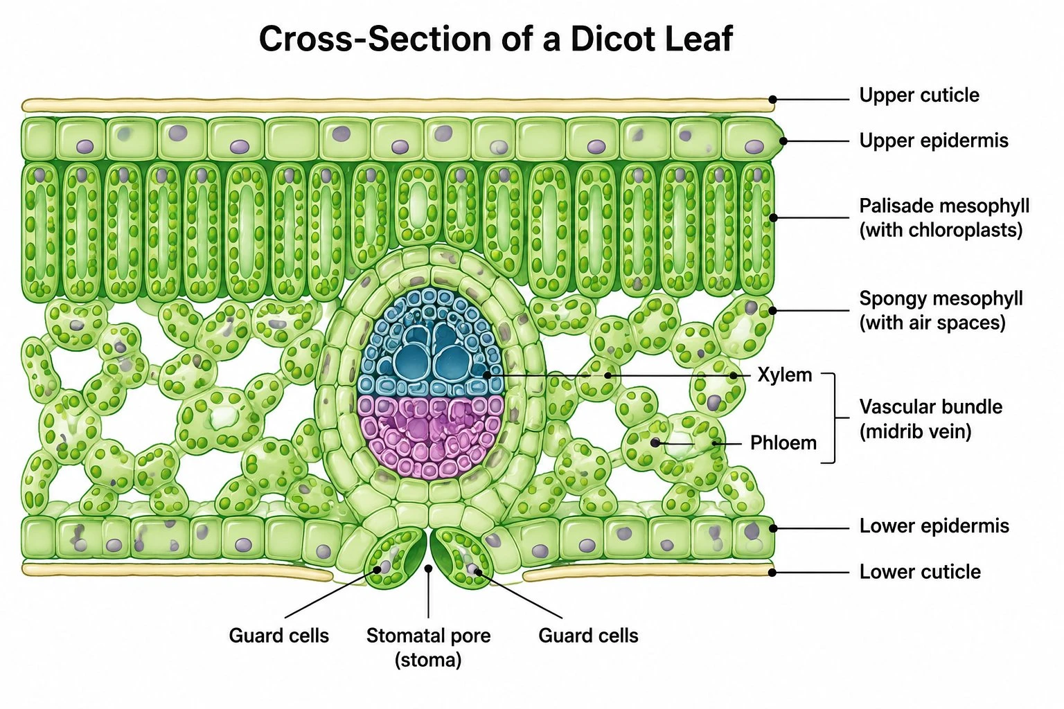
Leaf Structure Diagram
Labeled leaf cross-sections showing the layers where plant cells do their work.
 Biology
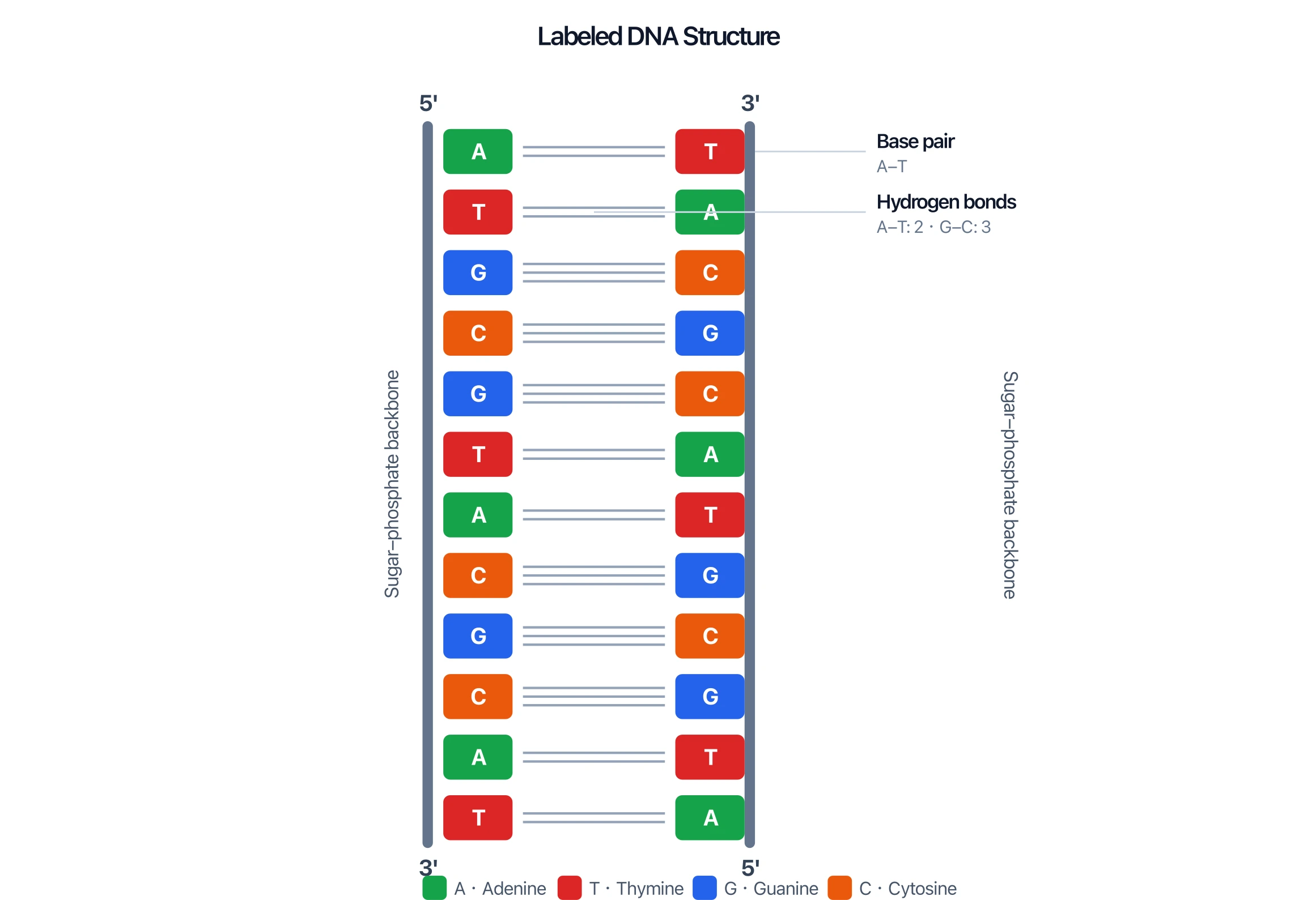
DNA Structure Diagram
Labeled DNA double-helix diagrams — the genetic material stored in the nucleus.