Tumor Microenvironment Diagram
Free labeled tumor microenvironment diagram, blank version, and hot vs cold figure for lecture slides and papers. Download, then customize. Check labels.
Free Tumor Microenvironment Diagrams
Every version below is free to view, download, and print. No sign-up. Check labels against your sources. Checked November 2026.
Labeled Tumor Microenvironment
Nine labels on this figure: Tumor cells, Cancer-associated fibroblast (CAF), Tumor-associated macrophage (TAM), Regulatory T cell (Treg), Myeloid-derived suppressor cell (MDSC), CD8+ T cell, Dendritic cell (DC), Blood vessel, and Extracellular matrix (ECM). There is no “Endothelial cells” label. Check each name against your sources.
Core diagrams
Immune phenotypes
These three files are free to view, download, and print. Custom generated figures follow the ConceptViz license. Read that page before a paper, a paid course, or commercial reuse.
Customize this diagram
Change cancer context, which cells are labeled, or hot versus cold. Describe it and generate your own. Then check every label.
Customize Your Tumor Microenvironment Diagram
Free to try ·
Your custom diagram will appear here
Ask for labeled overview, blank version, hot vs cold, or a cancer-specific stroma
What the tumor microenvironment is
The NCI Dictionary of Cancer Terms defines the tumor microenvironment as “the normal cells, molecules, and blood vessels that surround and feed a tumor cell.” A tumor can change that neighborhood, and the neighborhood can change how the tumor grows and spreads (NCI Dictionary, term tumor-microenvironment). NCI’s Tumor Biology and Microenvironment research pages add the working parts you will see on a teaching map: stromal composition, cell–cell and cell–matrix interactions, abnormal physiology, and heterogeneous tumor cells. Bidirectional signaling between tumor cells and host tissue can promote survival and aggressive behavior. These diagrams are maps for a lecture or a review figure, not a diagnosis and not a substitute for your methods section. Check every label against the source you are teaching from.
Cells and stroma: what each compartment is doing
- Tumor cells: NCI notes that they release cytokines and growth factors onto stroma, and that cell–matrix and exosome signaling can support survival, proliferation, motility, and invasion.
- Cancer-associated fibroblast (CAF): abundant in the TME, often at infiltrating edges (PMC8039952). They remodel ECM, can promote angiogenesis, and can exclude T cells by an ECM barrier, CXCL12 that misdirects CTLs, and TGFβ and IL-6.
- Tumor-associated macrophage (TAM): an immunosuppressive myeloid population. PMC8039952: TAMs can exclude T cells by regulating ECM, promote abnormal angiogenesis via VEGF and MMP9, and, under CSF-1/CSF-1R, skew toward an M2-like phenotype.
- Regulatory T cell (Treg): listed with TAMs and MDSCs as an immunosuppressive population common in cold tumors (PMC8039952). CAF-derived TGFβ can push naive CD4 T cells toward Tregs.
- Myeloid-derived suppressor cell (MDSC): grouped with TAMs and Tregs as immunosuppressive cells in cold tumors (PMC8039952).
- CD8+ T cell: the effector the hot-versus-cold contrast is about. CTLs recognize peptide–MHC, form an immune synapse, and can kill through perforin and granzyme and through Fas/FasL (PMC8039952).
- Dendritic cell (DC): professional antigen-presenting cells. They acquire tumor antigens, migrate to lymph nodes, present a peptide–MHC class I complex to T cells, and supply B7 (CD80/CD86) costimulation (PMC8039952). BATF3 DCs also make CXCL9 and CXCL10 that recruit CXCR3+ CD8 T cells.
- Blood vessel: the labeled figure uses this name, not “Endothelial cells.” NCI treats tumor-induced vasculature as the supply of oxygen and metabolites, with abnormal endothelial–pericyte interactions, flow, and permeability. PMC8039952: T cells must adhere to endothelium (P- and E-selectin, ICAMs, VCAMs); anergic or ETBR-high endothelium can block that step.
- Extracellular matrix (ECM): NCI calls it a major TME component — proteins, polysaccharides, and growth factors — that can stiffen and, as desmoplasia, physically bar endothelial, immune, and fibroblast subtypes.
- None of these one-line roles is complete biology. Phenotypes mix inside one tumor and between primary and metastasis. Use the map to remember names; use your paper for the functions you claim.
Hot vs cold tumors
Reviews of immune checkpoint inhibitors classify tumors by where CD8 T cells sit (Liu & Sun, PMC8039952, following the immune-inflamed / excluded / desert split). Immune-inflamed, also called “hot,” has high T-cell infiltration, increased interferon-γ signaling, PD-L1 expression, and often high tumor mutational burden, and tends to respond better to checkpoint blockade. Immune-excluded and immune-desert are grouped as “cold.” In excluded tumors, CD8 T cells stop at the invasion margin. In desert tumors they are missing from the tumor and its edge. Cold tumors in that review also tend toward low mutational load, low MHC class I, low PD-L1, and immunosuppressive TAMs, Tregs, and MDSCs, and they rarely respond to checkpoint inhibitor monotherapy. Driving T cells in is described as a cycle: antigen release, dendritic-cell presentation, priming, trafficking through blood, crossing vessels, recognition, and killing — which releases more antigen. Desert tumors fail early (priming). Excluded tumors fail at homing and entry. Inflamed tumors can still fail at killing because of checkpoints and exhaustion. The two-column figure on this page shows Hot tumor versus Cold tumor; the cold panel is the excluded pattern (T cells at the stroma edge). Desert is explained here, not drawn as a third column. A real lesion can be mixed, and the pattern can change after treatment. Do not label a patient’s tumor “hot” from this figure.
How to customize this for your project
Download the labeled map if you need a generic solid-tumor neighborhood. Download the blank version if you want numbered leader lines without names. Download hot vs cold if your slide is about checkpoint blockade and T-cell location. Then use the customize box when your project is not the generic cartoon: a dense-stroma pancreatic-style margin, a T-cell-rich melanoma-style interior, vessels drawn larger, or labels limited to the names your lecture uses. Say the cancer type, which cells to show, which to hide, labeled versus numbered blanks, and the view (overview vs margin close-up). Keep the prompt to cell and stroma names — do not put a journal name, a brand, or a paper title in the prompt. After you generate, check every spelling against this page’s labeled list and against the paper you are illustrating. Generated files are drafts. They are not a claim that a journal will accept the figure, and they are not a claim that the biology is complete.
Sources and what to verify
Definition: NCI Dictionary of Cancer Terms, tumor microenvironment (https://www.cancer.gov/publications/dictionaries/cancer-terms/def/tumor-microenvironment). Stroma, ECM, vessels, plasticity: NCI Tumor Biology and Microenvironment Research, updated 18 June 2026 (https://www.cancer.gov/about-nci/organization/dcb/research-portfolio/tbmr). Cell roles, DCs, and hot/cold: Liu YT, Sun ZJ. Turning cold tumors into hot tumors by improving T-cell infiltration. Theranostics. 2021;11(11):5365-5386. PMC8039952. If a label on our file disagrees with your atlas or your paper, trust the primary source and tell us. Checked November 2026.
Frequently Asked Questions
Related Biology Tools
 Research
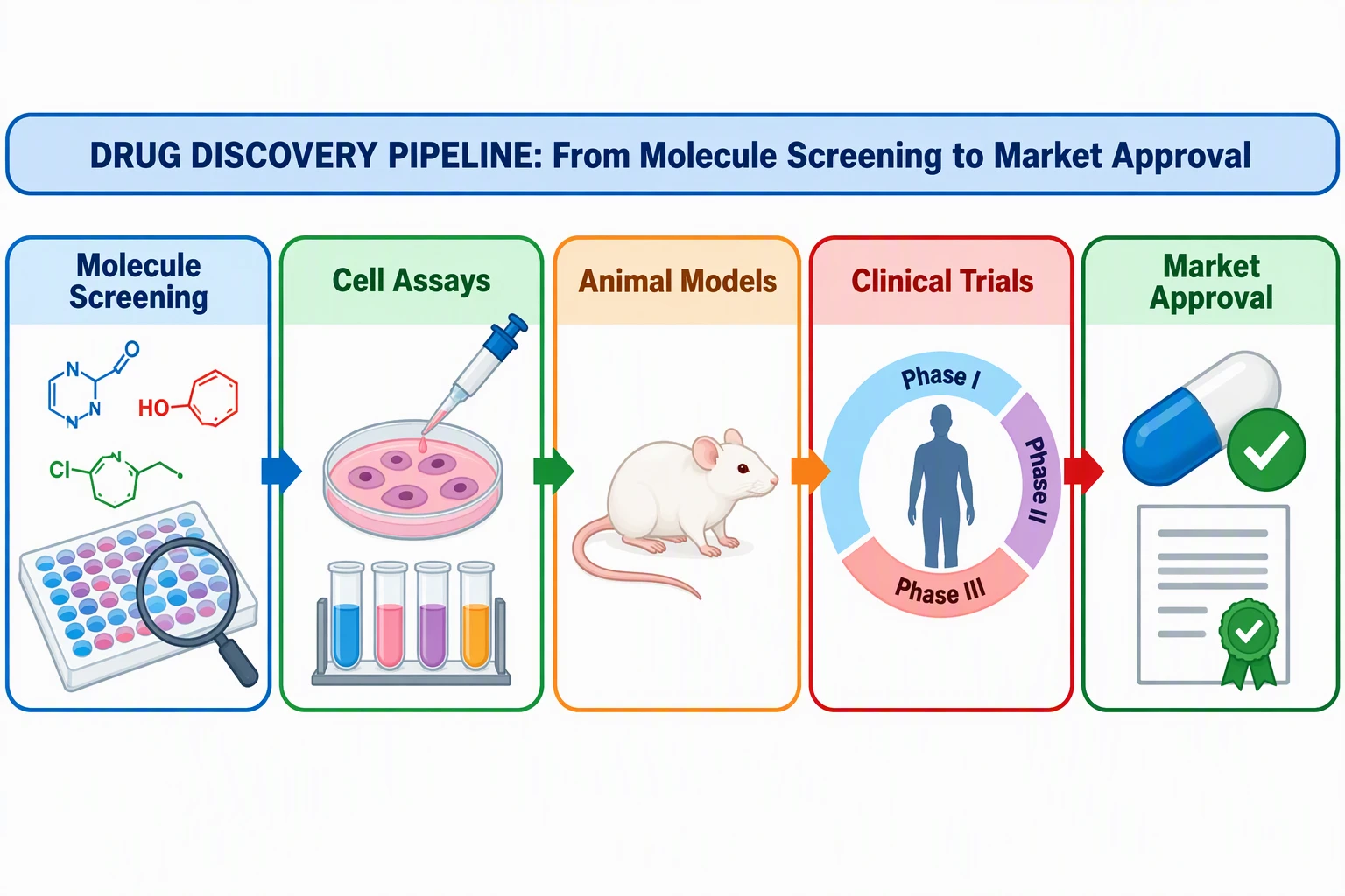
Graphical Abstract Maker
Draft a single-panel study arc when the TME map is one panel in a paper summary.
 Research
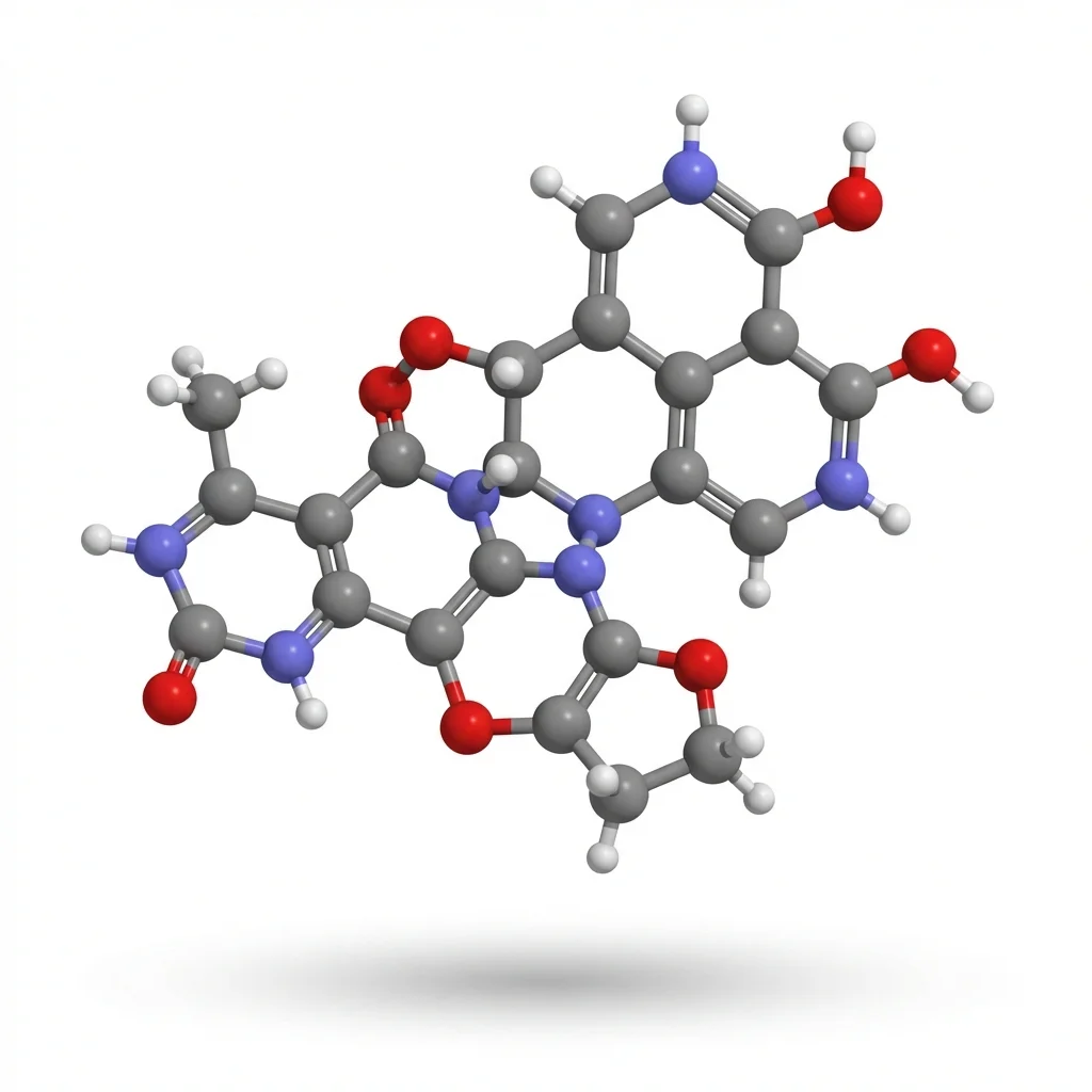
AI Scientific Image Generator
Custom scientific illustrations when the TME pack is not the right view.
 Medical
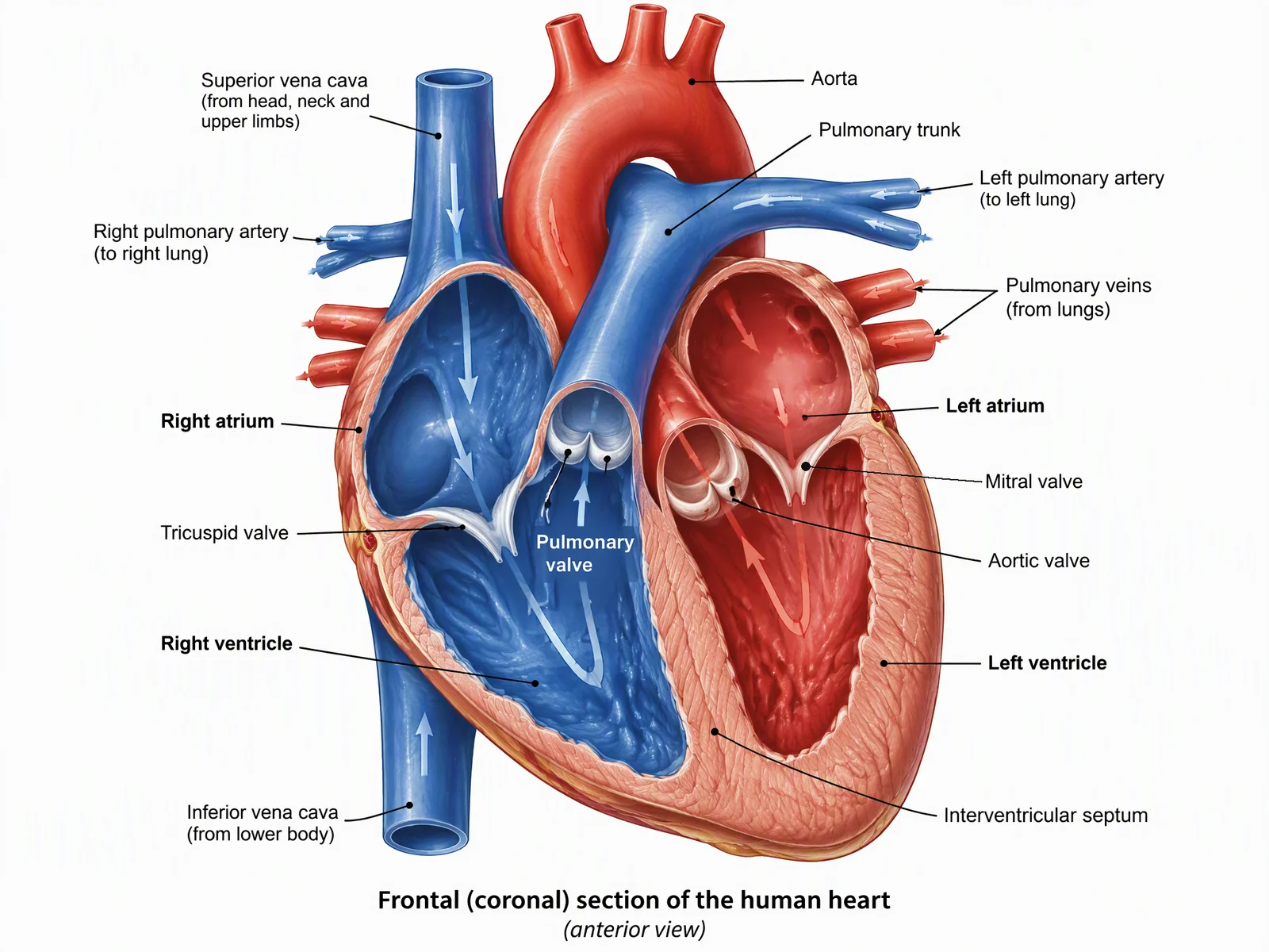
 Illustration Generator
Organ and tissue views that can sit next to a TME neighborhood map.
 Biology
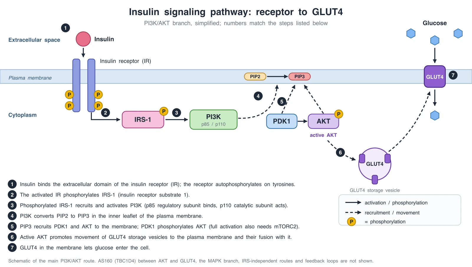
Signaling Pathway Diagram Generator
Intracellular pathways (for example TGF-β/SMAD) that CAFs and tumor cells use.
 Biology
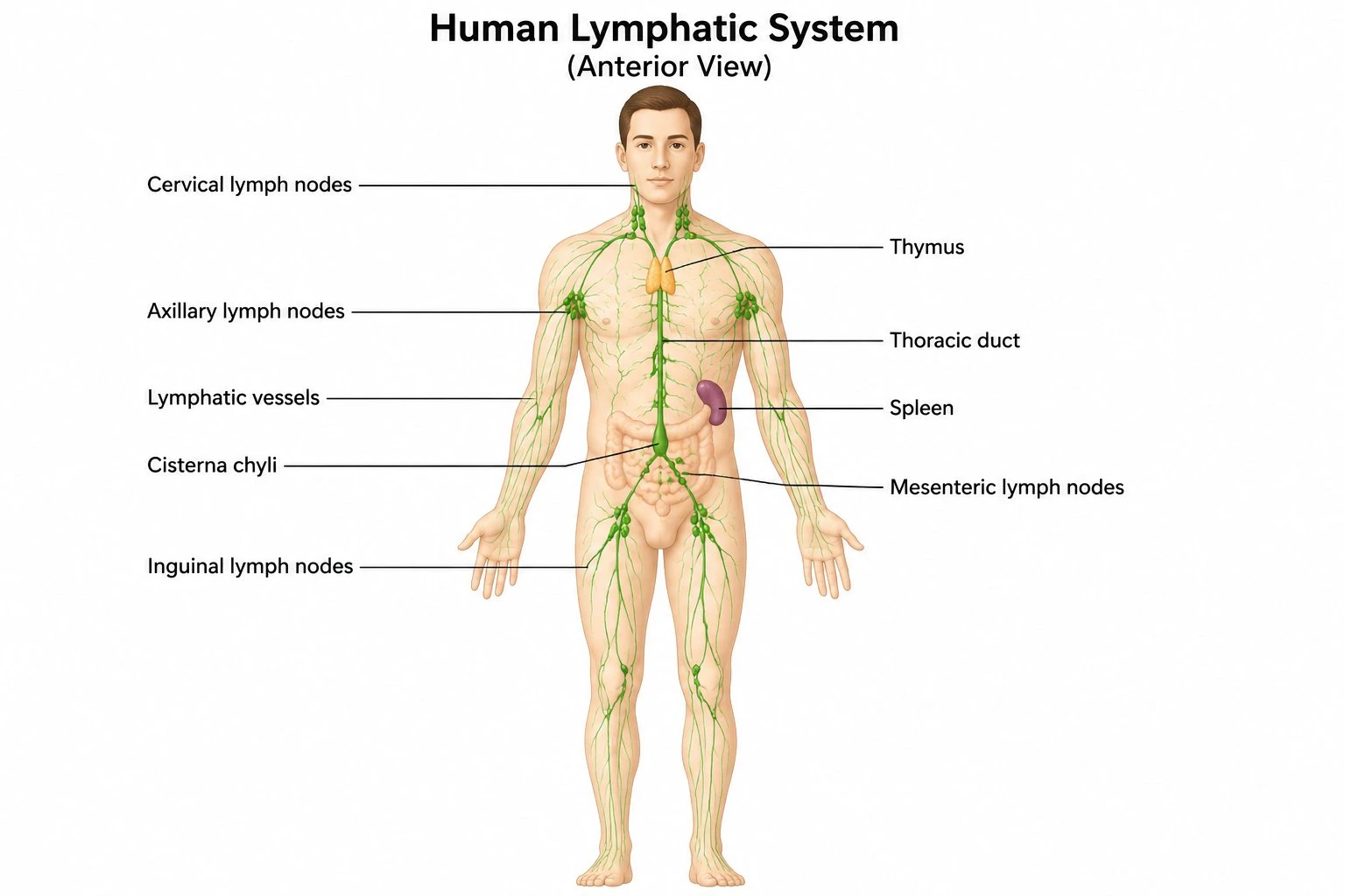
Lymphatic System Diagram Generator
Host lymphatic anatomy that pairs with tumor–lymphatic research notes on NCI’s TME pages.
 Biology
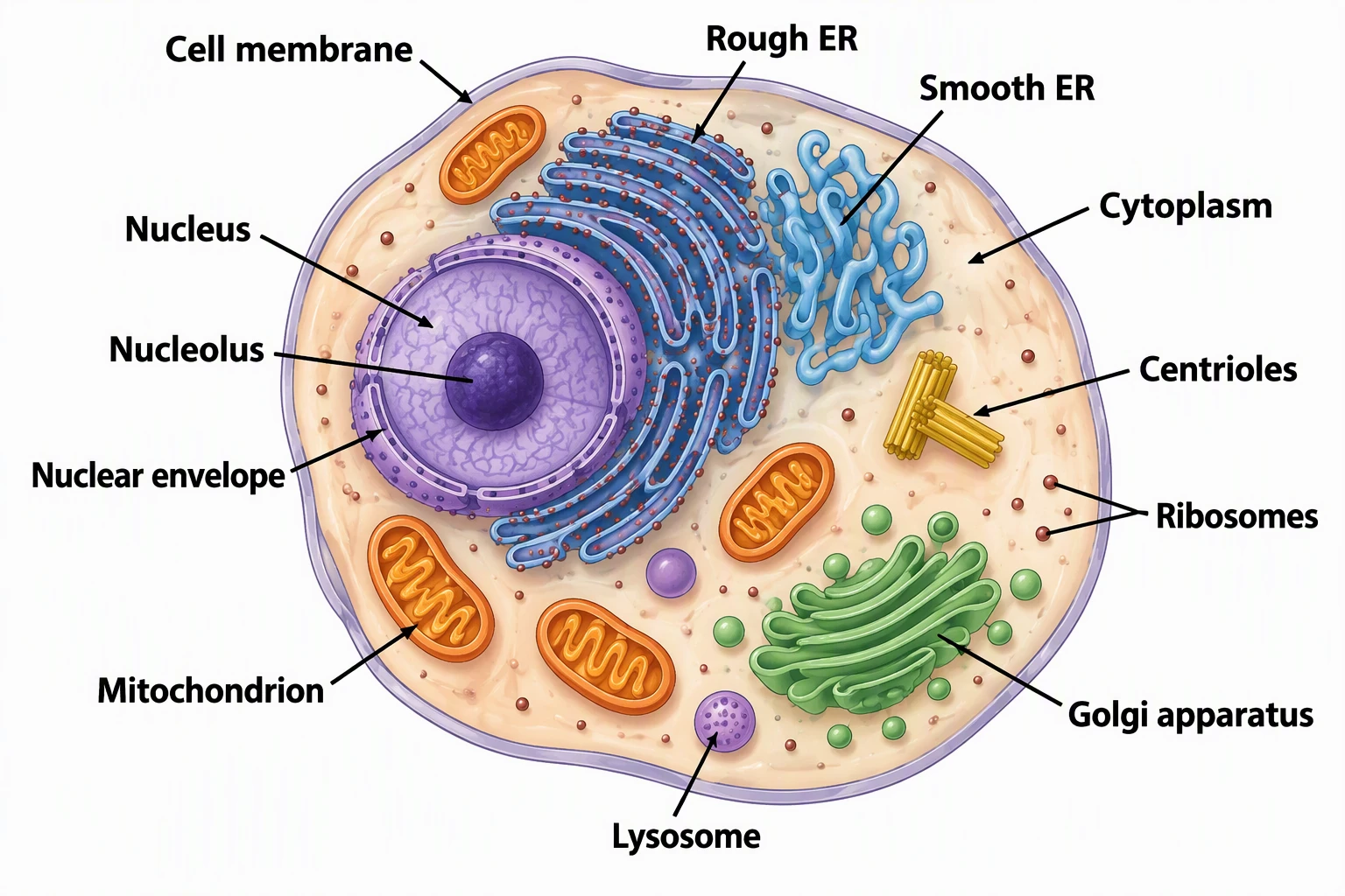
Animal Cell Diagram Generator
A single-cell labeled map when you need organelles, not a tumor neighborhood.
 Biology
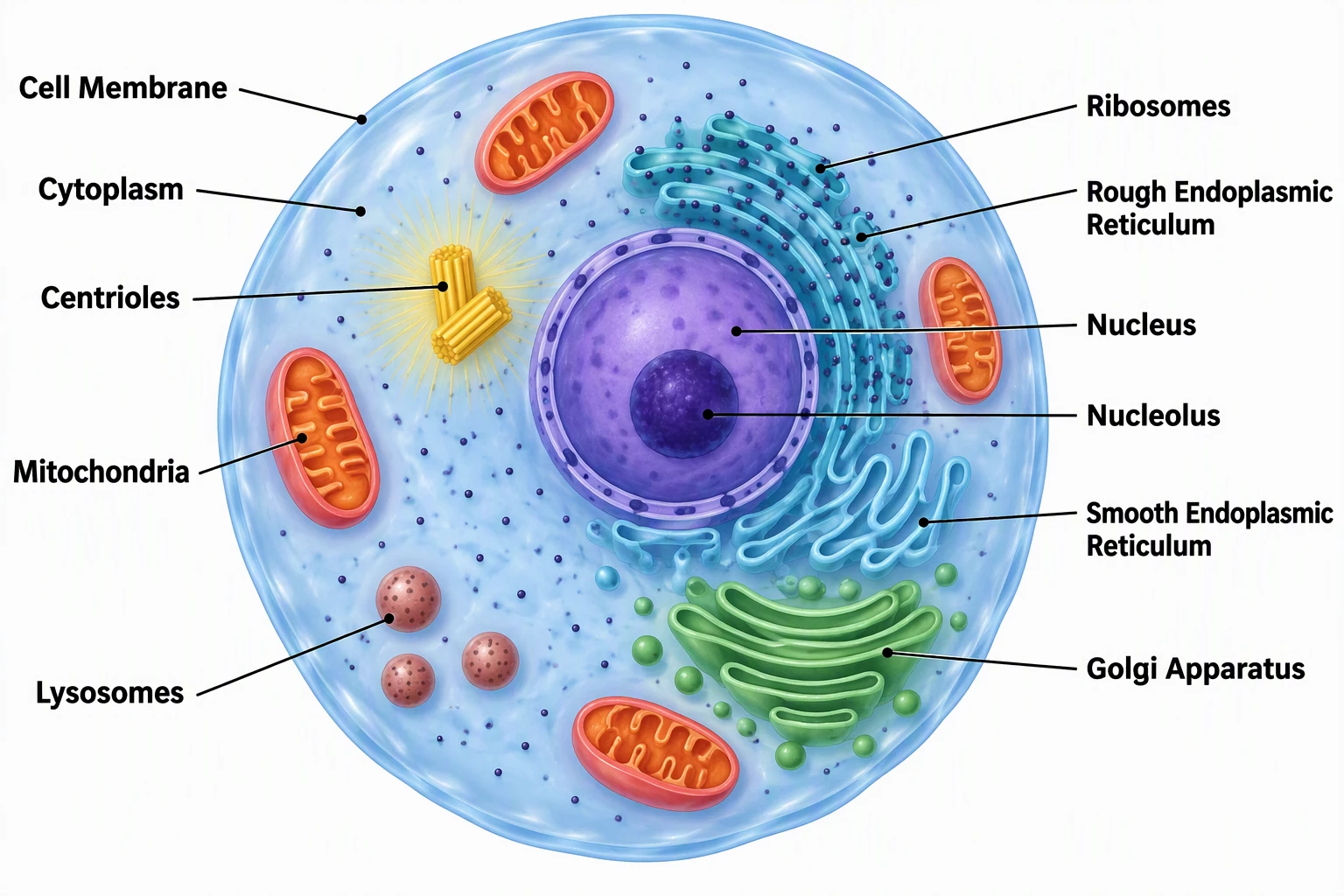
 Drawing Generator
Broader biology illustrations when the TME pack is too specific.