Lymphatic System Diagram Generator for Labeled & Blank Diagrams
Create a clearly labeled lymphatic system diagram in seconds. Show lymph nodes, lymphatic vessels, the spleen, thymus, tonsils, bone marrow, the thoracic duct, and cisterna chyli — or generate a blank, unlabeled version for worksheets and quizzes. Free to use.
Lymphatic System Diagram Generator
Free to try ·
Your lymphatic system diagram will appear here
Describe what you need and click Generate
Lymphatic System Diagram Examples
Labeled diagrams of lymph nodes, lymphatic vessels, lymphatic organs, and the lymph flow pathway
Full Lymphatic System
The complete map — major lymph node groups, green lymphatic vessels, thoracic duct, and key organs all labeled in anterior view.
Lymph Node Structure
Inside a lymph node — capsule, germinal centers, paracortex, medulla, and afferent versus efferent vessels labeled.
Simple Lymphatic System for Kids
A simplified version with large labels — perfect for elementary-level health and science lessons.
Lymphatic Organs
The four key lymphatic organs — thymus, spleen, tonsils, and bone marrow — with each organ's immune role called out.
How Lymph Flows
Step-by-step pathway from tissue fluid to lymph capillary, through lymph nodes, up the thoracic duct, and back into the bloodstream.
Blank Lymphatic System Worksheet
An unlabeled version with numbered blank lines — ready to print as a quiz or fill-in worksheet.
What is the lymphatic system?
The lymphatic system is a network of vessels, nodes, and organs that runs alongside the circulatory system and performs three main jobs: draining excess tissue fluid back into the bloodstream, absorbing dietary fats from the small intestine, and defending the body against infection. Unlike blood, lymph — the pale fluid inside the lymphatic vessels — moves in one direction only, driven by muscle contractions and one-way valves rather than a pump. Lymph nodes scattered along the vessels filter the fluid and trap pathogens before they can spread. This generator draws the complete lymphatic network with every structure labeled.
The primary lymphatic organs
- Bone marrow: the birthplace of all blood and immune cells, including the B cells and T cells that power adaptive immunity. Red bone marrow in flat bones such as the sternum and pelvis is the main production site.
- Thymus: a bilobed organ in the upper chest where immature T cells (thymocytes) migrate from the bone marrow and mature into fully functional T lymphocytes. The thymus is largest in childhood and gradually shrinks after puberty.
The secondary lymphatic organs
- Lymph nodes: small bean-shaped filters stationed along lymphatic vessels throughout the body. Immune cells inside the node detect and destroy pathogens, cancer cells, and debris carried in the lymph. Major clusters sit in the cervical (neck), axillary (armpit), and inguinal (groin) regions.
- Spleen: the largest lymphatic organ, located in the upper-left abdomen. Its red pulp filters old red blood cells from the blood; its white pulp is a hub of immune activity where lymphocytes encounter blood-borne antigens.
- Tonsils: rings of lymphatic tissue at the back of the throat and nasal passage (palatine, pharyngeal, and lingual tonsils) that intercept pathogens entering through the mouth and nose.
- Mucosa-associated lymphoid tissue (MALT): lymphatic tissue embedded in the lining of the gut, lungs, and other mucosal surfaces, providing local immune surveillance at the body's entry points.
How lymph flows: vessels, ducts, and valves
Lymph begins as interstitial fluid — the watery fluid that leaks out of blood capillaries and bathes every cell. About three litres of this fluid accumulate in the tissues each day and must be returned to the blood. Blind-ended lymph capillaries absorb the fluid, and one-way valves inside the larger lymphatic vessels prevent backflow. The vessels converge into two main ducts: the thoracic duct, which drains the lower body and left side of the upper body and empties into the left subclavian vein; and the right lymphatic duct, which drains the right side of the head, neck, and arm and empties into the right subclavian vein. Muscle movement, breathing, and arterial pulsation push lymph along.
The lymphatic system and immunity
When pathogens enter the body, antigen-presenting cells (such as dendritic cells and macrophages) capture them, break them down, and carry fragments via lymphatic vessels to the nearest lymph node. Inside the node, B cells and T cells recognise the antigen. B cells produce antibodies; cytotoxic T cells hunt down and destroy infected cells. Memory cells created during this response persist for years, explaining why a second exposure to the same pathogen produces a faster, stronger immune reaction — the basis of vaccination. Because the lymphatic system links virtually every tissue to the immune network, it is also the main route by which cancer cells spread (metastasis), which is why clinicians examine lymph nodes when staging cancer.
Labeled vs blank diagrams for worksheets and quizzes
A fully labeled diagram is ideal for teaching and study notes, while a blank, unlabeled version is what you need for worksheets, handouts, and quizzes. With this tool you can generate either: describe "a labeled lymphatic system diagram" to get every structure named, or ask for "a blank lymphatic system diagram with numbered blank lines" to get a fill-in version. Black-and-white line art works best for printing, and a numbered blank diagram doubles as both the quiz and — with labels added back — the answer key.
How to generate a labeled lymphatic system diagram
- Describe what you need in plain English — the full lymphatic system, just the lymph nodes and vessels, a close-up of lymph node structure, the lymphatic organs, or the flow pathway.
- Specify the view and labels you want, for example "anterior full-body view, lymphatic vessels in green, label the cervical, axillary, and inguinal node groups, thymus, spleen, and thoracic duct."
- Choose labeled for teaching or unlabeled (blank) for a worksheet or quiz, then generate the diagram.
- Review the result for accuracy, regenerate or refine the prompt if needed, and download the image to use in a slide, handout, or study guide.
Frequently Asked Questions
Related Biology Tools
 Biology
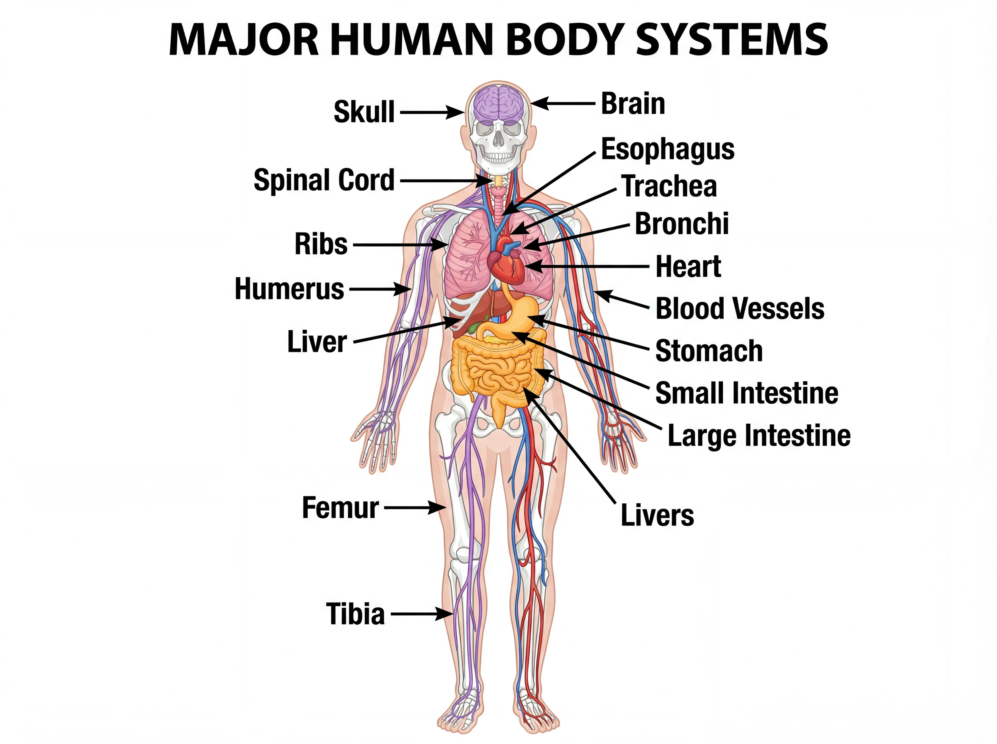
Human Body Systems Diagram Generator
Draw labeled diagrams of the body's organ systems, from skeletal to circulatory.
 Biology
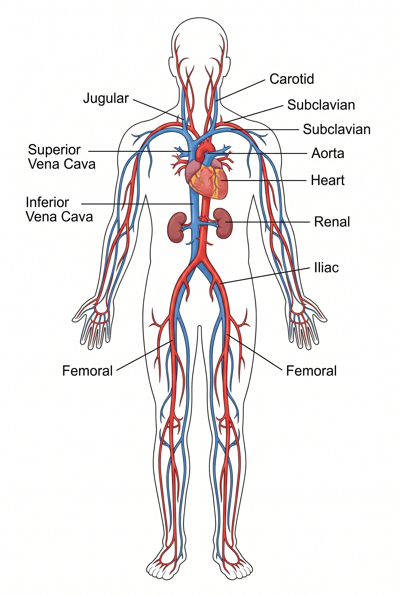
Circulatory System Diagram Generator
Create labeled diagrams of the heart, arteries, veins, and pulmonary and systemic circulation.
 Biology
Nervous System Diagram Generator
Generate labeled diagrams of the central and peripheral nervous systems, neurons, and reflex arcs.