cGAS-STING Pathway Diagram Generator dsDNA to interferon
Generate cGAS-STING pathway diagrams from text: cytosolic dsDNA, cGAMP, STING trafficking, TBK1, IRF3, NF-κB and type I interferon. Draft figures for papers.
Create Your cGAS-STING Pathway Diagram
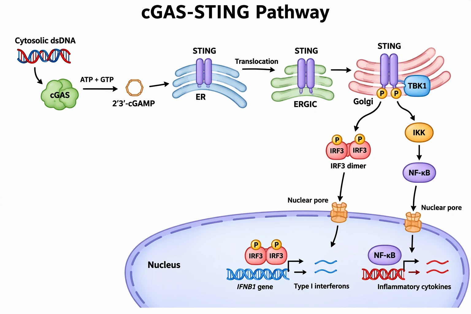 Example: Full pathwayView full size
Free to try ·
cGAS-STING Pathway Diagram Examples
Four AI drafts of the pathway, trafficking, drug targets, and DNA sources, with known flaws noted. AI diagrams need expert review.
cGAS-STING pathway, dsDNA to type I interferon
AI-generated. The order follows the review literature: cytosolic dsDNA, cGAS, 2′3′-cGAMP, STING at the ER, movement through the ERGIC to the Golgi, TBK1 and phosphorylation at the Golgi, the phosphorylated IRF3 dimer entering the nucleus through its own nuclear pore to drive type I interferon genes, and a separate NF-κB branch through a second pore to inflammatory cytokine genes. STING stays in the membrane throughout. Simplified: STING is drawn as a single dimer at each stop, and IKK is shown as one step between STING and NF-κB.
STING trafficking from the ER to the Golgi
AI-generated. Shows the route STING takes from the ER through COPII vesicles and the ERGIC to the Golgi, where an oligomer recruits TBK1 and IRF3, and its clearance in the lysosome. Simplified: STING is not drawn inside the transport vesicles or the ERGIC, the vesicles are plain circles, the oligomer is shown as four copies, and the TBK1 and IRF3 binding sites on the STING tail are not drawn in detail.
Where drugs act on the cGAS-STING pathway
AI-generated. Marks the three common intervention points: cGAS inhibitors, STING agonists (cGAMP mimetics) for cancer immunotherapy and vaccine adjuvants, and STING inhibitors for inflammatory disease. Simplified: the molecule icons are decorative and are not real structures, and the drug classes are not named.
What activates cGAS: sources of cytosolic DNA
AI-generated. Viral or bacterial DNA, mitochondrial DNA leakage, and micronuclei from DNA damage all feed cGAS, while the nuclease TREX1 degrades cytosolic DNA. Simplified: STING is drawn on a Golgi-like shape without the ER-to-Golgi step, and there is no scale between organelles.
This page is a focused companion to the signaling pathway diagram generator. To show where a drug acts on this pathway, use the mechanism of action diagram generator.
What a cGAS-STING pathway diagram has to show
The cGAS-STING pathway senses DNA in the cytosol and turns it into an interferon response. A correct figure shows the sensor (cGAS), the second messenger (2′3′-cGAMP), the adaptor (STING) and where it sits, the kinase (TBK1), the transcription factors (IRF3 and NF-κB), and the output. The compartment matters: STING starts in the ER membrane and signals after it moves through the ERGIC to the Golgi.
The reference order of the pathway
- Cytosolic double-stranded DNA binds cGAS, which activates its enzyme and uses ATP and GTP to make 2′3′-cGAMP.
- 2′3′-cGAMP binds STING, an ER membrane protein, which changes shape and oligomerizes.
- STING leaves the ER in COPII vesicles and moves through the ERGIC to the Golgi.
- At the Golgi the STING C-terminal tail recruits TBK1. TBK1 phosphorylates STING (Ser366 in humans), creating a motif that recruits IRF3, and TBK1 then phosphorylates IRF3.
- Phosphorylated IRF3 dimerizes, enters the nucleus, and works with NF-κB to drive type I interferon and inflammatory cytokine genes.
- STING also activates NF-κB; reviews differ on how much this needs TBK1 (IKKε can compensate) and the C-terminal tail. The signal ends when STING is degraded in lysosomes.
Where this pathway is used in research
- Innate immunity and infection: sensing of viral and bacterial DNA.
- Tumor immunity: STING agonists are being developed as cancer immunotherapy and vaccine adjuvants.
- Autoinflammatory disease: gain-of-function STING mutations (SAVI) and Aicardi-Goutières syndrome link the pathway to disease, and cGAS and STING inhibitors are being studied.
- Sources of cytosolic DNA: mitochondrial DNA leakage, micronuclei after DNA damage, and failure of nucleases such as TREX1.
How to prompt for an accurate pathway figure
- Give the chain in order: dsDNA, cGAS, 2′3′-cGAMP, STING, TBK1, IRF3, type I interferon.
- Name the compartments: cytosol, ER, ERGIC, Golgi, nucleus, and say that STING moves between them.
- State the step type: binds, synthesizes, translocates, phosphorylates, dimerizes.
- Ask for the NF-κB branch separately if you want it, and say whether it is drawn as dependent on TBK1.
- Check every label and arrow against a primary review before use. The reference sources below are a starting point.
Frequently Asked Questions
Reference sources (checked November 2026)
Related Biology Tools
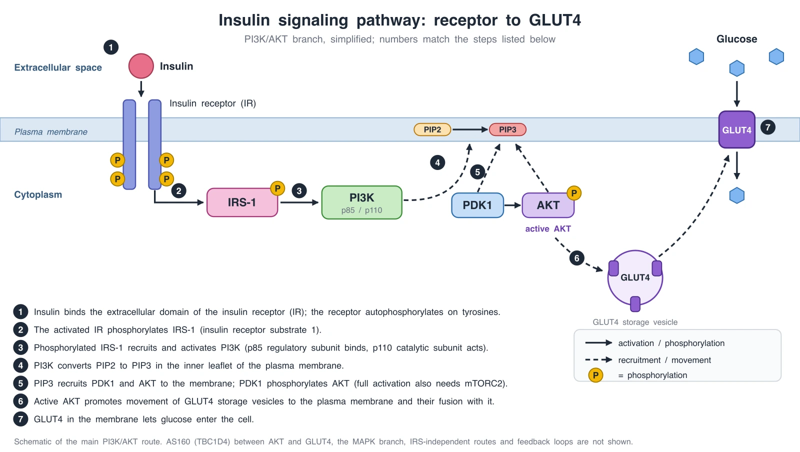
Signaling Pathway Diagram Generator
The general pathway hub: insulin, TGF-β/SMAD, and MAPK/ERK with membrane and nucleus layers.
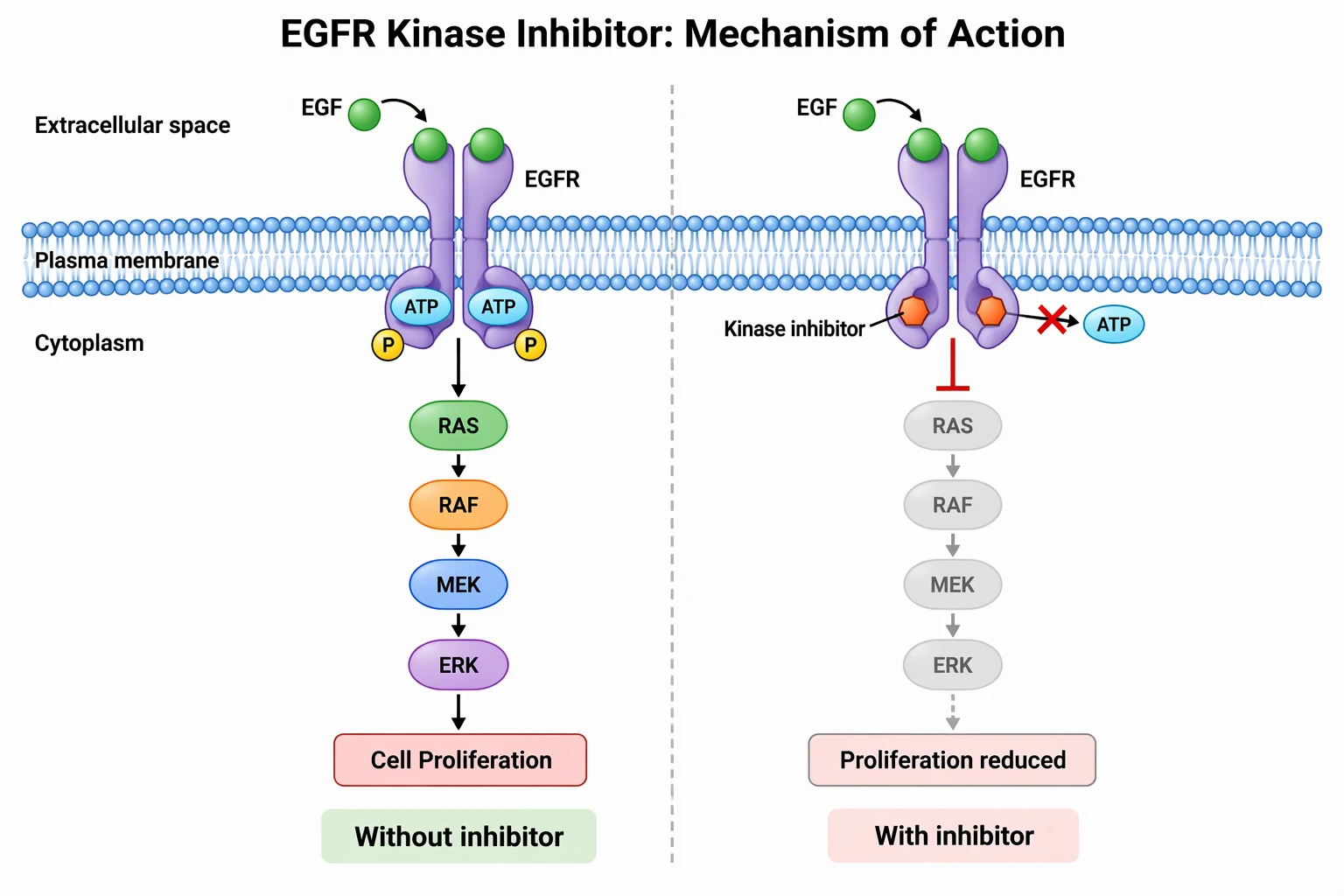
Mechanism of Action Diagram Generator
Show where a drug engages its target: inhibitors, antibodies, ADCs, and degraders.
AI Scientific Image Generator
Describe any lab or biology figure and generate a clean illustration.