Tooth Diagram Labeled, Blank & Printable
Free tooth diagrams — labeled anatomy, blank worksheets, simple kids versions, tooth types, and coloring. Download or print; no sign-up.
Free Tooth Diagrams
Every version below is free to view and download — no sign-up.
Labeled Tooth Anatomy
Full cutaway — enamel, dentin, pulp, cementum, root canal, gum, and bone labeled.
Core diagrams
Structure & focus
Print & practice
Test yourself: tooth anatomy
A quick self-test — questions reshuffle each time, with a review of anything you miss at the end.
What is the hardest substance covering the crown of a tooth?
Need a custom version?
Change the grade level, which parts are labeled, or focus on a molar — describe it and generate your own.
Customize Your Tooth Diagram
Free to try ·
Your custom tooth diagram will appear here
Ask for a labeled cutaway, blank worksheet, or tooth types chart
What is a tooth diagram?
A tooth diagram is a labeled cutaway of a tooth showing the hard outer layers and the soft living tissue inside. Most school diagrams show a permanent tooth in longitudinal section: enamel and dentin in the crown, pulp in the center, cementum on the root, and the root canal leading into the jaw. Use a labeled version to learn the names, or a blank version as a worksheet.
Main parts of a tooth
- Enamel: the hard outer layer of the crown — the hardest substance in the body.
- Dentin: the hard tissue under the enamel that forms most of the tooth’s bulk.
- Pulp: soft tissue with blood vessels and nerves in the pulp chamber and root canals.
- Cementum: covers the root and helps anchor the tooth via the periodontal ligament.
- Crown and root: crown is above the gum; root is embedded in the alveolar bone.
Types of teeth
Humans have incisors (cutting), canines (tearing), premolars, and molars (grinding). A tooth-types chart helps students match shape to function before zooming into a single cross-section. Wisdom teeth are third molars that appear later in some people.
How to use these free diagrams
Introduce anatomy with the fully labeled tooth, then check understanding with the blank worksheet. The simple kids version supports elementary health units; the types chart and molar focus support middle school. The coloring page is a low-prep center activity. All assets are free to view, download, and print with no sign-up.
Frequently Asked Questions
Related Biology Tools
 Medical
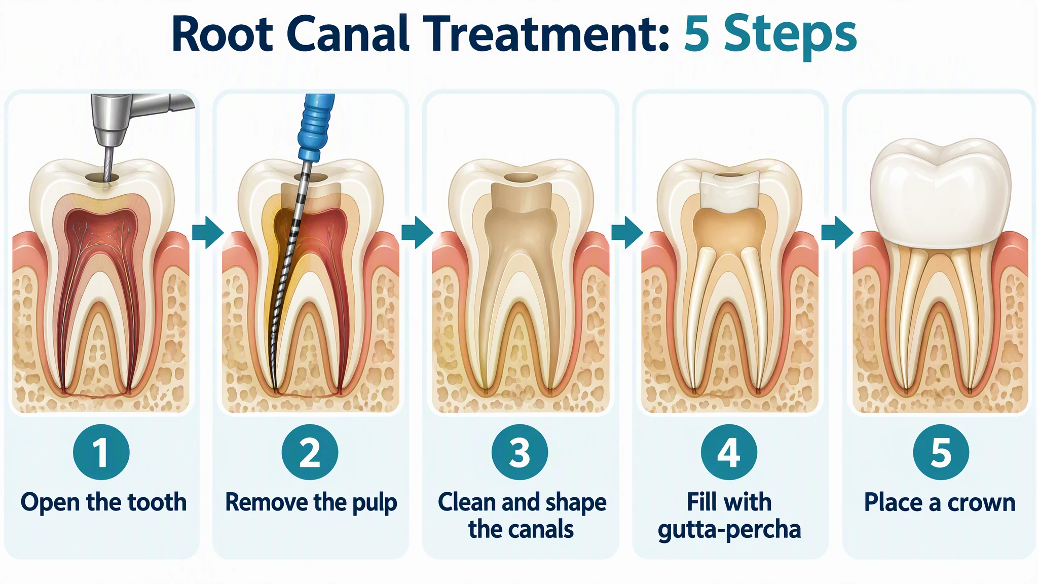
Root Canal Procedure Diagram Generator
Root canal step diagrams, infected tooth cutaways, and canals by tooth type for patient handouts.
 Biology
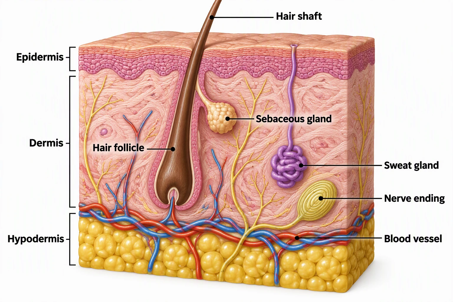
Skin Diagram
Free skin layers diagrams — epidermis, dermis, and hypodermis labeled.
 Medical
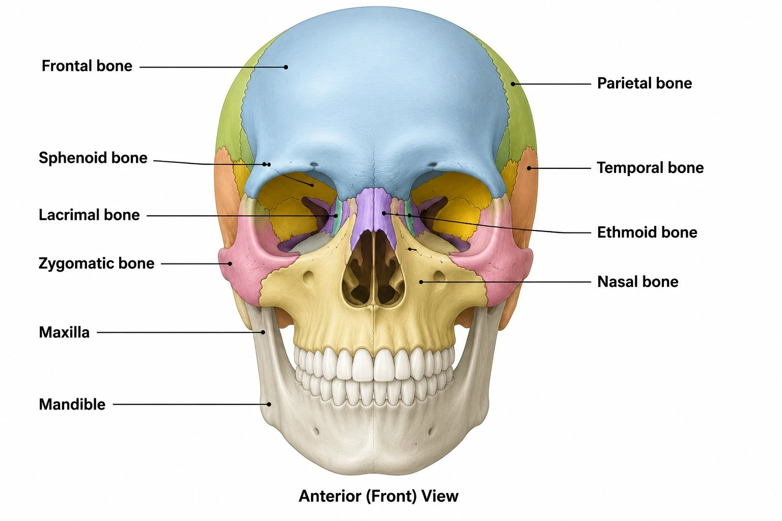
Skull Diagram
Labeled skull views for head and jaw anatomy units.
 Biology
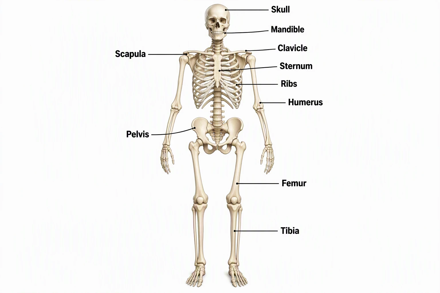
Skeletal System Diagram
Full skeleton maps including the skull and jaw.
 Biology
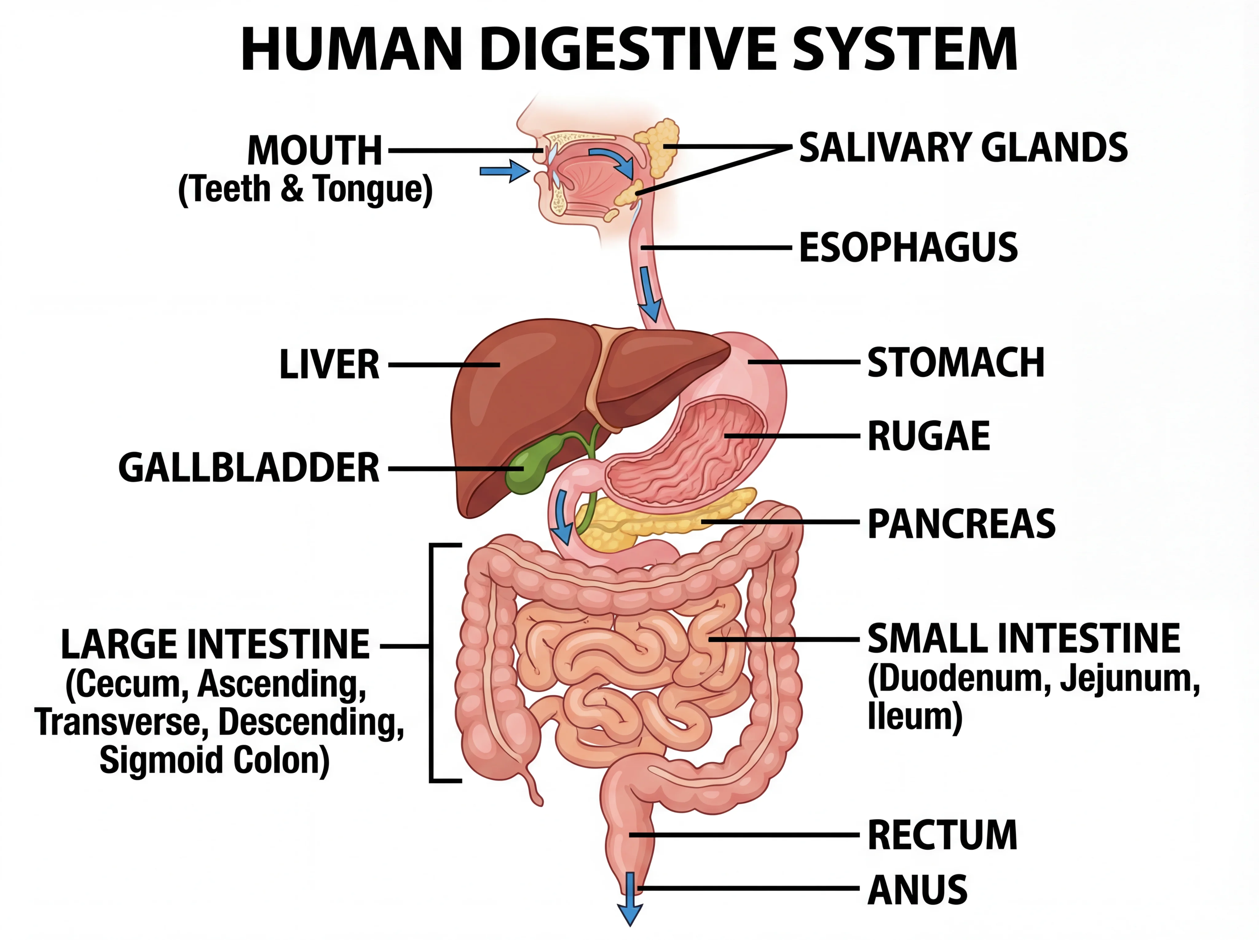
Digestive System Diagram
Mouth-to-gut maps that pair with dental health lessons.
 Biology
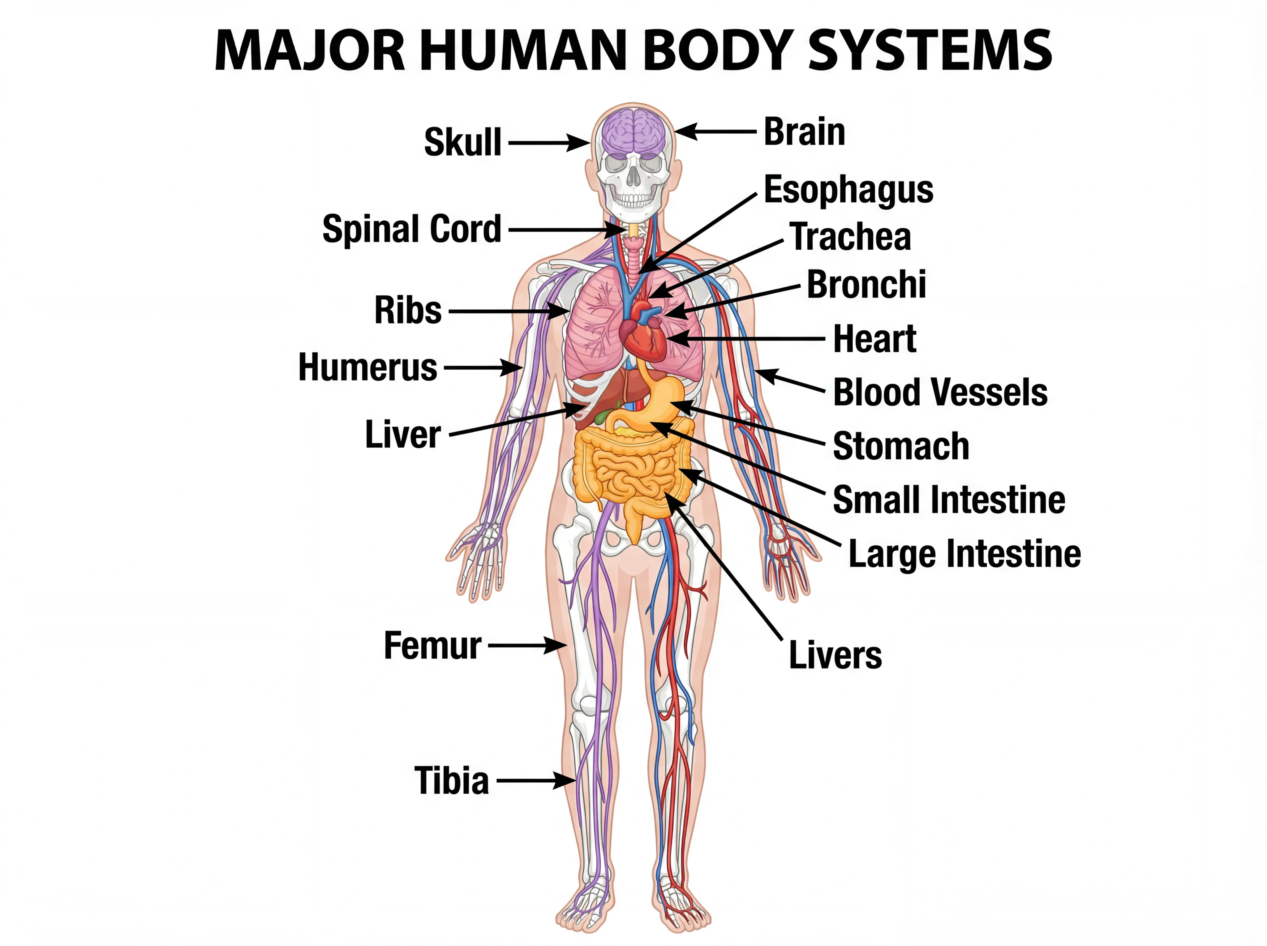
Human Body Systems
Overview diagrams of major body systems.
 Biology
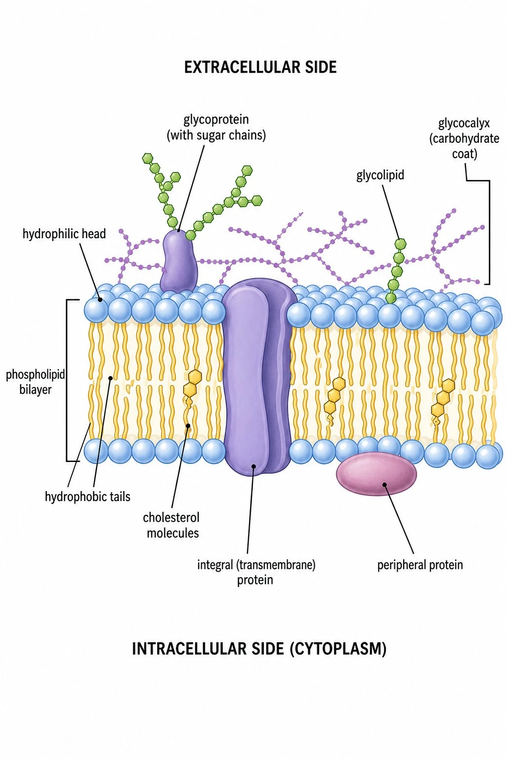
Cell Membrane Diagram
Fluid mosaic model for cell surface structure units.