Skull Diagram Generator Labeled Human Skull Diagrams
Create labeled skull diagrams with AI. Show the cranial bones (frontal, parietal, temporal, occipital, sphenoid, ethmoid), facial bones (mandible, maxilla, zygomatic, nasal, lacrimal), sutures (coronal, sagittal, lambdoid), foramen magnum, and orbits — anterior and lateral views — for anatomy class. Download as PNG.
AI Skull Diagram Generator
Free to try ·
Your skull diagram will appear here
Describe the view and bones to label
Skull Diagram Examples
Labeled diagrams of cranial and facial bones, skull sutures, and printable worksheets
Labeled Skull — Anterior View
Every cranial and facial bone named in a clean front-facing diagram — the standard view for anatomy study.
Labeled Skull — Lateral View
The skull from the side — all visible bones and the major sutures labeled for anatomy class.
Cranial vs Facial Bones
Cranial bones in blue, facial bones in green — an instant visual for learning the two groups.
Skull Sutures Diagram
The four major skull sutures labeled — coronal, sagittal, lambdoid, and squamous — in superior and lateral views.
Simple Skull for Kids
A friendly, simplified skull diagram with the five main bones labeled — great for elementary or middle-school science.
Blank Skull Worksheet
Numbered blank lines, no labels — print it and fill in as a quiz or study activity.
What does a skull diagram show?
A labeled skull diagram shows the individual bones of the human skull and how they fit together. The skull is divided into two regions: the neurocranium (braincase), which encloses and protects the brain, and the viscerocranium (facial skeleton), which forms the face. Key features shown in anterior and lateral views include the bones, the suture lines where bones meet, the orbits (eye sockets), the nasal cavity, the foramen magnum at the base, and the mandible (lower jaw). This generator creates clear, labeled skull diagrams for biology, anatomy, and health classes.
The eight cranial bones
- Frontal bone: forms the forehead and the roof of the eye sockets.
- Parietal bones (×2): form the top and sides of the braincase; meet at the sagittal suture.
- Temporal bones (×2): form the lower sides and base of the skull and house the structures of the middle and inner ear.
- Occipital bone: forms the back and base of the skull and contains the foramen magnum, the large opening through which the brainstem passes.
- Sphenoid bone: a butterfly-shaped bone at the base of the skull that articulates with all other cranial bones.
- Ethmoid bone: a small bone between the eyes that forms part of the nasal cavity roof and the medial wall of the orbits.
Key facial bones
- Mandible: the lower jaw — the only freely movable bone of the skull.
- Maxillae (×2): form the upper jaw, the floor of the orbits, and the front of the hard palate.
- Zygomatic bones (×2): the cheekbones, which also form part of the lateral orbit wall.
- Nasal bones (×2): form the bridge of the nose.
- Lacrimal bones (×2): the smallest facial bones; form part of the medial orbit wall near the tear ducts.
- Other facial bones include the palatine bones, inferior nasal conchae, and vomer.
The major skull sutures
Sutures are immovable fibrous joints where cranial bones meet. The four main sutures are: the coronal suture (frontal bone meets the two parietals), the sagittal suture (runs along the top of the skull between the two parietal bones), the lambdoid suture (parietals meet the occipital bone at the back), and the squamous suture (each temporal bone meets the adjacent parietal bone on the side). In infants the sutures are separated by soft spots called fontanelles, which allow the skull to flex during birth and close by around age two.
Key skull landmarks
Beyond individual bones, labeled skull diagrams often highlight important anatomical landmarks. The foramen magnum is the large hole at the skull base through which the spinal cord connects to the brainstem. The orbits are the bony cavities that house and protect the eyeballs. The mental foramen is a small hole in the mandible that lets the mental nerve and blood vessels reach the chin. The temporal fossa and zygomatic arch are important landmarks for the muscles of chewing (mastication). The mastoid process behind the ear and the styloid process are projection sites for muscles and ligaments.
Anterior view vs lateral view
The anterior (front) view is best for seeing the facial bones — the orbits, nasal cavity, zygomatic bones, maxillae, and mandible — and for counting and locating the bones by position. The lateral (side) view reveals the overall shape of the braincase, shows the temporal and occipital bones that are hidden in the front view, and makes the suture lines (coronal, squamous, lambdoid) and the zygomatic arch easy to see. Asking for both views side by side gives the most complete picture for a study guide or presentation.
Tips for generating a clear skull diagram
Specify the view (anterior, lateral, superior, or both), the bones you want labeled, and the level of detail. For a class worksheet, add "white background, clean label lines, educational anatomy style." For a comparison, ask for color-coding (e.g., cranial bones in blue, facial bones in green). For a blank quiz sheet, ask for "numbered blank lines, no labels on the bones, black-and-white line art suitable for printing."
Frequently Asked Questions
Related Biology Tools
 Biology
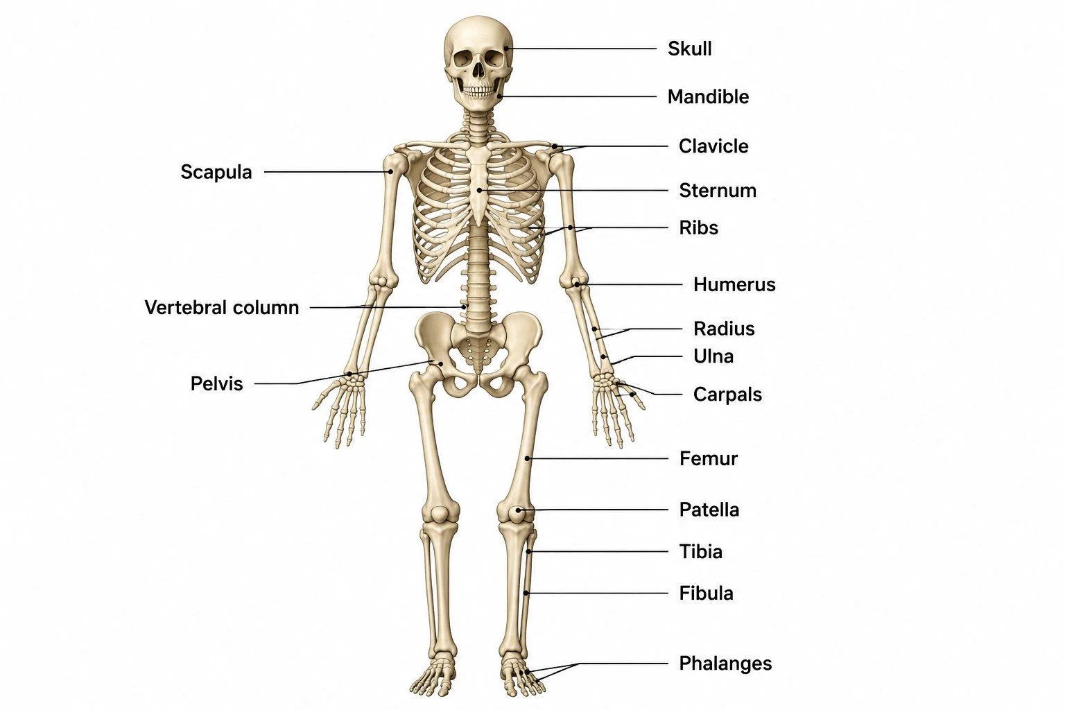
Skeletal System Diagram Generator
Create labeled diagrams of the full human skeleton — all 206 bones, axial vs appendicular divisions, and joint types.
 Biology
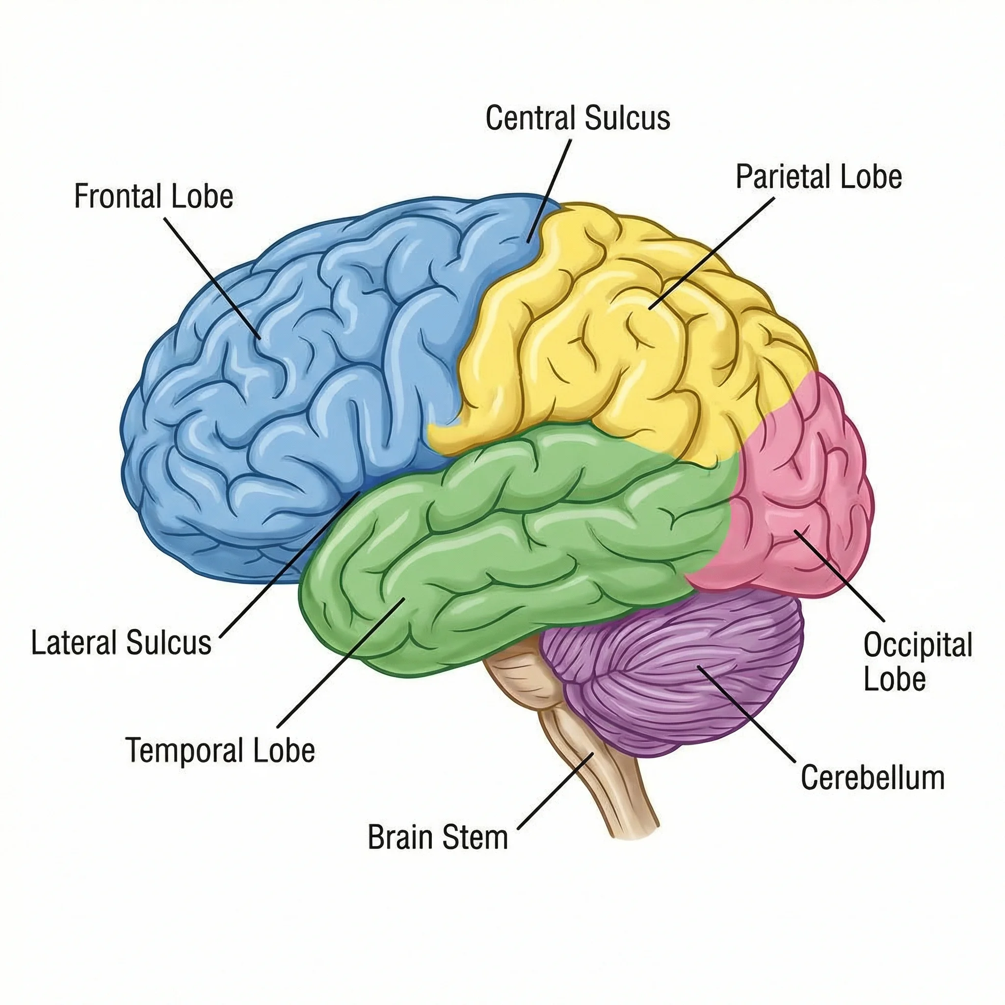
Brain Diagram Generator
Create labeled brain diagrams showing lobes, regions, and structures of the human brain.
 Biology
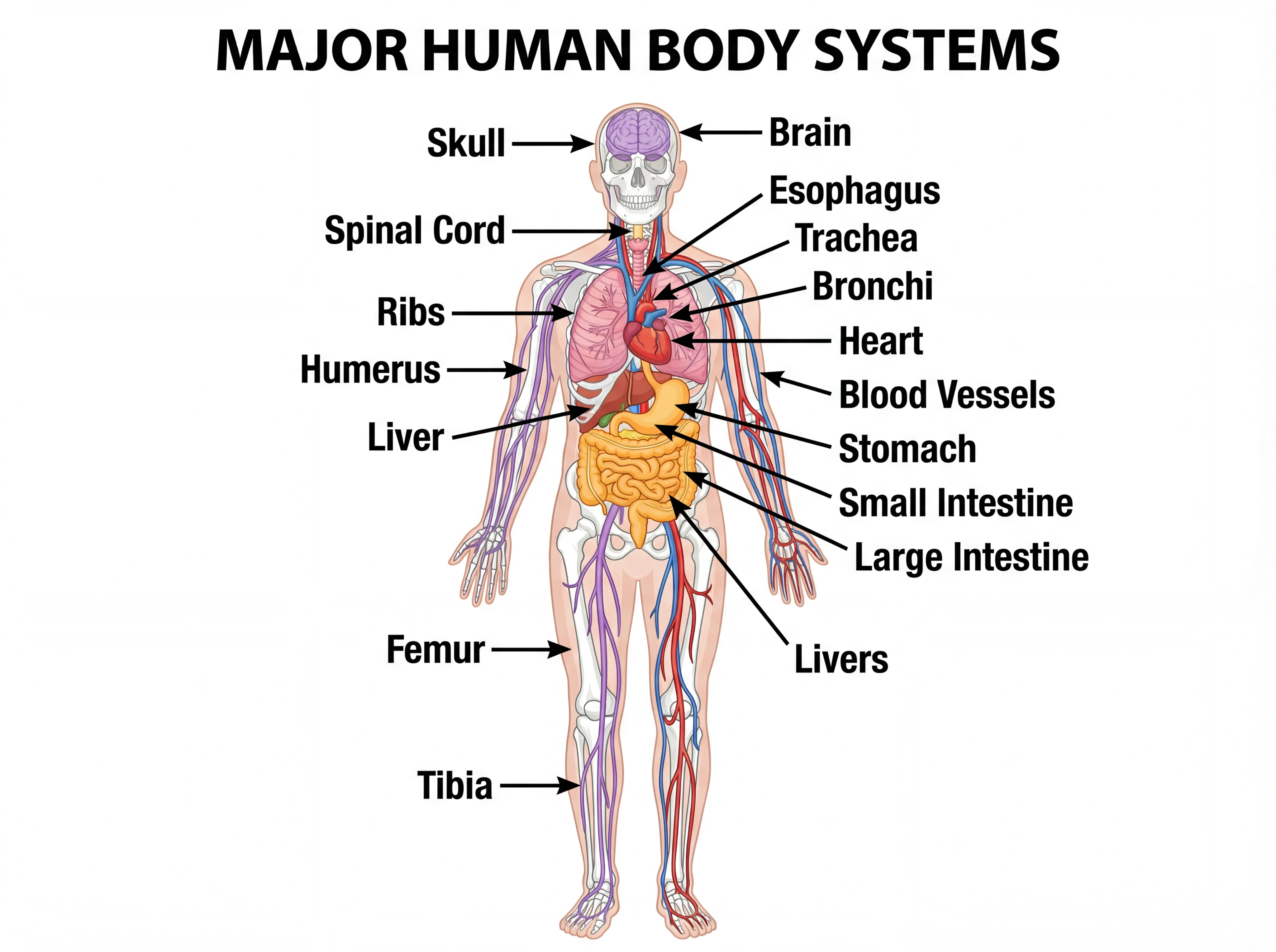
Human Body Systems Diagram Generator
Draw labeled diagrams of all the body's organ systems — skeletal, muscular, nervous, circulatory, and more.