Cell Membrane Diagram Labeled, Blank & Printable
Free cell membrane (plasma membrane) diagrams — fluid mosaic labeled, blank, simple, transport, and coloring. Download or print; no sign-up.
Free Cell Membrane Diagrams
Every version below is free to view and download — no sign-up. Covers cell membrane and plasma membrane study needs.
Labeled Fluid Mosaic Model
Full fluid mosaic view — bilayer, proteins, cholesterol, and sugar chains labeled for study and slides.
Core diagrams
Structure & focus
Print & practice
Test yourself: cell membrane
A quick self-test — questions reshuffle each time, with a review of anything you miss at the end.
In a phospholipid, which part faces water on the outside of the bilayer?
Need a custom version?
Change the grade level, which parts are labeled, or the transport focus — describe it and generate your own.
Customize Your Cell Membrane Diagram
Free to try ·
Your custom cell membrane diagram will appear here
Ask for a labeled fluid mosaic, blank worksheet, or transport focus
What is a cell membrane diagram?
A cell membrane diagram (also called a plasma membrane diagram) shows the thin boundary that surrounds every living cell. Because the real membrane is only a few nanometers thick and constantly moving, diagrams use a cutaway fluid mosaic view: a phospholipid bilayer with proteins, cholesterol, and sugar chains drawn large enough to label. Use a fully labeled version to learn the parts, or a blank version as a worksheet or quiz.
Parts of the fluid mosaic model
- Phospholipid bilayer: two layers of phospholipids with hydrophilic heads facing water and hydrophobic tails facing each other.
- Integral (transmembrane) proteins: embedded proteins that may form channels, carriers, or receptors across the membrane.
- Peripheral proteins: loosely attached to one face of the membrane, often for signaling or structure.
- Cholesterol: steroid lipids that help buffer membrane fluidity in animal cells.
- Glycoproteins and glycolipids: lipids or proteins with sugar chains on the extracellular side, forming the glycocalyx for cell recognition.
Cell membrane vs plasma membrane
In most biology courses the terms cell membrane and plasma membrane mean the same structure — the outer bilayer of the cell. “Plasma membrane” emphasizes that it bounds the cytoplasm (protoplasm). Plant cells still have a plasma membrane; the rigid cell wall sits outside it and is a separate structure.
Transport across the membrane
- Simple diffusion: small nonpolar molecules move down their concentration gradient through the bilayer.
- Facilitated diffusion: channel or carrier proteins help polar or charged substances move down their gradient without ATP.
- Active transport: pumps move substances against their gradient using energy (often ATP).
- Osmosis: water moves across the membrane toward higher solute concentration (through the bilayer or aquaporins).
How to use these free diagrams
Post the labeled fluid mosaic diagram while introducing vocabulary, then hand out the blank version of the same layout for students to label from memory. The simple version works for upper elementary or as a warm-up; the transport overview links structure to function. The coloring page is a low-prep station activity. All files are free to view, download, and print with no account required.
Frequently Asked Questions
Related Biology Tools
 Biology
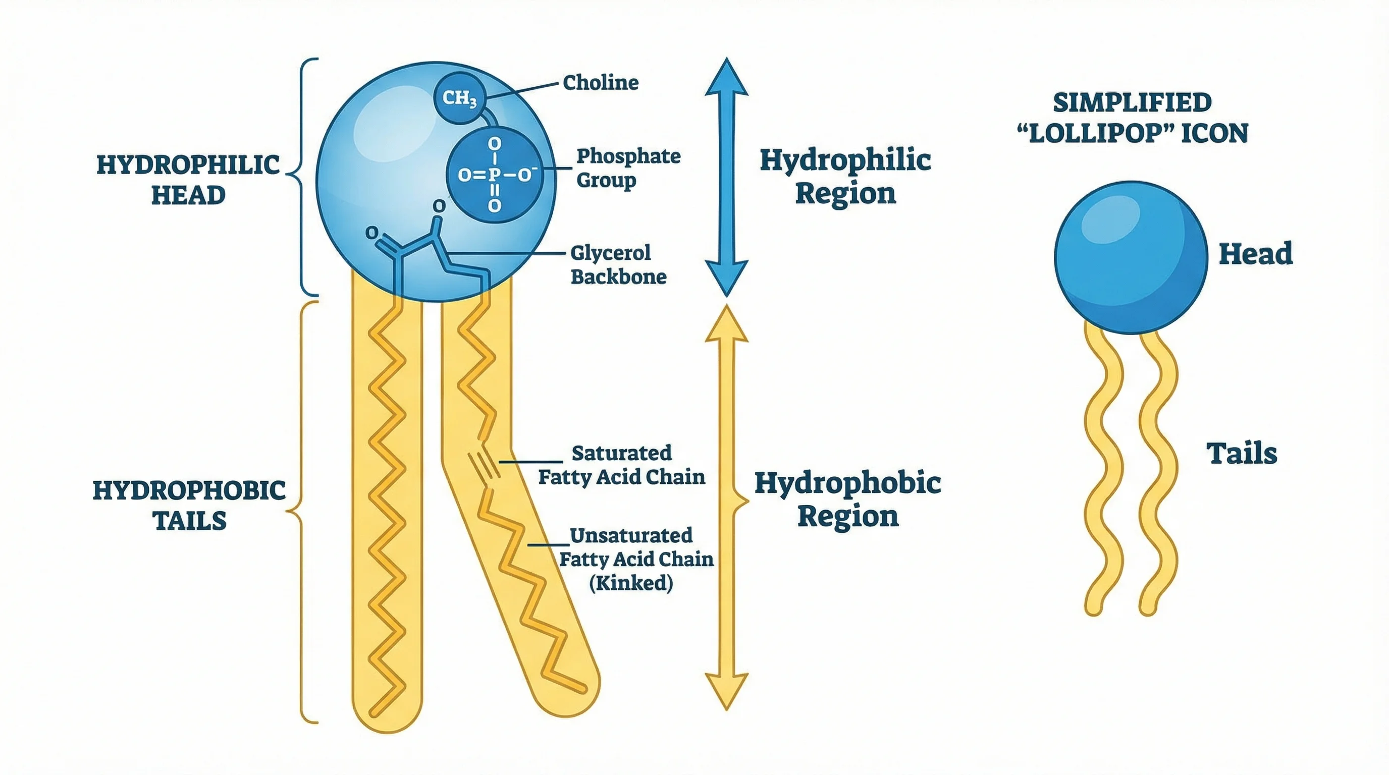
Phospholipid Diagram
Focus on phospholipid structure, bilayers, and membrane lipids.
 Biology
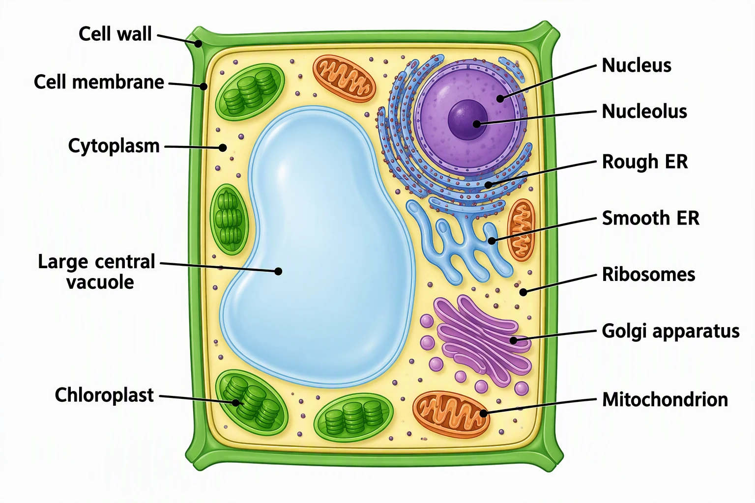
Plant Cell Diagram
Labeled plant cell with wall, membrane, chloroplasts, and vacuole.
 Biology
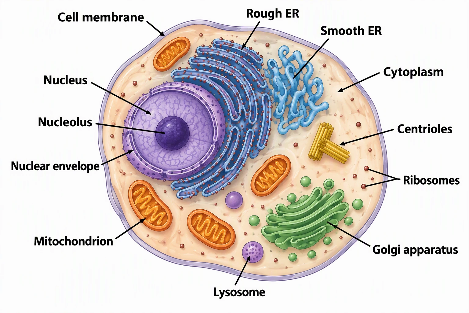
Animal Cell Diagram
Labeled animal cell organelles including the cell membrane.
 Biology
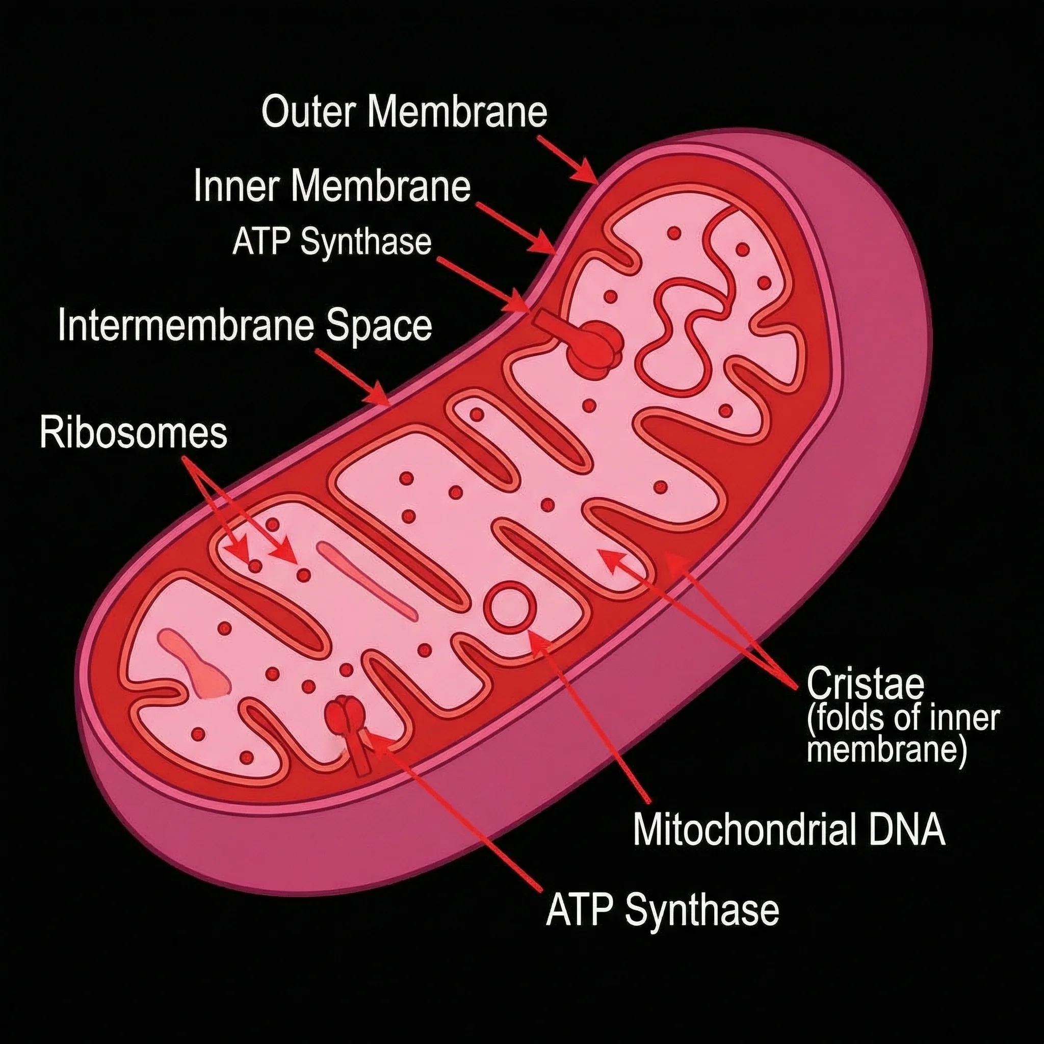
Mitochondria Diagram
Labeled mitochondria — energy organelles bounded by membranes.
 Biology
Animal vs Plant Cell
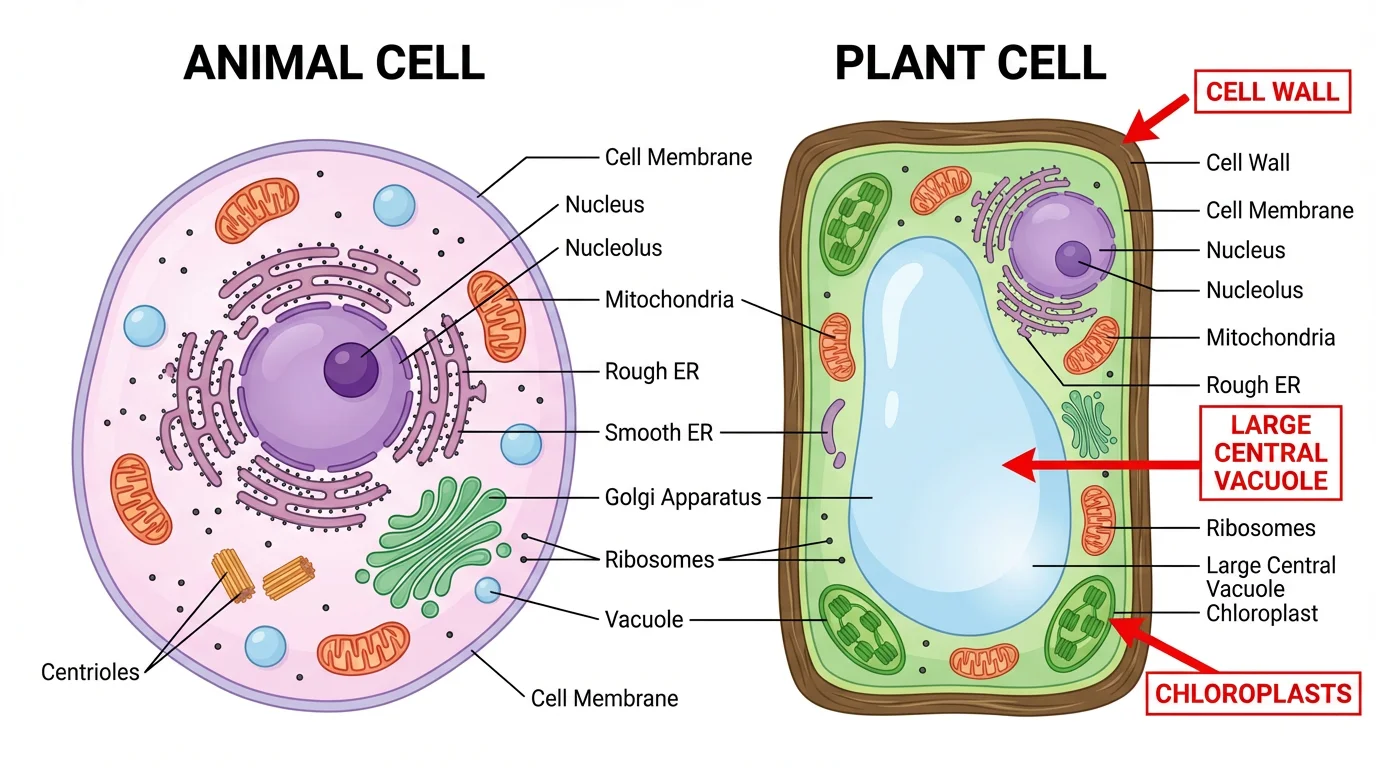
Side-by-side comparison of membrane, wall, and organelle differences.
 Biology
Chloroplast Diagram
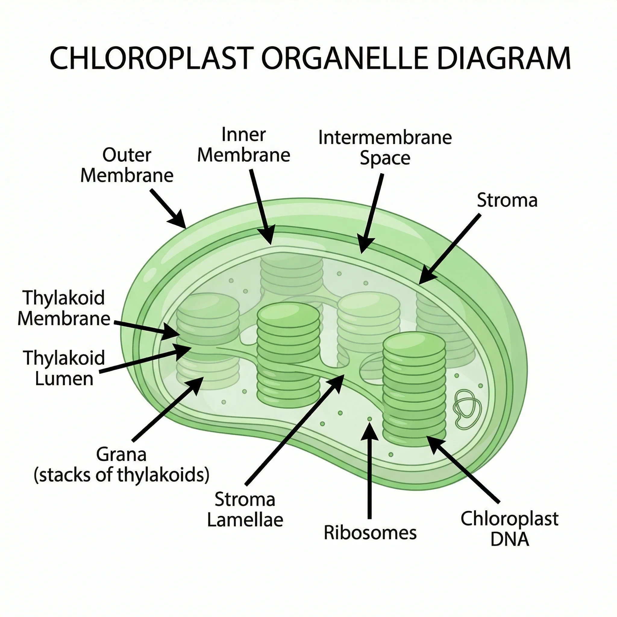
Labeled chloroplast membranes and thylakoid stacks for photosynthesis.