Root Canal Procedure Diagram Generator
Make root canal step diagrams, tooth cutaways, and before and after pictures for patient handouts. Steps checked against AAE. Clinician review needed.
Create Your Root Canal Diagram
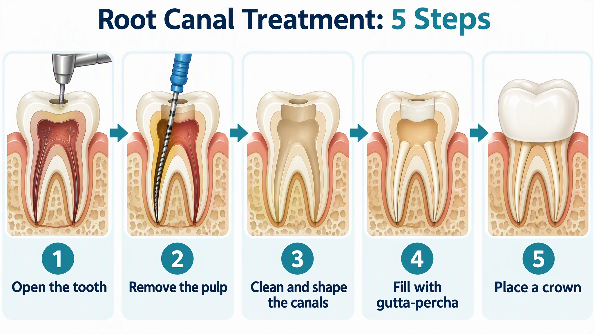 Example: Root canal treatment in 5 stepsView full size
Preview is free on this page ·
Your root canal diagram will appear here
AI diagrams are drafts. Check the tooth layers, the number of canals, and every label against your source, and have a clinician review before use with patients. No real patient data.
Root Canal Diagram Examples
Four AI diagrams, each checked against an official source and with known flaws noted. No real patients. A clinician should review every picture before patient use.
Root canal treatment in 5 steps (AI diagram)
AI-generated from the prompt shown, and all the text came out as written. We checked the order against the American Association of Endodontists (AAE) patient page "Root Canal Explained" (checked October 2026): the endodontist opens the crown, clears the pulp from the chamber and canals, cleans and shapes the canals, fills them with gutta-percha and cement, closes the opening with a temporary filling, and the patient returns to a general dentist for a crown or other restoration. Known flaws: AAE lists four steps and we split its second and third into five panels; the picture leaves out the first AAE step, the x-ray, local anesthetic and the dental dam, and panel 4 shows a white block closing the opening, which could be read as the temporary filling but is not labeled; the gutta-percha is drawn white, where it is normally pink or orange; panel 5 shows the crown sitting over the whole tooth, with no tooth preparation and no core or post under it, which a real crown on a treated tooth may need; the molar is drawn with one canal in each root, where molars usually have three or four; panel 3 does not clearly show empty canals, the inside of the tooth looks tan; panel 1 shows a drill at the opening and panel 2 a hand file, both simplified; AAE says molars and premolars generally need a crown while incisors and canines often do not, so step 5 does not apply to every tooth. Our first try asked for a front tooth, drew a molar anyway and filled only the top of the canal in step 4, so we asked again for a lower molar and this is the second try.
Inside an infected tooth (AI cutaway)
AI-generated from the prompt shown, and the seven labels came out spelled as written on the first try. The layers run in the right order from outside in: enamel, dentin, then the pulp chamber that continues into the root canals. AAE describes the pulp as the soft tissue inside the root canal that is treated when it becomes inflamed or infected (checked October 2026). Known flaws: each root shows one thin canal with colored lines for vessels and nerves, which is a simplification, because a lower molar usually has three canals; only one root tip carries the infection, drawn as a yellow-green ball, which is a symbol and not what an infection looks like on an x-ray; the cementum on the root is not labeled; the label "Root canal" points at one canal only, and the pulp chamber is drawn red throughout, which suggests more inflammation than every infected tooth has.
Before and after root canal treatment (AI comparison)
AI-generated from the prompt shown, and the words "Before", "After", "Infected pulp" and "Canals filled and crown placed" all came out as written. Our first try asked for an upper front tooth and the AI drew a molar anyway, so we asked for a lower molar with two roots and this is the second try. Known flaws: each root shows one canal, where a lower molar usually has three; the infection is drawn at the left root tip only, as a tan ball with a red glow, which is a symbol and not what an infection looks like on an x-ray; the "After" crown is wider than the original tooth and covers it down to the gum, and the root fillings show as pale shapes with no change in the gum or bone; the picture shows the tooth cleaned and restored and does not say how long it will last or that a tooth can need retreatment. AAE says most teeth that have had root canal treatment can last a lifetime (checked October 2026); we did not write that into the picture. Use it to show the idea, not an outcome.
Canals by tooth type (AI comparison)
AI-generated from the prompt shown, and every word came out as written. The counts are typical patterns from cone-beam CT scans of living patients (checked October 2026; links below): front teeth usually have one root and one canal, premolars have one or two, and molars three or four. The picture shows a front tooth with one canal, a premolar with one canal that splits near the tip, and a lower molar with three canals, two in the front root and one in the back root. Known flaws: the three teeth are not drawn to the same scale and only one example of each type is shown; upper molars often have four canals and are not drawn; some lower front teeth have two canals, and the reported share varies a lot between studies (about 30 percent in one, under 1 percent in another), and the picture says "Usually 1 canal" for front teeth but does not mention that; the premolar split is one of several patterns. The footer says "Typical patterns. Every tooth differs." because counts vary with the tooth and the person.
Need the anatomy of a healthy tooth first? Use our free Tooth Diagram page. For other body parts try the Medical Illustration Generator, for charts and posters the Medical Infographic Maker, and for skin care handouts the Dermatology Patient Education Illustrations page. Sources, checked October 2026: AAE Root Canal Explained, AAE reasons for root canal treatment, AAE root canal pain and comfort, CBCT study of root canal systems of permanent teeth, and CBCT root and canal morphology study.
What this page is for
This page is for dentists, endodontists, dental hygienists, front desk and patient coordinators, and dental or dental hygiene students who need a root canal picture for a handout, a consent conversation, a waiting-room poster, or a lecture slide. You describe the picture and the exact wording, and the generator drafts it: a step diagram, a tooth cutaway, a before and after pair, or a canals-by-tooth chart. It is a drafting tool. It is not a library of approved patient materials, and nothing on this page is a diagnosis or medical advice. A clinician should review every picture before it reaches a patient.
How this differs from our tooth and medical pages
- Our Tooth Diagram page is a free anatomy resource: labeled tooth layers, blank worksheets, and tooth types for biology classes. Use it when you need the anatomy of a healthy tooth. This page is about treatment: the order of the steps, what is removed and what is placed, and how an infected tooth differs from a treated one.
- The Medical Illustration Generator draws general anatomy and procedures for teaching. The Medical Infographic Maker lays out facts as charts and posters with a source line. Use this page when the picture you want is a tooth with a root canal procedure in it, with the step labels in plain words.
The steps as the AAE patient page describes them
- The American Association of Endodontists (AAE) page "Root Canal Explained" lists four stages. First, the endodontist takes an x-ray, gives local anesthetic, and once the tooth is numb places a protective sheet called a dental dam. Second, the endodontist makes an opening in the crown of the tooth and clears the pulp from the chamber and canals with tiny tools. Third, the canals are cleaned and shaped, then filled with a rubber-like material called gutta-percha plus cement to seal them, and a temporary filling usually closes the opening. Fourth, the patient returns to a general dentist for a crown or other restoration. Checked October 2026.
- The same page says treatment takes one or two appointments, each lasting roughly 90 minutes, and that molars and premolars generally need a crown for chewing while incisors and canines often do not. The AAE pain page says that with local anesthesia you may feel pressure or movement but should not feel pain, that mild soreness for a few days is normal, and that severe pain or pain lasting more than a few days is a reason to call the endodontist. Checked October 2026.
- The AAE page "Reasons for Root Canal Treatment" lists deep decay, repeated dental procedures on a tooth, a faulty crown, a crack or chip, and trauma, and says untreated pulp inflammation or infection can lead to an abscess. Checked October 2026.
- Our five-panel example splits the AAE second and third stages and leaves out the numbing and the dental dam. If your handout needs those, write them into the prompt as extra panels.
How many canals does a tooth have?
- The number depends on the tooth. In cone-beam CT scans of living patients, upper and lower front teeth (incisors and canines) most often have one root with one canal; premolars have one or two canals, with the upper second premolar and the upper molars reported as the most variable; lower molars usually have two roots and three canals, and upper molars usually have three roots with four canals common, especially in first molars. Some teeth have extra or curved canals, and the numbers vary between studies and populations. Checked October 2026.
- This is why a step diagram drawn on one tooth type can mislead. Say which tooth you are drawing in the prompt, and check the result: a molar drawn with one canal per root is a simplification, and a front tooth drawn with three canals is wrong.
How to make a root canal handout you can defend
- Pick one message and one audience, such as "adult patients, what happens at the appointment" or "dental hygiene students, canal anatomy".
- Take the steps from a named source such as the AAE patient pages or your own practice protocol, and write them into the prompt as exact labels in order.
- Say which tooth to draw: an upper front tooth, a premolar, or a lower molar. AI often draws a molar when asked for a front tooth, so look at the roots and canals.
- Ask for a calm, simple style without blood, and keep any infection symbol small.
- Add the source, the date you checked it, and "For education only" as footer text.
- Generate, then read every word and check the tooth layers, the number of canals, and the order of the steps against your source. If one thing is wrong, generate again.
- Have a dentist or endodontist review the final picture and wording before it goes to a patient.
Accuracy and what AI gets wrong
The four pictures on this page were generated from the prompts shown, and each caption lists the flaws we found. The words came out right on every try. The anatomy was looser than the words: our first step diagram drew a molar when we asked for a front tooth and filled only the top of the canal, our first before and after try also drew a molar, canals are drawn as one line per root, and an infection is a colored ball. Other common problems are the wrong number of canals for the tooth, steps out of order, a crown drawn on a tooth that would not get one, and symbols that look more serious than the situation. Treat each picture as a draft. It does not diagnose, it does not give medical advice, and it must be reviewed by a qualified clinician before use with patients.
Keep it calm
Patients who read a root canal handout are often anxious. Prefer soft colors, no blood, and plain words such as "open the tooth" and "clean and shape the canals". AAE says most patients report that having a root canal feels similar to getting a routine filling, but that is a claim for the clinician to make in their own words, so we left it out of the pictures.
Privacy and use with patients
Do not enter patient names, dates of birth, record numbers, x-rays, or real patient results. Describe the tooth in general terms. Check your practice's policy on AI tools and on what may go into patient materials, and follow its review and sign-off rules.
Free trial and downloads
Start free with 2 image credits, valid for 7 days, no card required. Free accounts download PNG or JPG.
Frequently Asked Questions
Related Medical Tools
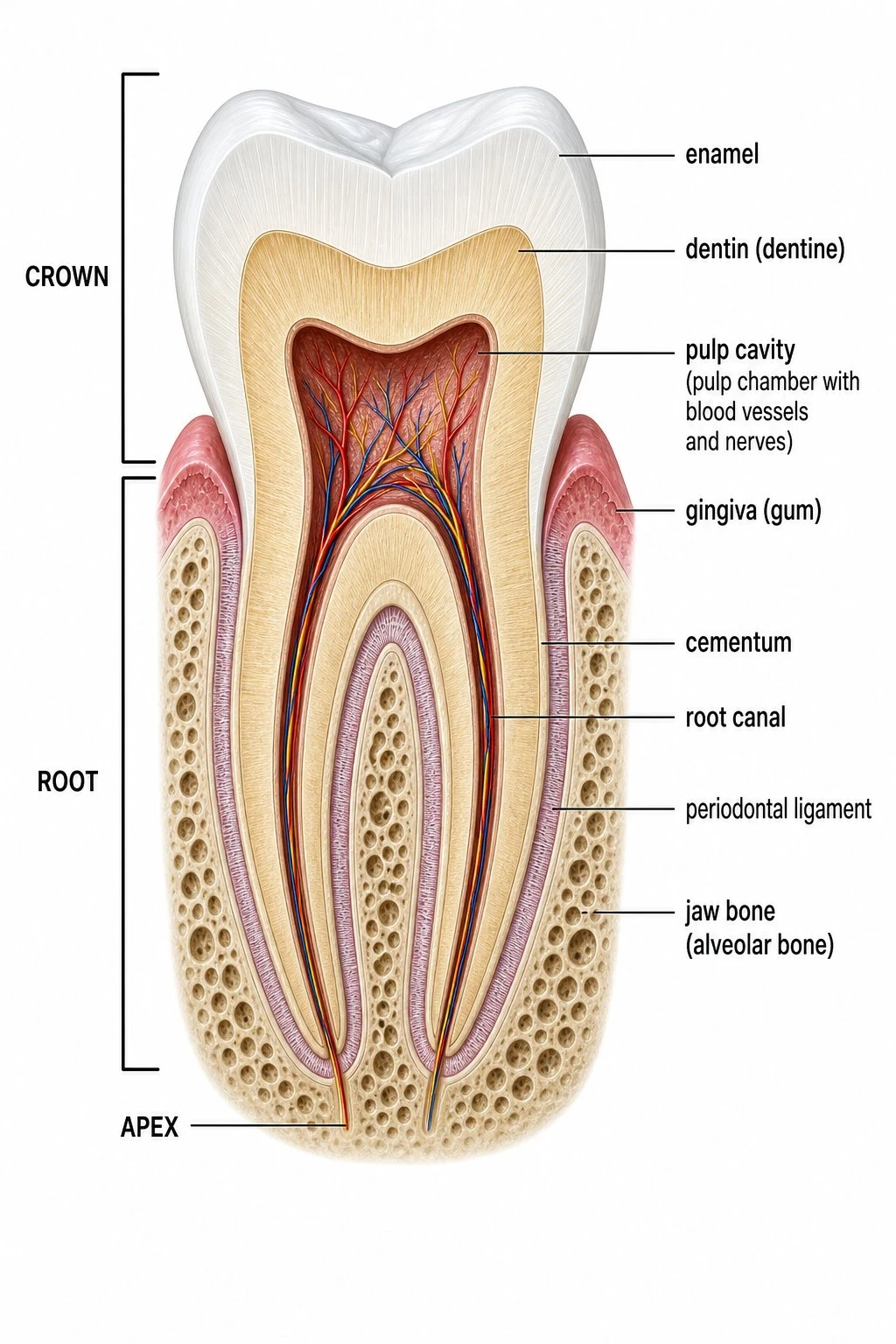 Biology
Tooth Diagram
Free tooth anatomy diagrams: labeled cutaway, blank worksheet, tooth types, and coloring pages.
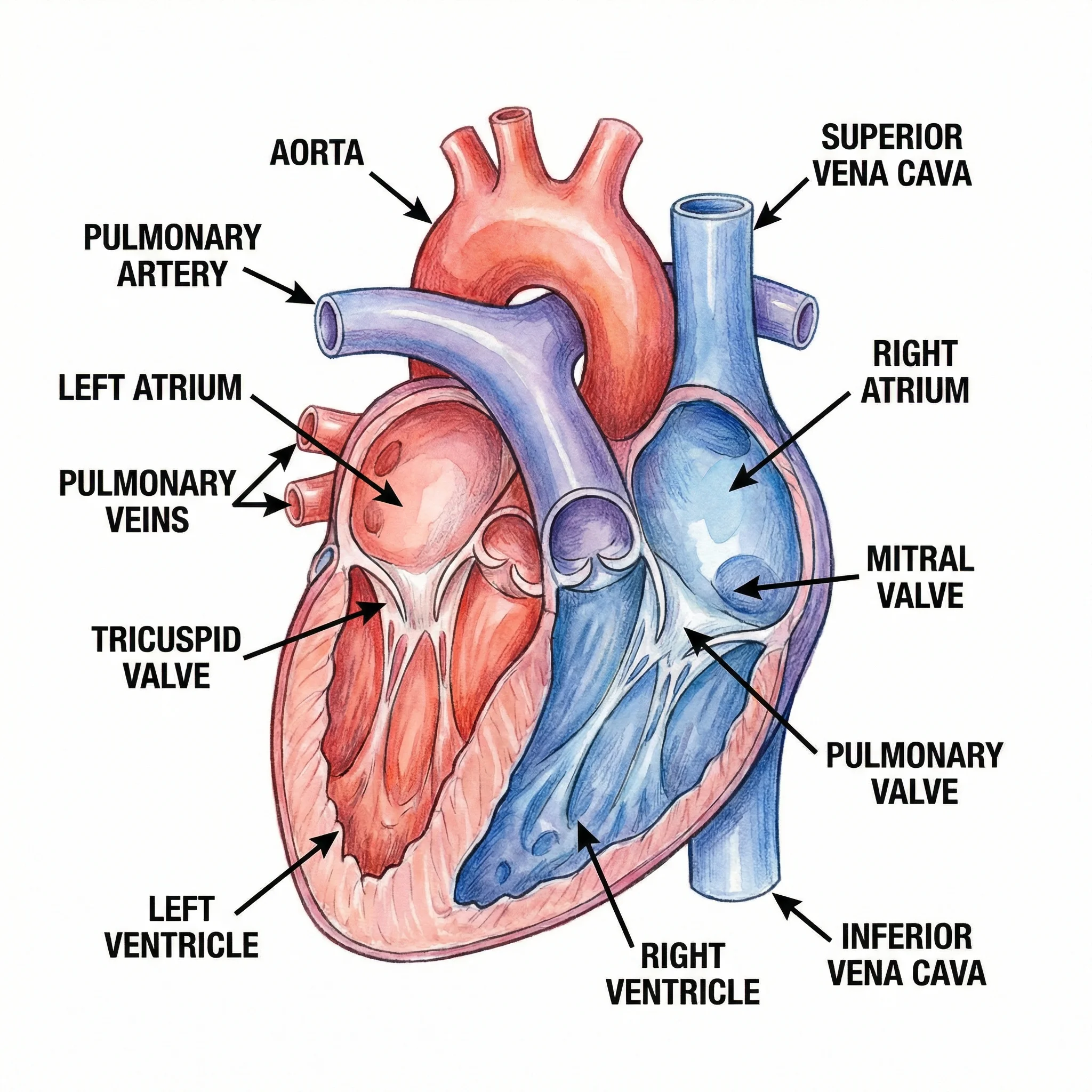 Medical
Medical Illustration Generator
Create labeled medical and anatomy illustrations for teaching and patient education.
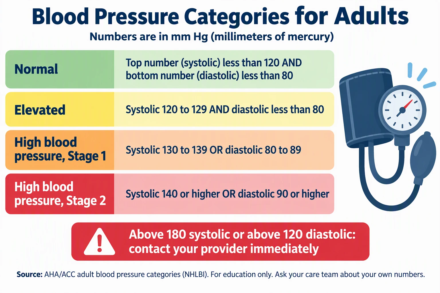 Medical
Medical Infographic Maker
Make medical and patient education infographics from facts you cite. Clinician review needed.
 Medical
Dermatology Patient Education Illustrations
Make skin condition and skin care illustrations for patient handouts, with diverse skin tones and plain-language labels.