Skin Diagram Layers Labeled & Printable
Free skin diagrams and skin layers diagrams — labeled, blank, simple, epidermis focus, and coloring. Download or print; no sign-up.
Free Skin Diagrams & Skin Layers Charts
Every version below is free to view and download — no sign-up. Built for “skin diagram” and “skin layers diagram” lessons.
Labeled Skin Layers Diagram
Full cross-section — epidermis, dermis, hypodermis, hair follicle, and glands labeled for study.
Core diagrams
Structure & focus
Print & practice
Test yourself: skin layers
A quick self-test — questions reshuffle each time, with a review of anything you miss at the end.
Which layer is the outermost layer of the skin?
Need a custom version?
Change the grade level, which parts are labeled, or focus on the epidermis — describe it and generate your own.
Customize Your Skin Diagram
Free to try ·
Your custom skin diagram will appear here
Ask for labeled layers, a blank worksheet, or a simple kids version
What is a skin diagram?
A skin diagram (or skin layers diagram) is a labeled cross-section of human skin. Because real skin is thin and layered, diagrams enlarge a cutaway so you can see the epidermis, dermis, and hypodermis (subcutaneous fat), plus structures such as hair follicles, sebaceous glands, and sweat glands. Use a fully labeled version to learn the parts, or a blank version as a worksheet or quiz.
The three main layers
- Epidermis: the outer layer; it has no blood vessels and forms a protective barrier. Surface cells are flattened and often dead (stratum corneum).
- Dermis: the thicker middle layer with collagen, blood vessels, nerve endings, hair follicles, and glands.
- Hypodermis (subcutaneous layer): fat and connective tissue that cushion the body and help with insulation.
Skin diagram vs skin layers diagram
Searchers use both phrases for the same classroom need: a clear cutaway of the layers. “Skin layers diagram” emphasizes epidermis / dermis / hypodermis; “skin diagram” may also include hair and glands. This page includes both styles so you can download the version that matches your lesson.
How to use these free diagrams
Teach with the labeled layers diagram, then assess with the blank version of the same layout. The simple three-layer poster works for elementary; the epidermis focus and functions poster support middle and high school. The coloring page is a low-prep station. All files are free to view, download, and print with no account required.
Frequently Asked Questions
Related Biology Tools
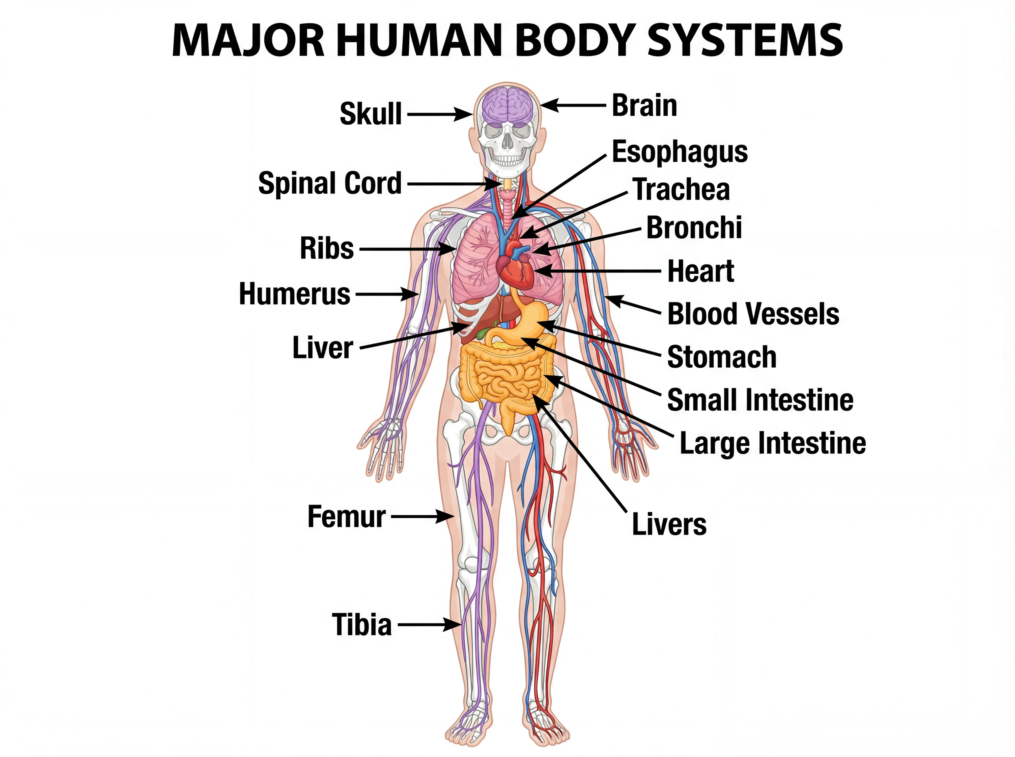 Biology
Human Body Systems
Overview diagrams of major body systems for full-body context.
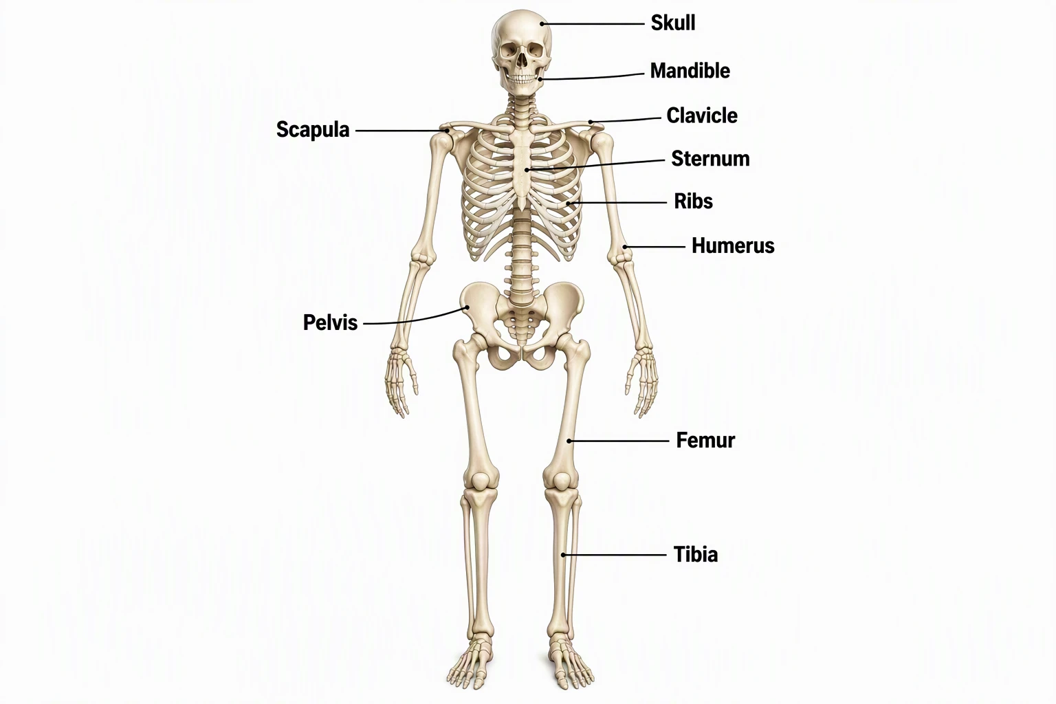 Biology
Skeletal System Diagram
Labeled skeleton maps that pair with integumentary units.
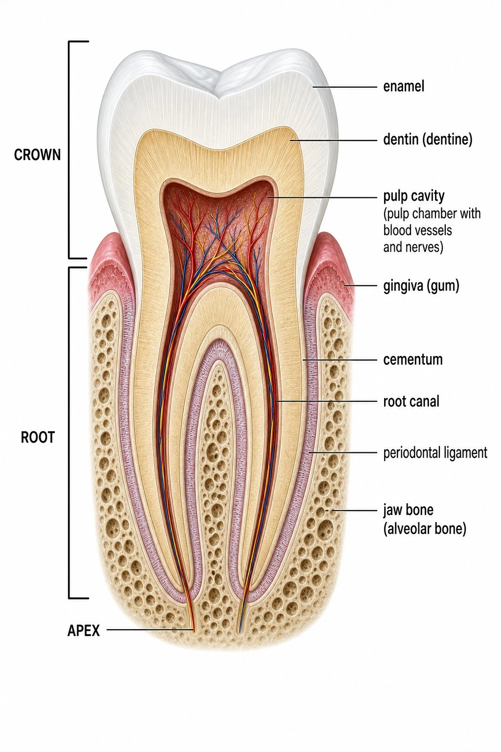 Biology
Tooth Diagram
Free labeled tooth anatomy diagrams — enamel, dentin, pulp, and root.
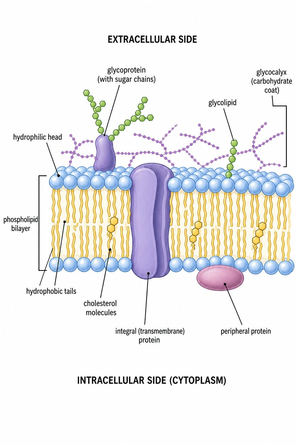 Biology
Cell Membrane Diagram
Fluid mosaic model — how cell surfaces act as barriers and gates.
 Biology
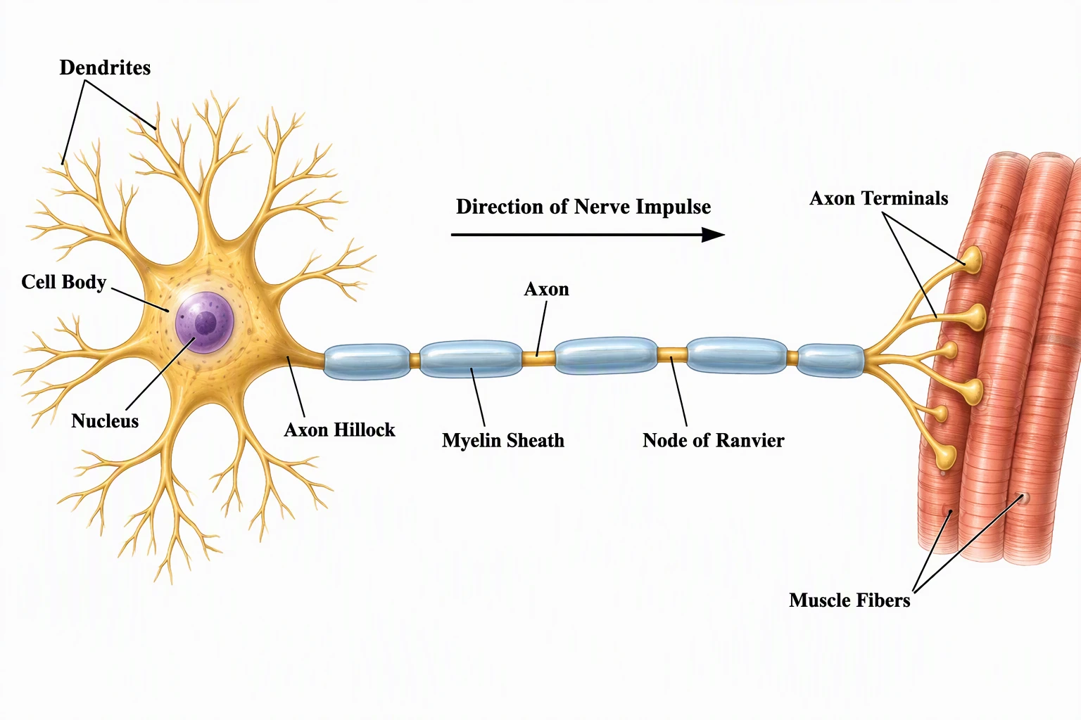
Nervous System Diagram
Brain and nerve maps for lessons on skin sensation.
 Medical
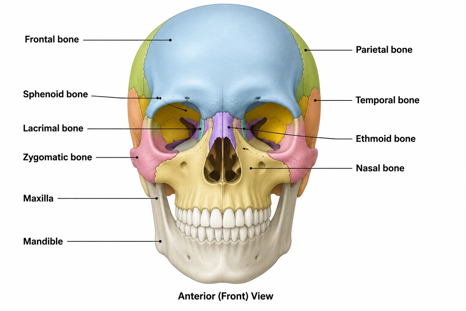
Skull Diagram
Labeled skull views for head and face anatomy units.