Digestive System Diagram Generator — Labeled & Blank
Create a digestive system diagram in seconds — labeled or blank. Generate a clearly labeled version showing the mouth, esophagus, stomach, small intestine, large intestine, liver, gallbladder, and pancreas, or a printable unlabeled diagram for worksheets and quizzes. Free to use.
Digestive System Diagram Generator
Free to try ·
Your digestive system diagram will appear here
Ask for a labeled, blank, or printable version of the diagram
Digestive System Diagram Examples
Labeled, blank, and printable digestive system diagrams
Labeled Digestive System
Every major organ labeled — the mouth, esophagus, stomach, small and large intestine, liver, gallbladder, and pancreas.
Stomach Cross-Section
A close-up of the stomach — cardia, fundus, body, pylorus, rugae, and the muscular wall layers.
Small Intestine & Villi
Zooms from the small intestine into the villi and microvilli where nutrients are absorbed.
Accessory Organs
The accessory organs — liver, gallbladder, and pancreas — and the ducts that feed the duodenum.
Digestion Process Flow
Traces the path of food, showing the mechanical and chemical digestion at each stage.
Blank Worksheet
A blank, unlabeled version with numbered lines — drop it straight into a worksheet or quiz.
What is the digestive system?
The digestive system is the group of organs that breaks food down into nutrients the body can absorb and use. It runs as one long tube — the gastrointestinal (GI) tract — from the mouth to the anus, supported by accessory organs that add enzymes and chemicals along the way. A digestive system diagram lays this out as a single picture so you can see every organ, the order they come in, and the path food takes. This generator draws that diagram for you in both labeled and blank versions — a labeled diagram for studying and a clean unlabeled one for worksheets and quizzes.
The organs of the digestive system, in order
- Mouth: chewing (mechanical digestion) breaks food into smaller pieces while saliva and salivary amylase begin breaking down starch.
- Esophagus: a muscular tube that pushes the swallowed food (bolus) down to the stomach using waves of muscle called peristalsis.
- Stomach: churns food with strong muscles and mixes it with gastric acid (HCl) and the enzyme pepsin, which begins breaking down protein.
- Small intestine: the duodenum, jejunum, and ileum — where most chemical digestion and nutrient absorption happen through tiny finger-like villi.
- Large intestine (colon): absorbs water and salts and forms the remaining waste; includes the cecum, ascending, transverse, descending, and sigmoid colon.
- Rectum and anus: store and then release the solid waste from the body.
- Accessory organs — liver, gallbladder, and pancreas: the liver makes bile, the gallbladder stores it, and the pancreas adds digestive enzymes; all feed into the small intestine.
The path food takes and what happens at each stage
Food follows one route: mouth, esophagus, stomach, small intestine, large intestine, rectum, and anus. At each stage two things happen — mechanical digestion (physically breaking food apart, such as chewing in the mouth and churning in the stomach) and chemical digestion (enzymes and acids breaking molecules into smaller units). Carbohydrates begin breaking down in the mouth, proteins in the stomach, and fats in the small intestine, where bile from the liver emulsifies them and pancreatic enzymes finish the job. Most absorption — nutrients passing into the blood — happens in the small intestine, while the large intestine reclaims water. A digestive system diagram with arrows makes this flow easy to follow and is one of the most common things students are asked to label.
Labeled vs. blank (unlabeled) diagrams
- Labeled diagram: every organ is named — ideal for studying, presentations, and answer keys.
- Blank / unlabeled diagram: the organs are drawn but the labels are left empty so students can fill them in — perfect for worksheets, quizzes, and homework.
- Printable coloring page: black-and-white line art with blank label lines, ready to print and hand out.
- Generate a labeled key and a matching blank version of the same diagram, so the quiz and the answer key always line up.
How to generate a labeled diagram from a description
- Describe the diagram you want — for example, "a labeled human digestive system" or "a blank digestive system for a quiz."
- Add any organs you want emphasized, such as the small intestine villi, the stomach cross-section, or the accessory organs (liver, gallbladder, pancreas).
- Ask for a labeled version, an unlabeled version, or a printable black-and-white coloring page.
- Generate the image, then download it to drop into a worksheet, slide, study guide, or printed handout.
Using digestive system diagrams in the classroom
Teachers use labeled diagrams to introduce the GI tract and accessory organs, then hand out blank or unlabeled versions for students to complete from memory — a quick way to check understanding. Detailed labeled cross-sections suit high school and introductory biology, while simple coloring pages work for younger grades. Because the diagrams are AI-generated, review them for accuracy before using them with students — check that the organs, their order, and the labels are correct, and treat the images as educational illustrations rather than medical or diagnostic references. Used that way, generating a labeled key and a matching blank diagram takes seconds instead of hunting for two images that happen to match.
Frequently Asked Questions
Related Biology Tools
 Biology
Human Body Systems Diagram Generator
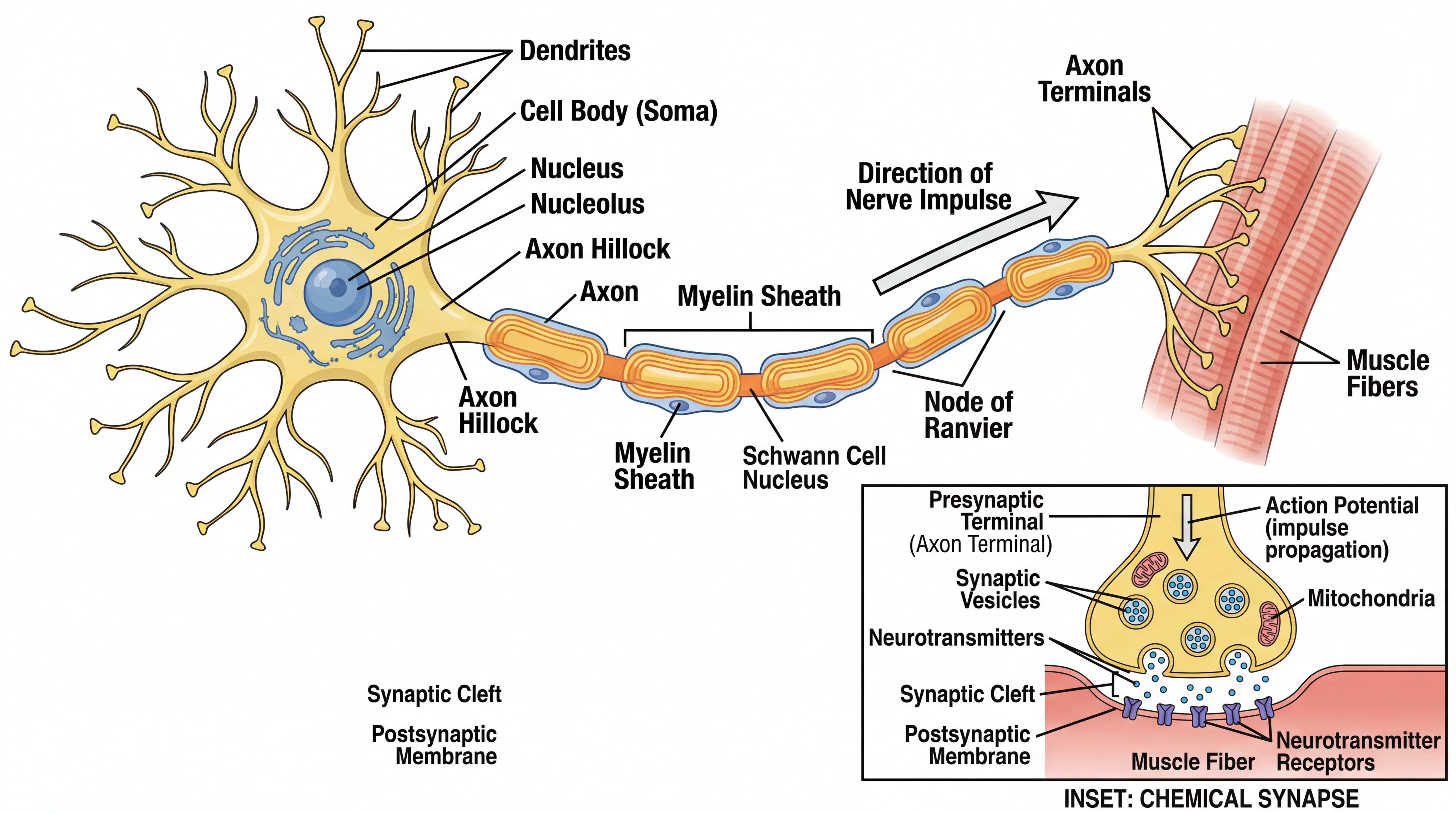
Create labeled diagrams of the body systems — circulatory, respiratory, nervous, and more.
 Biology
Anatomical Drawing Generator
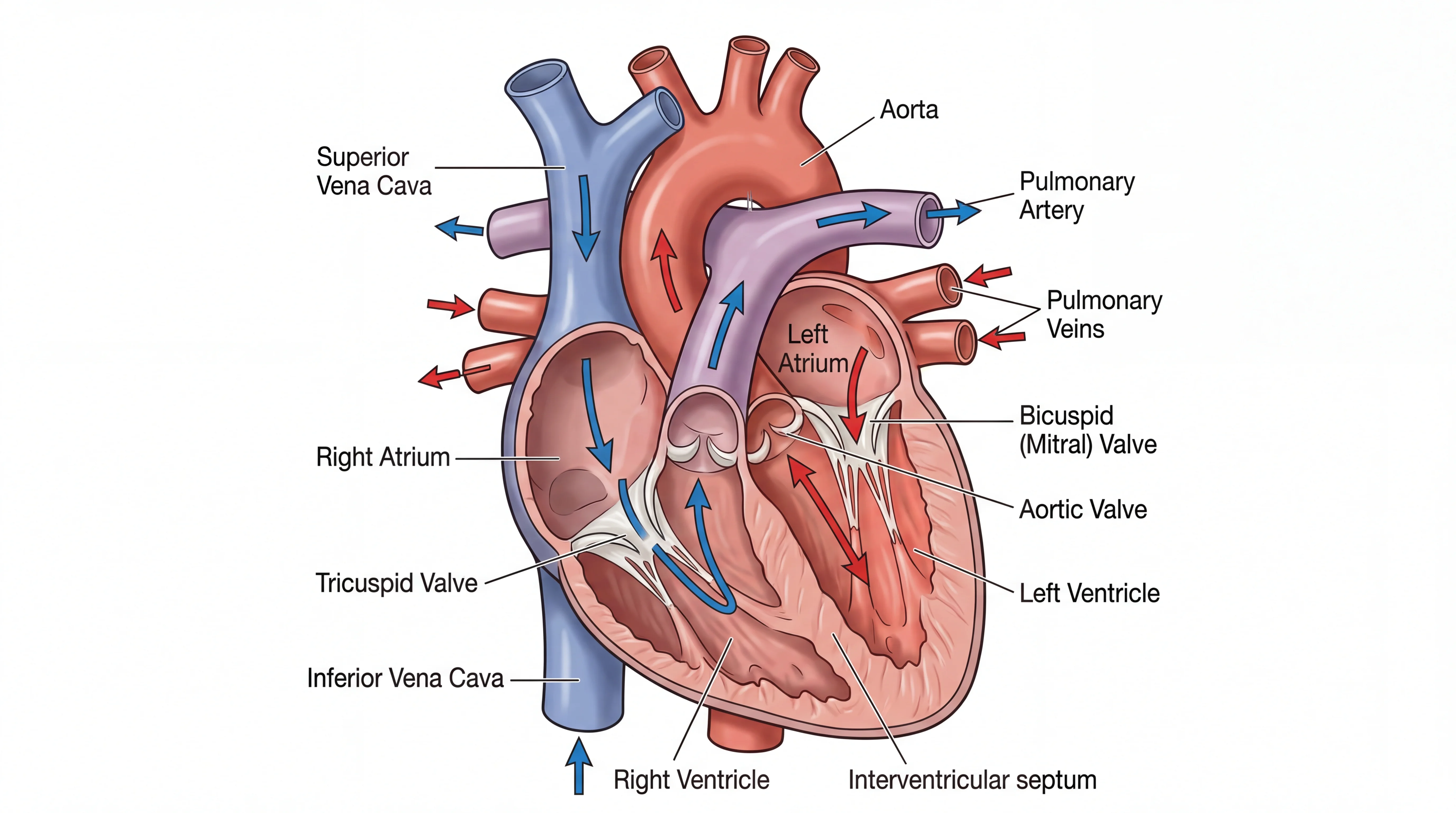
Generate labeled anatomical drawings of organs and body structures for study and review.
MedicalMedical Illustration Generator
Make clean medical illustrations of organs, systems, and procedures from a description.