Skull Diagram
Free skull diagrams: labeled anterior and lateral views, blank worksheet, simple version, and coloring page. Printable; free download, no sign-up.
Free Skull Diagrams
Every version below is free to view, download, and print. No sign-up.
Labeled Skull (Anterior View)
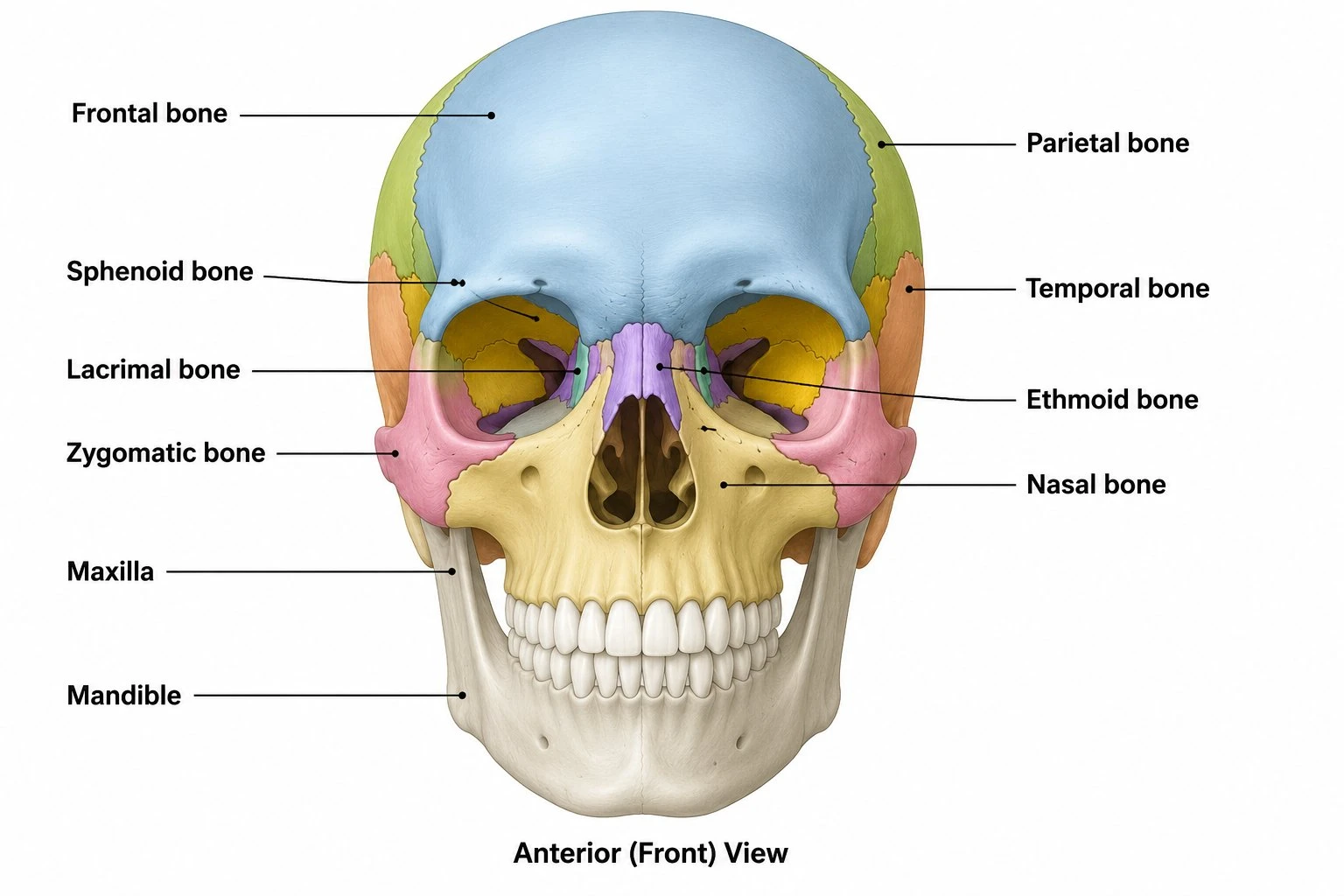
A front view of the skull with ten bones named: Frontal, Parietal, Temporal, Sphenoid, Ethmoid, Nasal, Lacrimal, and Zygomatic bones, plus the Maxilla and Mandible.
Core diagrams
Structure & focus
Print & practice
Test yourself: skull bones
A quick self-test. Questions reshuffle each time.
The mandible is…
Customize this diagram
Change view, grade level, or which bones and sutures are labeled. Describe it and generate your own.
Customize Your Skull Diagram
Free to try ·
Your custom diagram will appear here
Ask for anterior, lateral, sutures, blank worksheet, or simple labels
How to read the anterior (front) view
Start with the labeled anterior skull. It names Parietal bone, Frontal bone, Temporal bone, Sphenoid bone, Nasal bone, Lacrimal bone, Zygomatic bone, Ethmoid bone, Maxilla, and Mandible. The forehead is Frontal bone. Parietal bone shows at the upper sides; Temporal bone is lower on the side of the head. Down the midline of the face you meet Nasal bone, then Maxilla (upper jaw), then Mandible (lower jaw). Zygomatic bone is the cheek. Lacrimal bone sits on the medial wall of the orbit. Sphenoid bone and Ethmoid bone appear in the orbit and between the eyes; they are easy to miss, so follow those two leader lines all the way to the structure, not to nearby bone. This view uses ten labels so the type stays large on a slide.
Why the lateral view is not optional
Occipital bone is at the back of the skull. An anterior drawing cannot show it well, so this pack includes a labeled lateral view. That view names Frontal bone, Parietal bone, Sphenoid bone, Temporal bone, Occipital bone, Nasal bone, Lacrimal bone, Zygomatic bone, Maxilla, and Mandible. Print anterior and lateral on facing pages so students can find Mandible and Zygomatic bone in both orientations. That is the fastest way to stop “the mandible is only on the front picture” mistakes. Squamous suture is not on this lateral bone list; it is on the sutures figure.
Sutures, then a facial close-up
The sutures figure labels Coronal suture, Sagittal suture, Lambdoid suture, Squamous suture, Frontal bone, Parietal bone, Temporal bone, and Occipital bone. Coronal suture joins Frontal bone to Parietal bone. Sagittal suture is the midline between the two parietal bones. Lambdoid suture joins the parietals to Occipital bone. Squamous suture joins Parietal bone to Temporal bone. The facial close-up then labels Orbit, Nasal bone, Lacrimal bone, Zygomatic bone, Maxilla, and Mandible. Use it when the lesson is “bones of the face” rather than “whole skull.”
Simple diagram, blank sheet, and coloring page
The simple diagram labels Cranium, Orbit, Nasal cavity, and Mandible. That is the right first slide for middle school or for a warm-up before a lab. The blank worksheet matches the ten-label anterior view: same leader-line geometry, numbers 1 to 10, no words. Project the labeled anterior view, hide it, and give students five minutes on the blank sheet. The coloring page is an outline front view with no text. Ask the class to color cranial bones one color and facial bones another so the two groups stay distinct on the page.
How to use these files in class
A reliable sequence is simple diagram, labeled anterior, labeled lateral, sutures, then the blank anterior sheet. Homework can be the coloring page plus the blank sheet on the reverse. All files are free to view, download, and print. Drop the anterior view into slides, or print letter or A4. This page is a skull close-up. For the rest of the skeleton, use the skeletal-system generator; for the hand and foot, use the packs linked below. Do not copy every skull name onto a full-body figure, and do not copy the whole skeleton onto these skull files.
What this pack does not try to be
These drawings are classroom maps of named bones and sutures. They are not a radiology atlas, not a surgical approach guide, and not a list of foramina. If you need a custom angle (basal view, labeled foramina, pediatric skull), use the customize box on this page or the skull generator linked in related tools. Keep the seven files here for the job they do: grab, print, label, and quiz.
Frequently Asked Questions
Related Anatomy Tools
 Medical
Hand Bones Diagram
Free labeled carpals, metacarpals, and phalanges.
 Medical
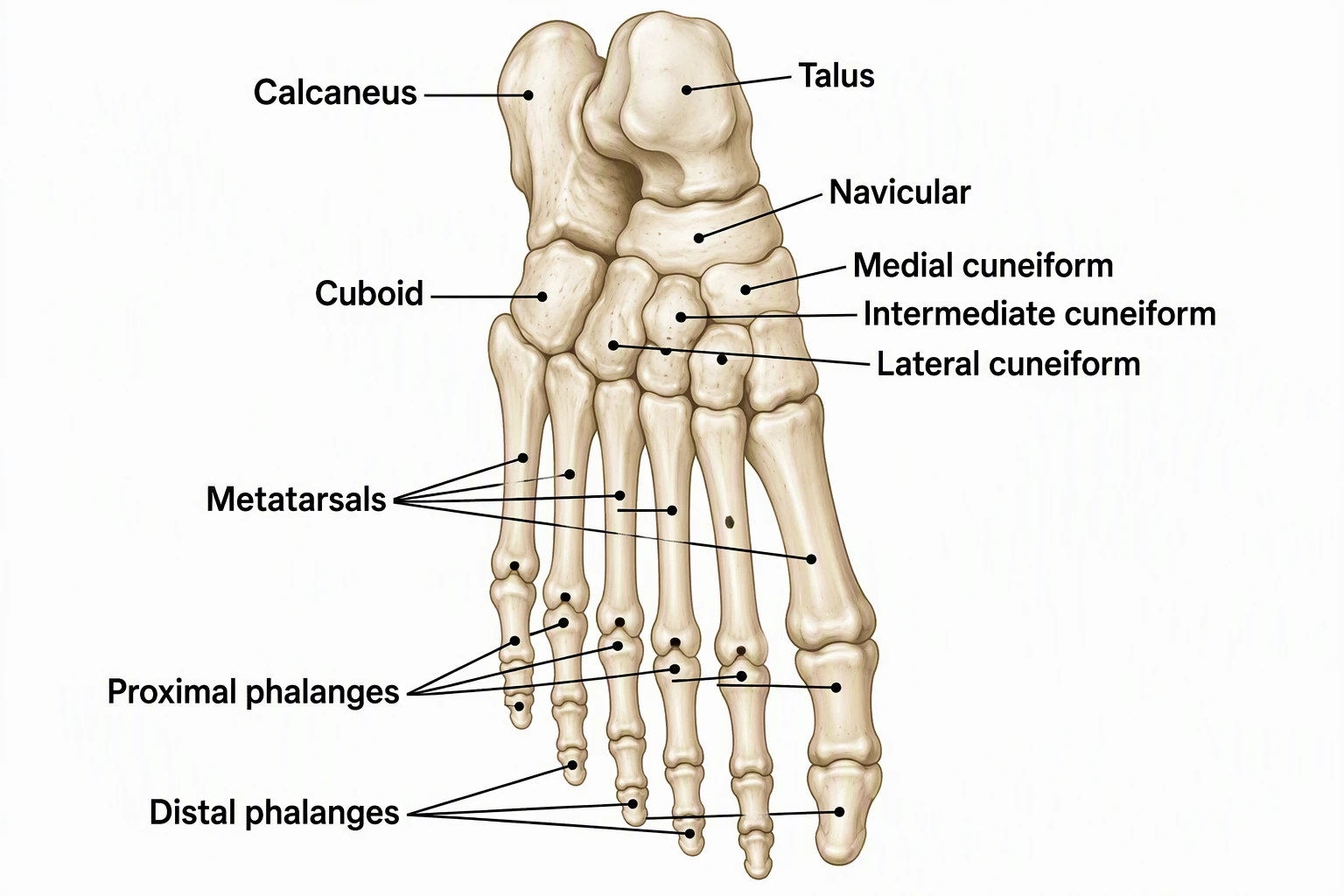
Foot Bones Diagram
Free labeled tarsals, metatarsals, and phalanges.
 Biology
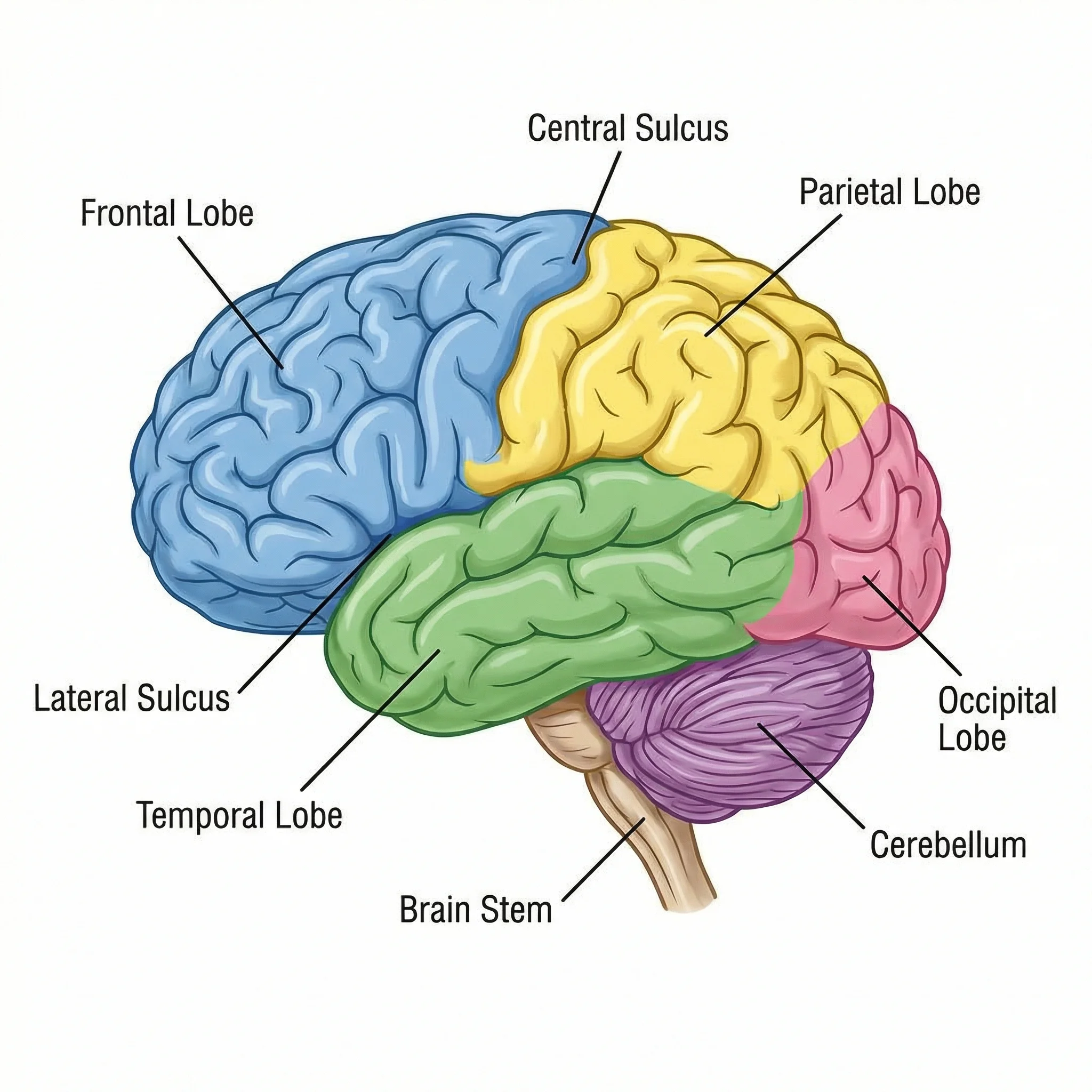
Brain Diagram Generator
Lobes and pathways that sit inside the cranial cavity.
 Biology
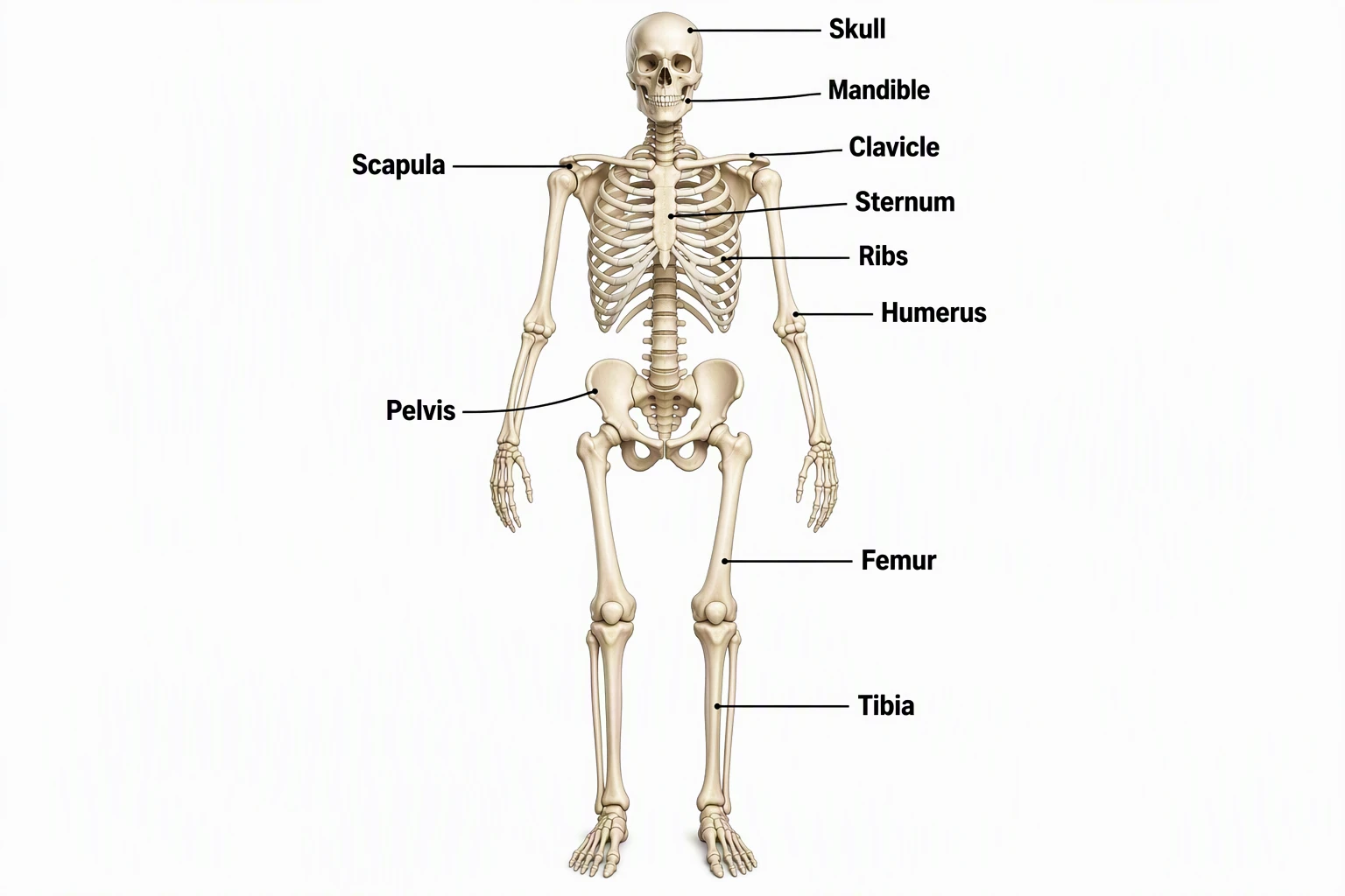
Skeletal System Diagram Generator
Full-body skeleton maps; zoom here for the skull.
 Medical
Skull Diagram Generator
Custom AI skull views when you need a layout this pack does not include.
 Biology
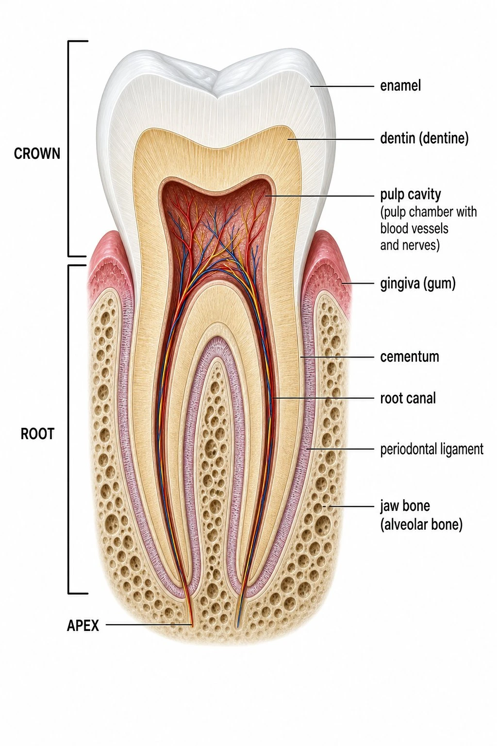
Tooth Diagram
Labeled tooth anatomy that pairs with a skull unit.