Sarcomere Diagram
Free sarcomere diagrams: labeled Z line, A band, and I band, blank worksheet, contraction, and coloring page. Printable; free download.
Free Sarcomere Diagrams
Every version below is free to view, download, and print. No sign-up.
Labeled Sarcomere
A sarcomere bracket from Z line to Z line, named Sarcomere, Z line (two), M line, Actin (thin filament), Myosin (thick filament), Titin (the gray spring), H zone, I band (two), and A band. Thin and thick filaments overlap at both ends.
Core diagrams
Structure & focus
Print & practice
Test yourself: sarcomere
A quick self-test. Questions reshuffle each time.
A sarcomere is the segment:
Customize this diagram
Change view, grade level, or which structures are labeled. Describe it and generate your own.
Customize Your Sarcomere Diagram
Free to try ·
Your custom diagram will appear here
Ask for labeled sarcomere, blank worksheet, contraction rows, or simple labels
How to read the labeled sarcomere
Start with the labeled sarcomere. It names Sarcomere (a bracket from Z line to Z line), Z line (two), M line, Actin (thin filament), Myosin (thick filament), Titin (the gray spring), H zone, I band (two), and A band. Thin and thick filaments overlap at both ends. A sarcomere is the segment between two Z lines. Actin (thin filament) attaches at each Z line. Myosin (thick filament) sits in the middle and meets the M line. The A band is the dark region of thick filaments; the I band is the light region of thin filaments only; the H zone is the middle of the A band with no thin-filament overlap. Follow every leader line to the band or filament it names.
Relaxed versus contracted
The contraction figure is two rows: Relaxed on top, Contracted below. Each row names Z line (two), I band (two), A band, and H zone. This figure has no M line and no titin. In the contracted row the Z lines are closer. The A band stays the same length (dashed lines align) because thick filaments do not shorten. The I band and the H zone get shorter because thin and thick filaments overlap more. Print this sheet beside the labeled sarcomere so students can find A band, I band, and H zone in both states. That is the sliding-filament story in one glance.
From muscle down to sarcomere
The hierarchy figure names Muscle, Fascicle, Muscle fiber, Myofibril, and Sarcomere as a zoom sequence. Start here if the class has not yet placed the sarcomere inside a fiber. A whole muscle is bundles of fascicles; a fascicle is bundles of fibers; a fiber is packed with myofibrils; a myofibril is a chain of sarcomeres. Project this figure for two minutes, then switch to the labeled sarcomere. Students mix “fiber” and “filament” less often after they have seen the zoom.
Cross-bridge cycle
The four-panel figure names Attachment, Power stroke, Detachment, and Re-cocking, plus Actin and Myosin head. ATP is labeled on the Detachment panel. The blue molecule on the Re-cocking panel is not labeled. Attachment is the myosin head binding actin. Power stroke is the head pulling the thin filament. Detachment needs ATP. Re-cocking resets the head. Teach this after students can point to Actin and Myosin on the labeled sarcomere. It is a cycle, not a one-way arrow; walk the class around the four panels twice.
Simple diagram, blank sheet, and coloring page
The simple diagram labels Z line (two), Actin, Myosin, and M line in large type, with no titin. That is the right first slide for middle school or for a warm-up before a lab. The blank worksheet matches the labeled sarcomere: same leader-line geometry, numbers 1 to 11, no words. Z line and I band each appear twice. Project the labeled view, hide it, and give students five minutes on the blank sheet. The coloring page is the same sarcomere in black-and-white outline, still labeled Sarcomere, Z line (two), M line, Actin (thin filament), Myosin (thick filament), Titin, H zone, I band (two), and A band. Ask the class to color thin filaments, thick filaments, and titin in three colors, then compare with the labeled view.
How to use these files in class
A reliable sequence is simple four-name diagram, hierarchy zoom, labeled sarcomere, contraction rows, cross-bridge cycle, then the blank sheet. Homework can be the coloring page plus the blank sheet on the reverse. All files are free to view, download, and print. Drop the labeled sarcomere into slides, or print letter or A4. This page is a myofibril close-up. Use the muscular system generator for whole-body muscle maps, and the action potential graph generator when the unit moves from contraction to membrane voltage. Keep each download set on its own page so labels stay short and readable.
Frequently Asked Questions
Pair this pack with the muscular system diagram generator for whole-body muscle maps, and the action potential graph generator when the unit moves from contraction to membrane voltage.
Related Anatomy Tools
 Biology
Muscular System Diagram Generator
Full-body muscle maps; zoom here for the sarcomere.
 Medical
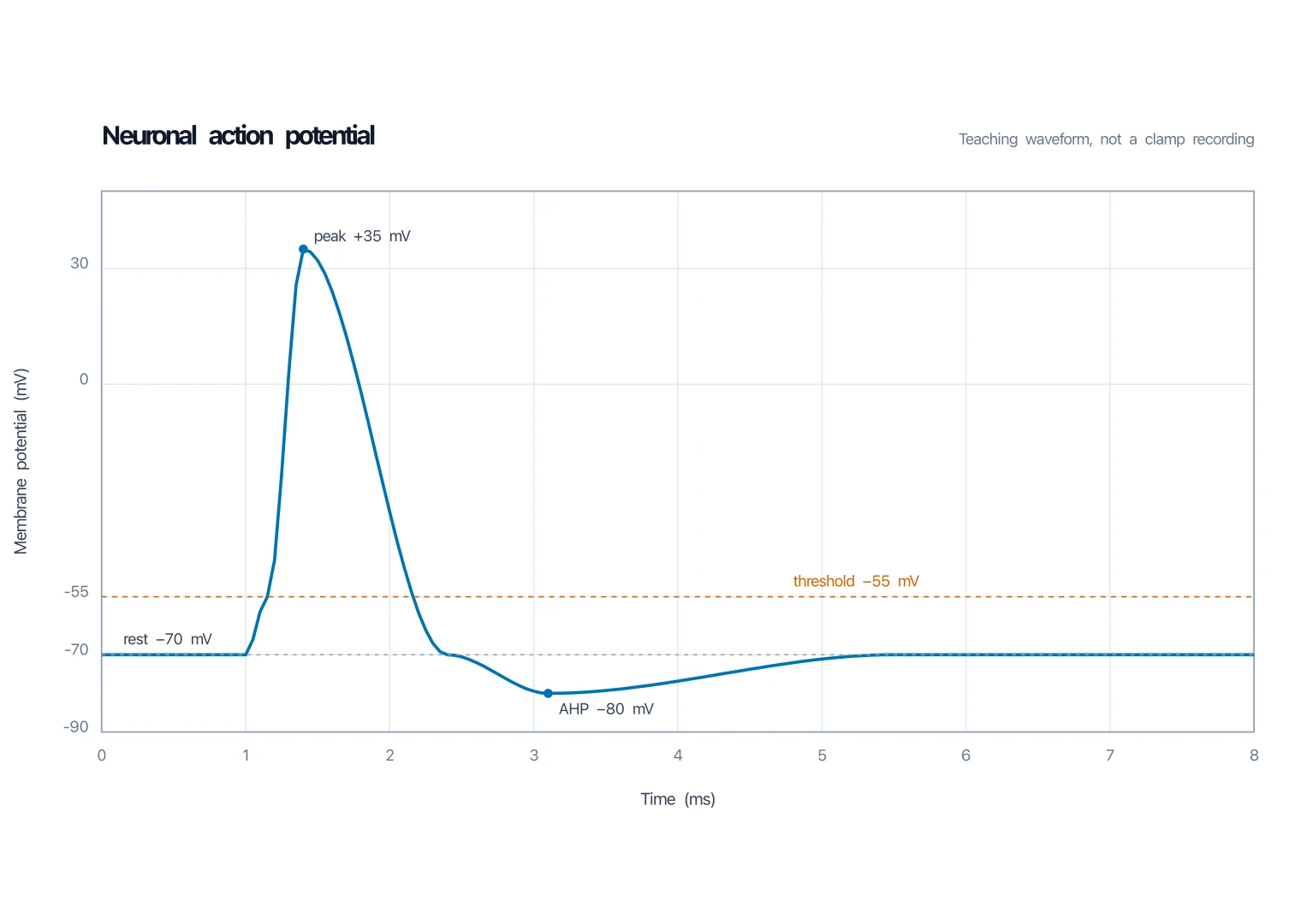
Action Potential Graph Generator
Membrane voltage versus time for the same unit.
 Biology
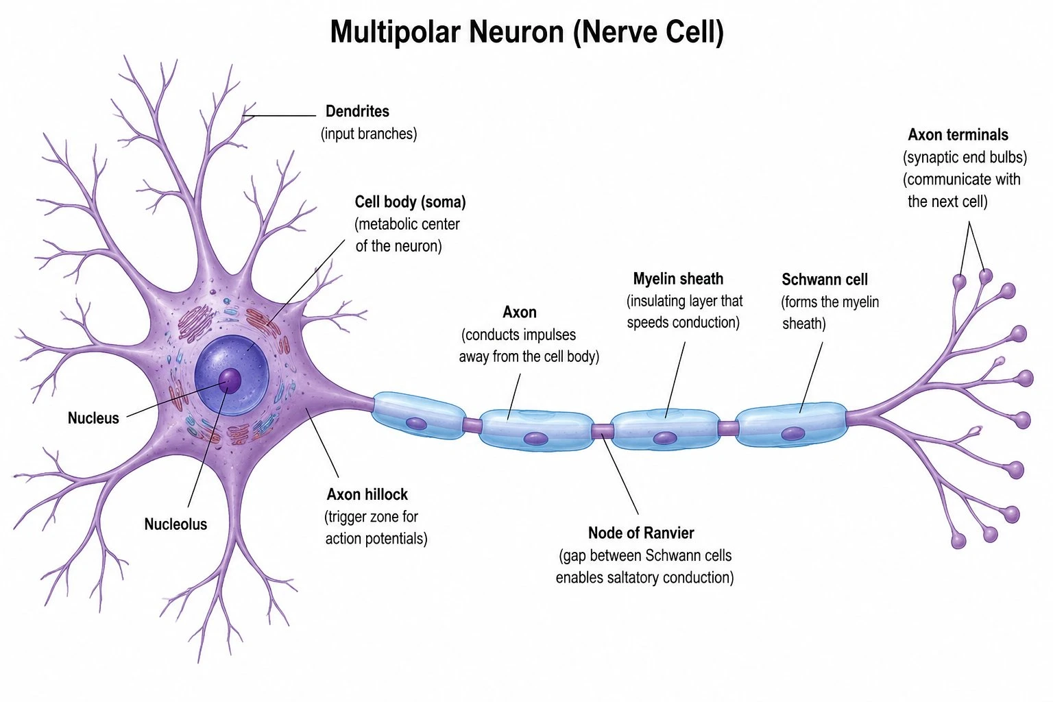
Neuron Diagram Generator
Nerve-cell maps that pair with a contraction unit.
 Biology
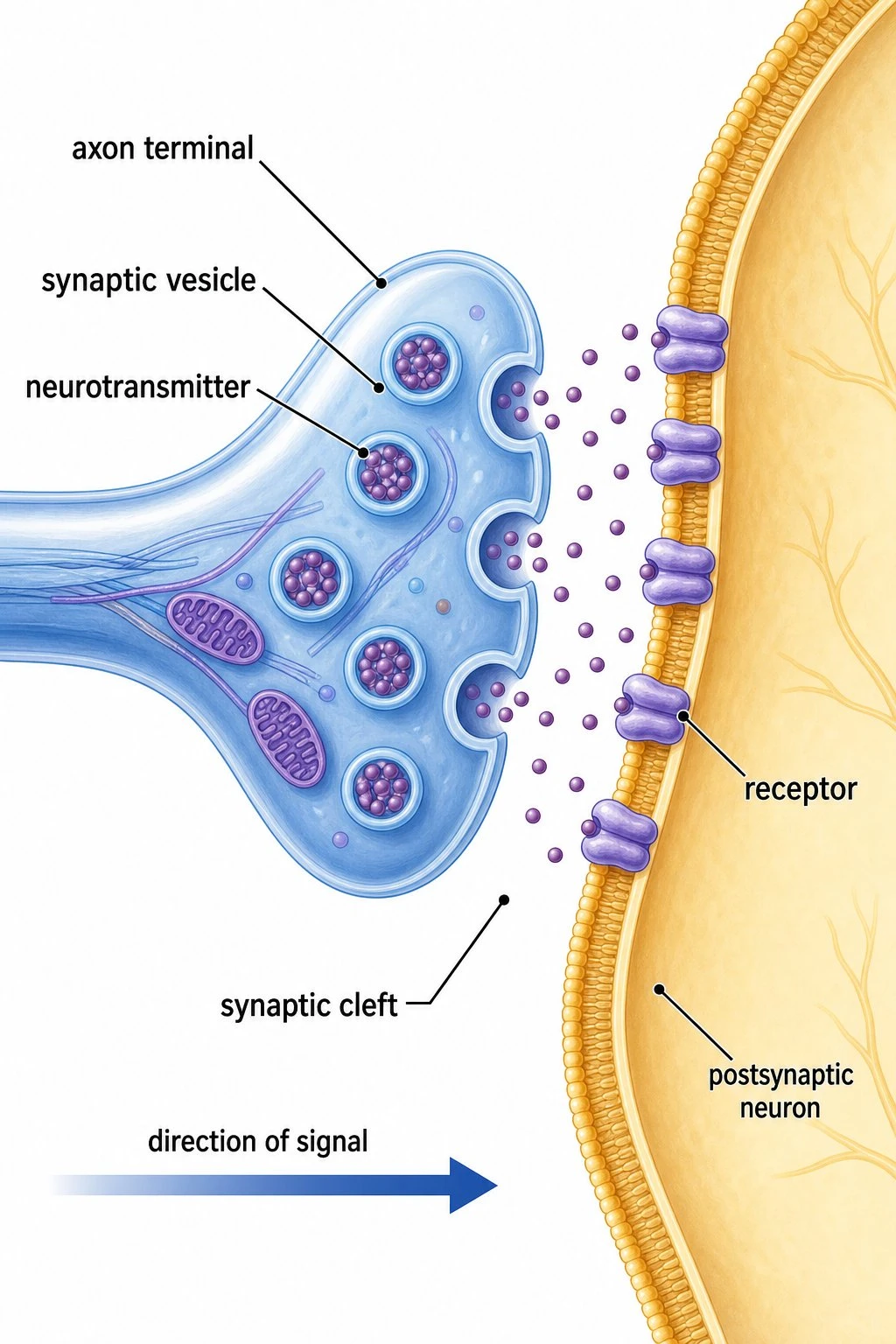
Synapse Diagram
Labeled chemical synapse for the same physiology week.
 Biology
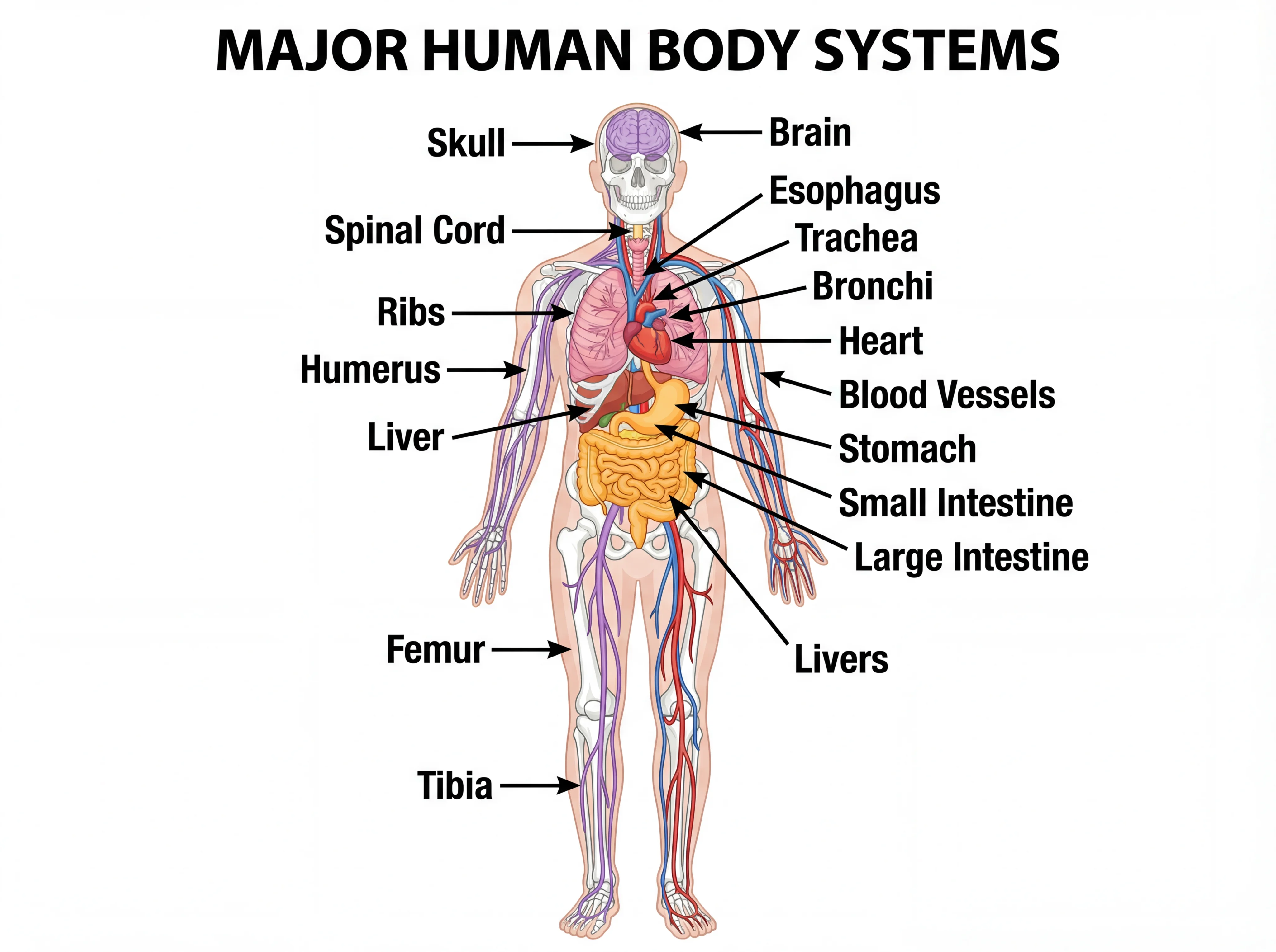
Human Body Systems
Overview diagrams of major body systems.
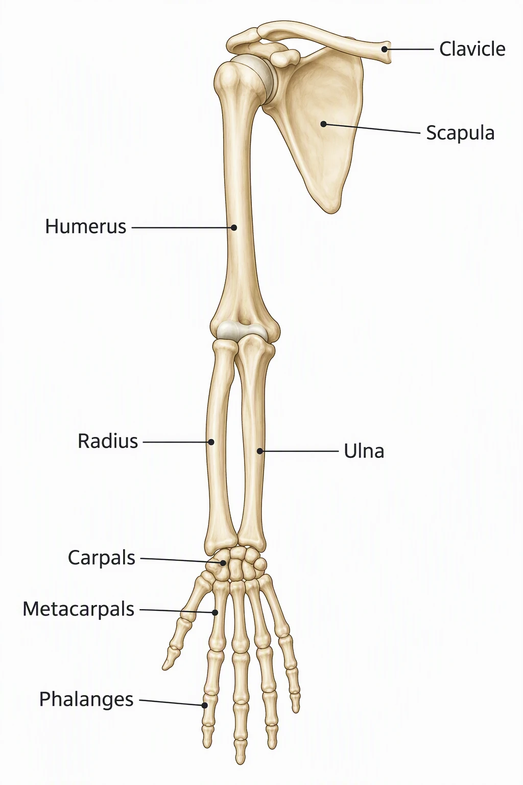 Medical
Bones of the Arm Diagram
Skeletal close-up for a combined muscle-and-bone week.