Neuron Diagram Generator Labeled Nerve Cell Diagrams
Create labeled neuron diagrams with AI. Show the dendrites, cell body (soma), nucleus, axon, myelin sheath, nodes of Ranvier, axon terminals, and Schwann cells — or how a nerve signal travels — for biology and neuroscience class. Download as PNG.
AI Neuron Diagram Generator
Free to try ·
Your neuron diagram will appear here
Describe the type and parts to label
Neuron Diagram Examples
Labeled diagrams of neuron parts, types, nerve signals, and blank worksheets
Labeled Neuron Diagram
A complete labeled diagram of a multipolar neuron with every major structure identified.
Simple Neuron for Kids
A clean, color-coded version with the four key parts — ideal for middle school students.
Neuron Types Comparison
The three main neuron types compared in one diagram — multipolar, bipolar, and unipolar.
How a Nerve Signal Travels
Step-by-step illustration of how an electrical signal travels from dendrite to axon terminal.
Myelin Sheath & Nodes of Ranvier
A zoomed-in view of the insulating myelin sheath and the exposed nodes that speed signal transmission.
Blank Neuron Worksheet
An unlabeled neuron outline with numbered blanks — ready to print as a quiz or worksheet.
What does a neuron diagram show?
A neuron diagram shows the structure of a nerve cell and its specialized parts for receiving, processing, and transmitting electrical signals. A standard labeled diagram includes the dendrites (input branches), the cell body or soma (which contains the nucleus and carries out cell functions), the axon hillock (where signals are initiated), the axon (the long cable that carries the signal), and at the far end, the axon terminals (which release neurotransmitters). Many diagrams also show the myelin sheath, Schwann cells, and nodes of Ranvier that wrap and insulate the axon to speed up signal conduction. This generator creates clear, labeled neuron diagrams for biology and neuroscience classes.
The main parts of a neuron
- Dendrites: branching extensions that receive incoming signals from other neurons and carry them toward the cell body.
- Cell body (soma): the metabolic center of the neuron, containing the nucleus and organelles that keep the cell alive.
- Axon hillock: the junction between the soma and the axon where an action potential is triggered if the incoming signal is strong enough.
- Axon: the long, slender projection that carries electrical impulses away from the cell body toward the axon terminals.
- Myelin sheath: a fatty insulating layer produced by Schwann cells (in the peripheral nervous system) or oligodendrocytes (in the central nervous system) that wraps the axon and greatly speeds up signal conduction.
- Nodes of Ranvier: tiny gaps in the myelin sheath where the electrical signal "jumps" (saltatory conduction), making transmission faster.
- Axon terminals: the branching endpoints of the axon that release neurotransmitter chemicals into the synapse to signal the next neuron or muscle cell.
Types of neurons
Neurons are classified by the number of processes extending from the soma. Multipolar neurons — the most common type, including motor neurons — have many dendrites and one axon. Bipolar neurons have one dendrite and one axon and are found in sensory organs like the retina and olfactory epithelium. Unipolar (pseudounipolar) neurons have a single process that splits close to the cell body; they carry sensory signals (touch, pain, temperature) from the body to the spinal cord. Knowing the type is important for any neuron diagram because each has a distinct shape.
How a nerve signal travels
A neuron receives signals through its dendrites. If the combined input at the axon hillock exceeds a threshold, an action potential fires: sodium ions rush into the axon, creating a wave of electrical charge that sweeps down the axon toward the terminals. In myelinated axons, the signal jumps between nodes of Ranvier (saltatory conduction), which dramatically increases speed. At the axon terminals, the arriving signal triggers the release of neurotransmitters across the synapse, binding to receptors on the next neuron or target cell — and the chain continues.
Tips for a clear neuron diagram
Choose the view that fits your lesson — a complete labeled diagram for study notes, a simplified version for younger students, a type-comparison for a unit on neuron classification, or a close-up of the myelin sheath for signal-speed topics. Specify which parts to label and whether you want a fully labeled or blank worksheet version. Generate a few options and download the one that best matches your level of detail.
Frequently Asked Questions
Related Biology Tools
 Biology
Nervous System Diagram Generator
Create labeled diagrams of the central and peripheral nervous system, including the brain and spinal cord.
 Biology
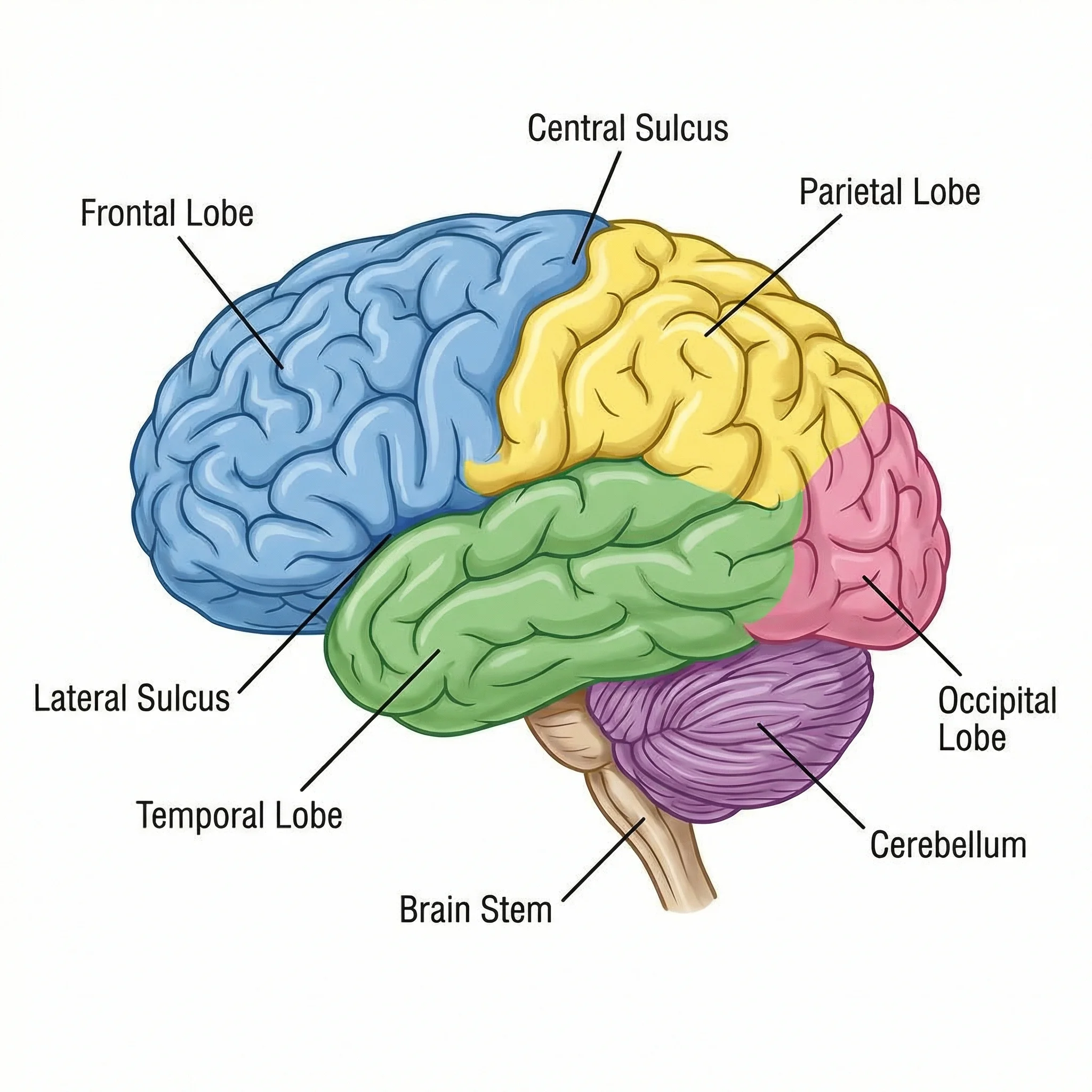
Brain Diagram Generator
Generate labeled brain diagrams showing lobes, regions, and key structures.
 Biology
Human Body Systems Diagram Generator
Create labeled diagrams of the nervous, circulatory, respiratory, digestive, and other body systems.