Muscular System Diagram Generator for Labeled & Blank Diagrams
Create a clearly labeled muscular system diagram in seconds. Show major muscles in anterior and posterior views, compare the three muscle types (skeletal, smooth, cardiac), or generate a blank diagram for worksheets and quizzes. Free to use.
Muscular System Diagram Generator
Free to try ·
Your muscular system diagram will appear here
Describe what you need and click Generate
Muscular System Diagram Examples
Labeled diagrams of anterior and posterior views, muscle types, major muscle groups, and blank worksheets
Full Muscular System — Anterior
Every major muscle named from the front — deltoid, pectorals, biceps, abs, quads, and more.
Muscular System — Posterior View
The muscles from behind — trapezius, lats, triceps, glutes, hamstrings, and calves labeled.
Simple Muscular System for Kids
A simplified, friendly diagram with big labels — perfect for elementary and middle school science.
Three Muscle Types
Skeletal, smooth, and cardiac muscle compared side by side with microscopic detail.
Major Muscle Groups
Color-coded muscle groups across the whole body — chest, back, shoulders, core, and legs.
Blank Muscular System Worksheet
Numbered blank lines, no labels — print and fill in as a quiz or anatomy study activity.
What is the muscular system?
The muscular system is the network of over 600 muscles that powers every movement the body makes. Muscles work by contracting — shortening to pull bones closer together — and then relaxing, allowing another muscle on the opposite side to pull back. This system does far more than move limbs: it also pumps the heart, moves food through the digestive tract, controls breathing, and maintains posture. This generator draws the muscular system with every major muscle clearly labeled so you can see exactly where each one sits and what it does.
The three muscle types
- Skeletal muscle: the voluntary muscles attached to bones by tendons. They are striated (striped under the microscope), under conscious control, and responsible for all deliberate movement — walking, lifting, chewing, and blinking. Each fiber is a long, cylindrical cell with multiple nuclei.
- Smooth muscle: the involuntary muscle found in the walls of hollow organs — the stomach, intestines, blood vessels, bladder, and uterus. It is not striated, has a single central nucleus in each spindle-shaped cell, and contracts slowly and rhythmically without conscious control.
- Cardiac muscle: found only in the heart. Like skeletal muscle it is striated, but like smooth muscle it is involuntary and contracts automatically. Cardiac cells (cardiomyocytes) are branched and connected by intercalated discs that allow electrical signals to spread rapidly, making the heart beat as one coordinated unit.
Major muscles of the anterior (front) view
Starting at the neck and working down: the sternocleidomastoid runs from the sternum to the skull and rotates the head. The trapezius covers the upper back and neck. The deltoid caps the shoulder. The pectoralis major covers the chest and pulls the arm across the body. The biceps brachii flexes the elbow. The rectus abdominis (the "six-pack") and the external obliques form the abdominal wall. The iliopsoas flexes the hip. The quadriceps group — rectus femoris, vastus lateralis, vastus medialis, and vastus intermedius — extends the knee. The tibialis anterior on the shin dorsiflexes the foot. The gastrocnemius and soleus form the calf.
Major muscles of the posterior (back) view
The trapezius spans the upper back and neck, elevating and retracting the scapula. The latissimus dorsi is the broad back muscle that extends and adducts the arm. The rhomboids and infraspinatus sit between and over the shoulder blades. The erector spinae group runs the length of the spine, maintaining an upright posture. The triceps brachii on the back of the upper arm extends the elbow. The gluteus maximus is the largest muscle in the body and extends the hip. The hamstrings — biceps femoris, semitendinosus, and semimembranosus — flex the knee and extend the hip. The gastrocnemius and the Achilles tendon anchor the calf to the heel.
How muscles create movement
- Antagonistic pairs: muscles work in opposing pairs. When the biceps brachii contracts to flex the elbow, the triceps brachii relaxes; when the triceps contracts to extend the elbow, the biceps relaxes. This push-pull arrangement is the fundamental principle behind joint movement.
- Origin and insertion: each skeletal muscle attaches at two points. The origin is the more stationary attachment (usually the proximal bone); the insertion is the attachment that moves (usually the distal bone). When the muscle contracts, the insertion moves toward the origin.
- Tendons and muscle tone: tendons are the tough connective tissue cords that attach muscles to bones. Even at rest, most skeletal muscles maintain a low level of contraction called muscle tone, which keeps the body ready to move and helps maintain posture.
Labeled vs blank diagrams for worksheets and quizzes
A fully labeled diagram is ideal for study notes, lectures, and handouts, while a blank, unlabeled version is what you need for student worksheets and quizzes. Ask for "a labeled muscular system diagram" to get every muscle named, or "a blank muscular system worksheet with numbered blank lines" to get a printable quiz sheet. Black-and-white line art works best for printing, and you can generate a matching labeled version to use as the answer key. You can also ask for simplified versions for younger students, or detailed regional close-ups — the shoulder, the arm, the leg — for deeper study.
How to generate a labeled muscular system diagram
- Describe what you need in plain English — the full muscular system, anterior or posterior view, the three muscle types, a specific muscle group such as the shoulder or leg, or a blank worksheet.
- Specify the view and labeling style you want, for example "anterior full-body view with every major muscle labeled" or "posterior view highlighting the back and hamstring muscles."
- Choose labeled for teaching or unlabeled (blank) for a worksheet or quiz, then generate the diagram.
- Review the result for accuracy, refine your description if needed, and download the image to use in a slide deck, handout, or study guide.
Frequently Asked Questions
Related Biology Tools
 Biology
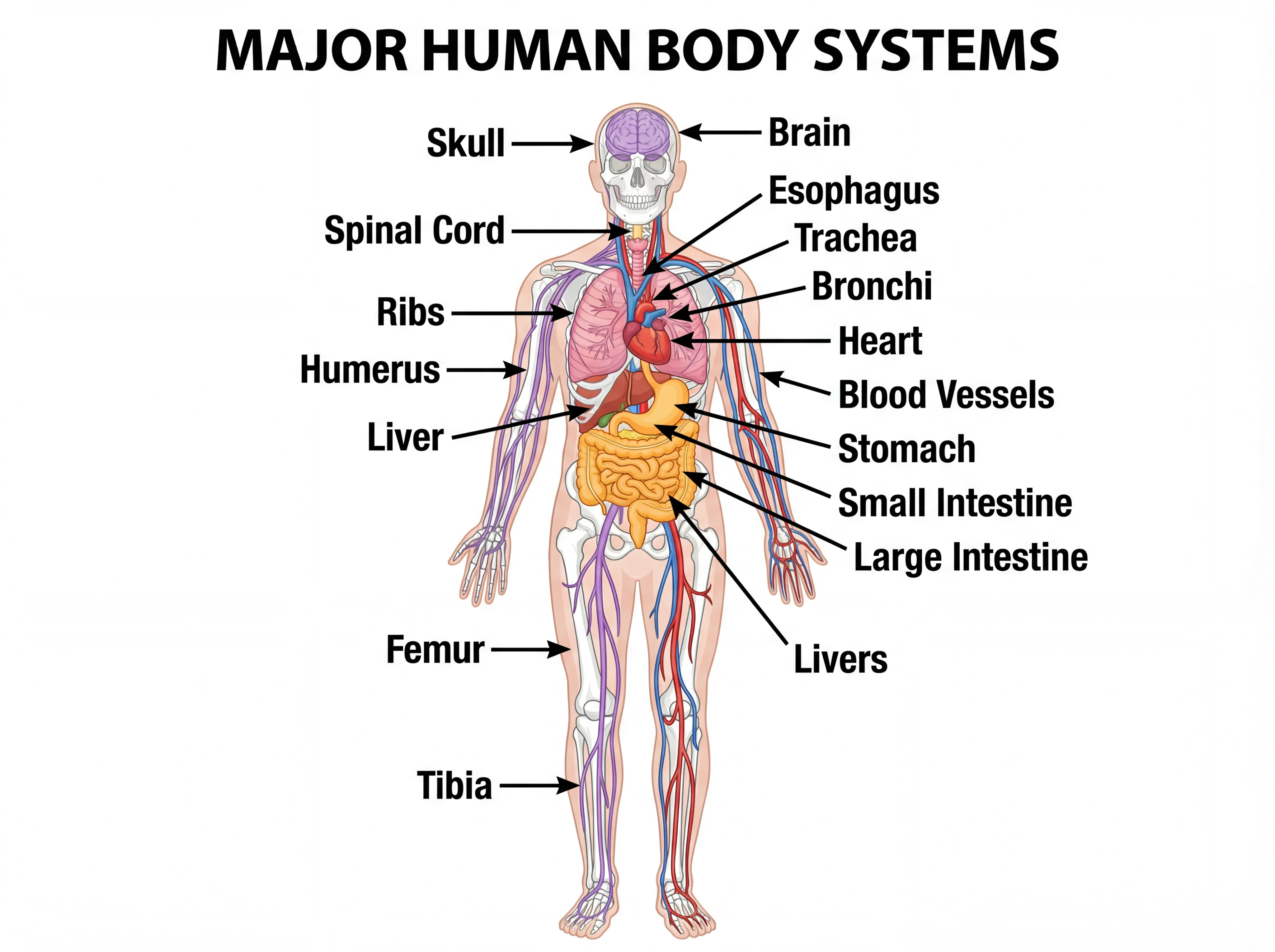
Human Body Systems Diagram Generator
Draw labeled diagrams of all the body's organ systems — skeletal, muscular, nervous, circulatory, and more.
 Biology
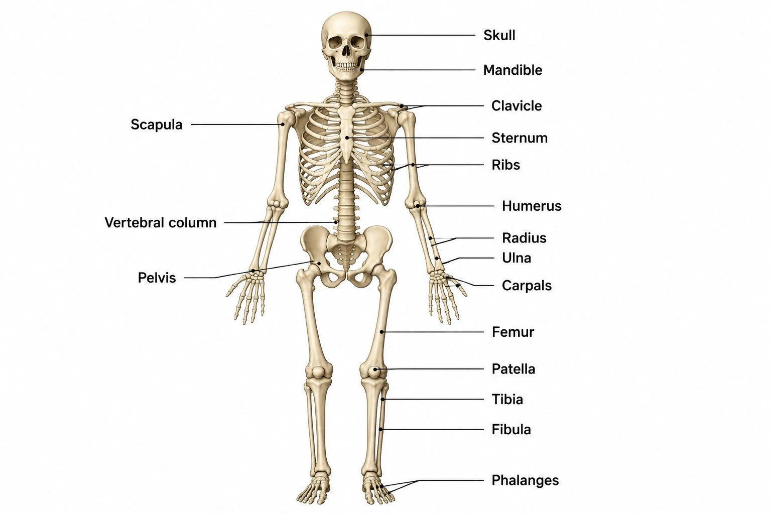
Skeletal System Diagram Generator
Create labeled skeletal system diagrams showing all 206 bones, the axial and appendicular divisions, and joint types.
 Biology
Nervous System Diagram Generator
Create labeled nervous system diagrams showing the brain, spinal cord, and major peripheral nerves.