Synapse Diagram Labeled, Blank & Printable
Free synapse diagrams — labeled chemical synapse, blank worksheet, simple kids version, signal steps, and coloring. Download or print; no sign-up.
Free Synapse Diagrams
Every version below is free to view and download — no sign-up. For the whole nerve cell, see the Neuron Diagram tool.
Labeled Chemical Synapse
Full junction view — vesicles, cleft, receptors, and direction of signal.
Core diagrams
Structure & focus
Print & practice
Test yourself: synapse
A quick self-test — questions reshuffle each time, with a review of anything you miss at the end.
In a chemical synapse, the message crosses the gap mainly as…
Need a custom version?
Change the grade level, which parts are labeled, or simplify the cleft — describe it and generate your own.
Customize Your Synapse Diagram
Free to try ·
Your custom synapse diagram will appear here
Ask for a labeled junction, blank worksheet, or simple kids version
What is a synapse diagram?
A synapse diagram shows the junction where one neuron passes a signal to another cell. Most school diagrams show a chemical synapse: the axon terminal holds vesicles of neurotransmitter, a tiny synaptic cleft separates the cells, and receptors sit on the postsynaptic membrane. This is different from a full neuron diagram, which shows dendrites, cell body, and axon at a larger scale. Use a labeled synapse for notes, or a blank version as a worksheet.
Parts of a chemical synapse
- Presynaptic terminal (axon ending): where the signal arrives and vesicles wait.
- Synaptic vesicles: small packets of neurotransmitter.
- Synaptic cleft: the gap the chemical crosses.
- Postsynaptic membrane and receptors: detect the neurotransmitter and continue the message.
- Direction: usually drawn left-to-right or top-to-bottom from sending cell to receiving cell.
Synapse vs whole neuron
Searchers looking for “synapse diagram” want the zoomed junction. “Neuron diagram” is the whole cell. Keep them as hub and spoke: this page owns the synapse; our Neuron Diagram tool covers the full cell with dendrites and axon. Cross-link both so students see structure at two scales.
How to use these free diagrams
Teach with the labeled chemical synapse, then assess with the blank worksheet. The simple version works for introductory middle school; the steps poster supports “how does the signal cross?” lessons. The neuron-context figure answers “where is this on the cell?” All files are free to view, download, and print with no sign-up.
Frequently Asked Questions
Related Biology Tools
 Biology
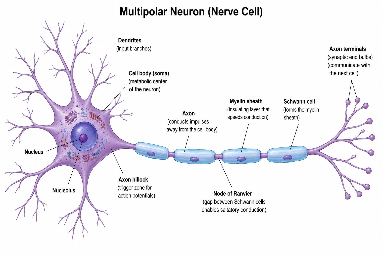
Neuron Diagram
Labeled whole neuron — dendrites, cell body, axon, and terminals.
 Biology
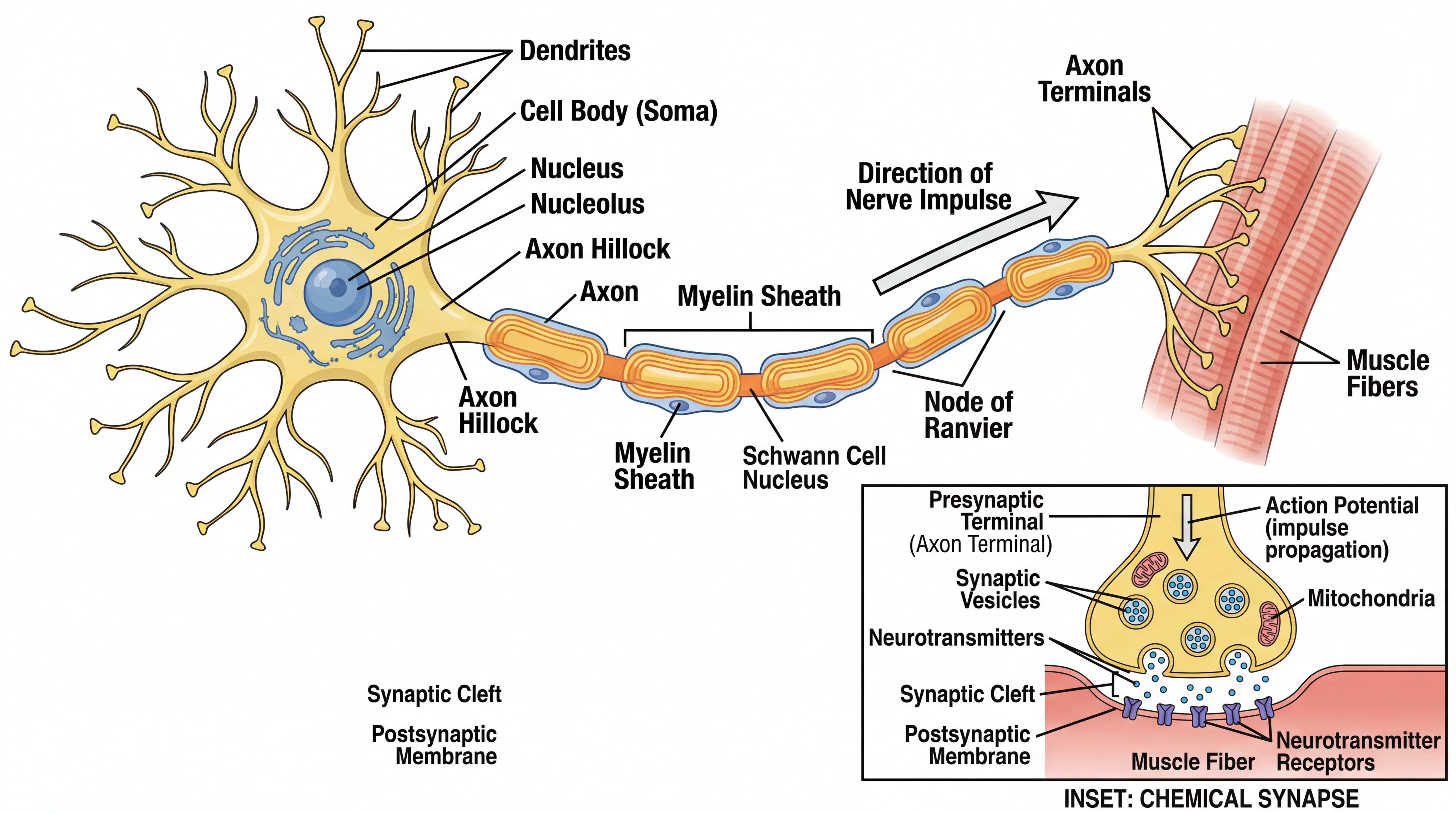
Nervous System Diagram
Brain, spinal cord, and nerve maps for system-level lessons.
 Biology
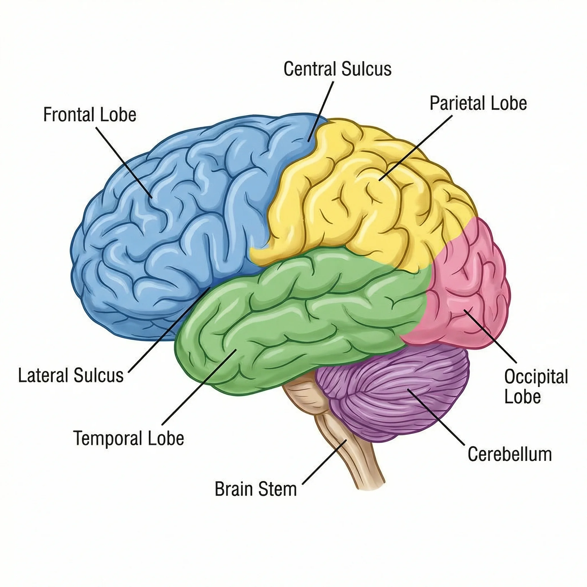
Brain Diagram
Labeled brain views for CNS units that use synapses heavily.
 Biology
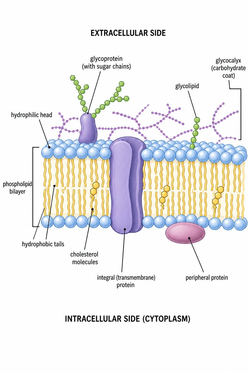
Cell Membrane Diagram
Fluid mosaic model — receptors sit in membranes at synapses.
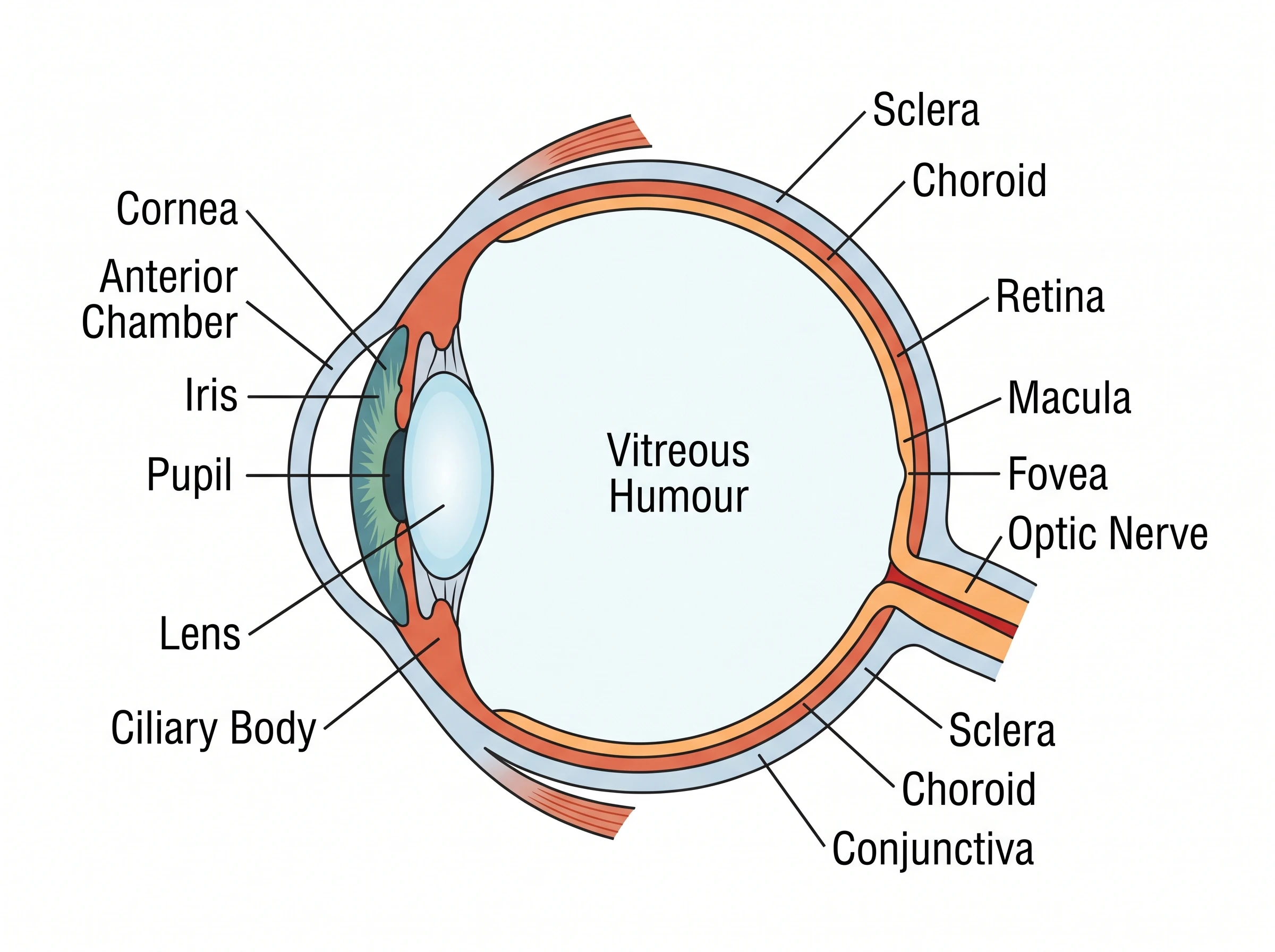 Medical
Eye Anatomy Diagram
Sensory pathway context for nervous system units.
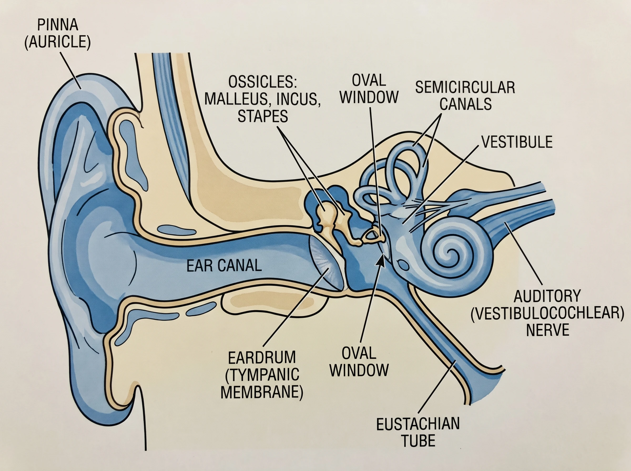 Medical
Ear Anatomy Diagram
Another sensory organ pathway that ends in neural signals.