Nephron Diagram Labeled, Blank & Printable
Free nephron diagrams — labeled, blank, simple, glomerulus focus, and coloring. View, download, or print; no sign-up.
Free Nephron Diagrams
Every version below is free to view and download — no sign-up.
Labeled Nephron Diagram
Full functional unit labeled — corpuscle, tubules, loop of Henle, and collecting duct.
Core diagrams
Structure & focus
Print & practice
Test yourself: nephron
A quick self-test — questions reshuffle each time, with a review of anything you miss at the end.
What is a nephron?
Need a custom version?
Change the grade level, which parts are labeled, or focus on the glomerulus — describe it and generate your own.
Customize Your Nephron Diagram
Free to try ·
Your custom nephron diagram will appear here
Ask for a labeled nephron, blank worksheet, or glomerulus focus
What is a nephron diagram?
A nephron diagram shows the functional unit of the kidney — the tiny tube that filters blood and adjusts water and solutes to form urine. Real nephrons are microscopic, so diagrams enlarge one unit: the glomerulus and Bowman's capsule (renal corpuscle), the proximal and distal convoluted tubules, the loop of Henle, and the collecting duct, often with afferent and efferent arterioles. Use a labeled version to learn the order of parts, or a blank version as a quiz.
Parts of the nephron
- Glomerulus: a ball of capillaries where blood is filtered under pressure.
- Bowman's (glomerular) capsule: the cup that catches filtrate around the glomerulus.
- Proximal convoluted tubule: reabsorbs much of the water, ions, and nutrients from filtrate.
- Loop of Henle: descends into the medulla and helps create the salt gradient for concentrating urine.
- Distal convoluted tubule: further adjusts ions and pH under hormonal control.
- Collecting duct: receives fluid from many nephrons and finalizes water reabsorption; urine then moves toward the pelvis.
Nephron in the kidney
Each human kidney contains roughly one million nephrons. Corpuscles sit in the cortex; loops of Henle and collecting ducts run into the medulla. Blood arrives via branches of the renal artery; filtered blood leaves via the renal vein; urine exits through the ureter. A “nephron in kidney” diagram links one unit to the whole organ — useful before zooming into structure alone.
How to use these free diagrams
Teach with the fully labeled nephron, then assess with the matching blank worksheet. The simple version introduces the filter → tube → loop idea for younger students. The glomerulus focus supports lessons on filtration pressure and arterioles. The kidney-context figure answers “where is this inside the organ?” All assets are free to view, download, and print with no sign-up.
Frequently Asked Questions
Related Biology Tools
 Biology
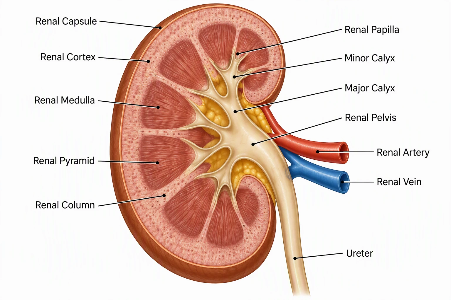
Kidney Diagram
Labeled kidney cross-sections, filtration overview, and worksheets.
 Biology
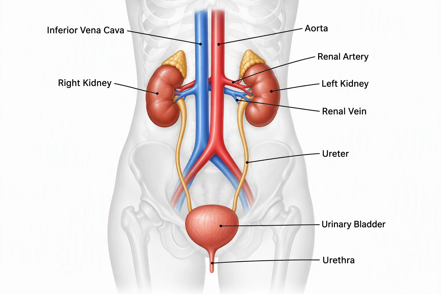
Urinary System Diagram
Full urinary tract — kidneys, ureters, bladder, and urethra.
 Biology
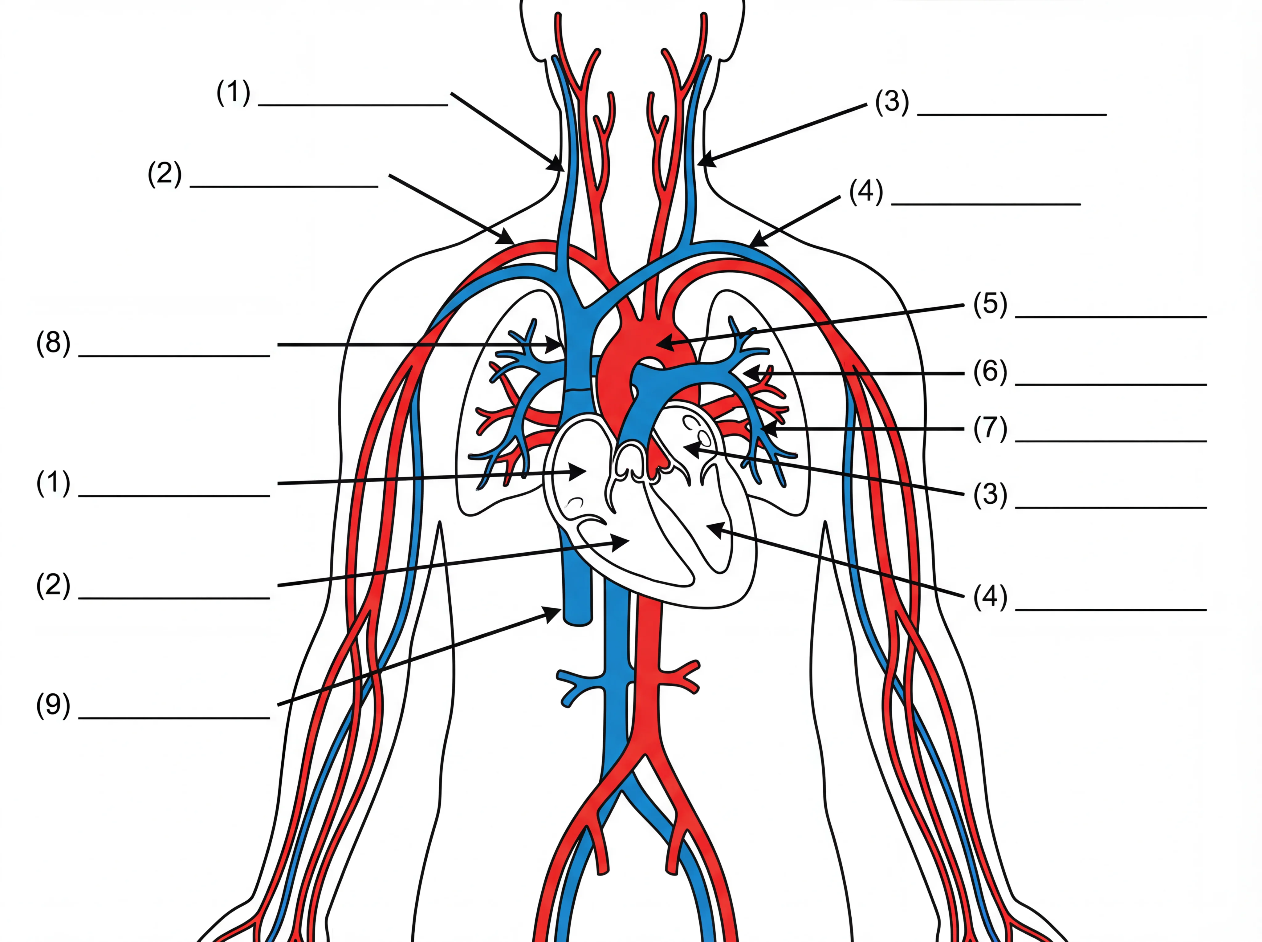
Human Body Systems
Overview diagrams of major body systems including excretion.
 Biology
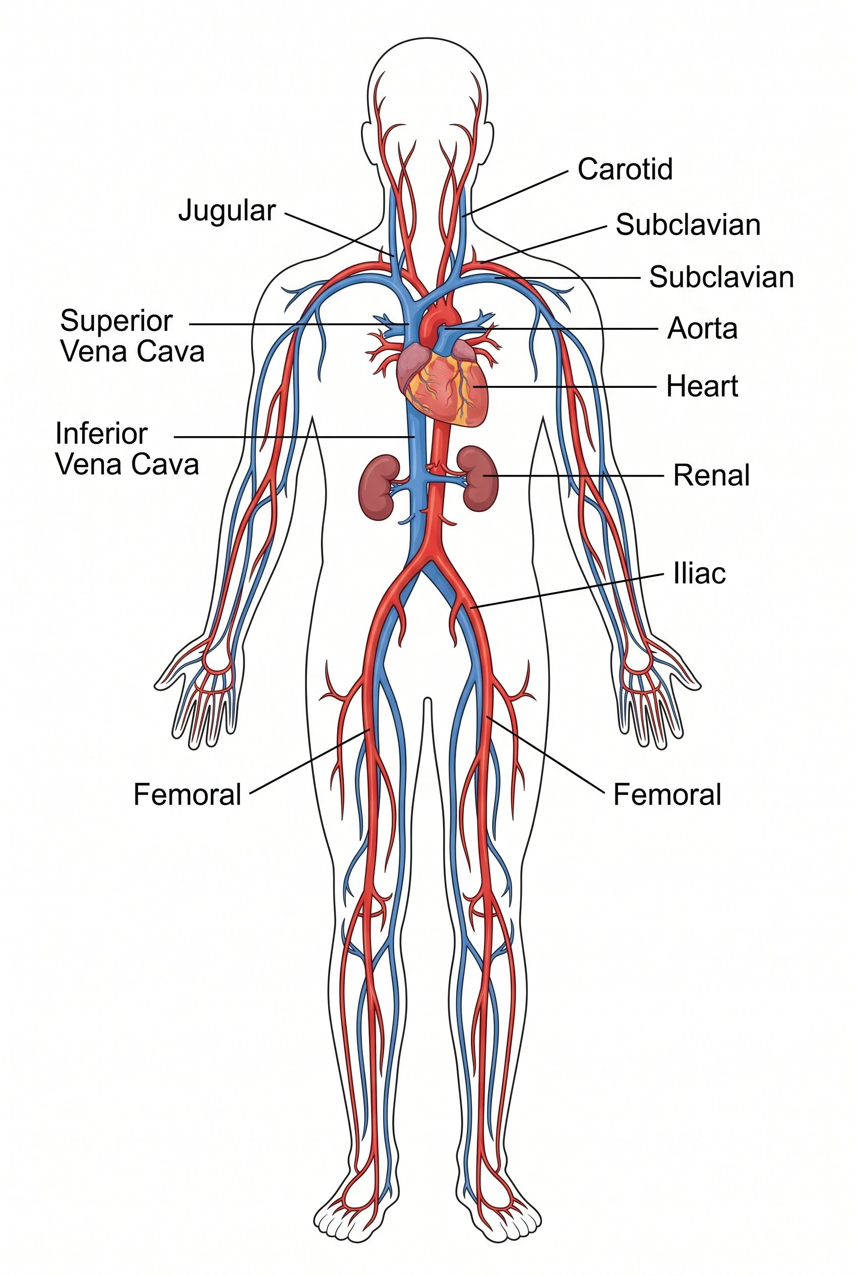
Circulatory System Diagram
Heart and vessel maps that connect to renal blood flow.
 Biology
Cell Membrane Diagram
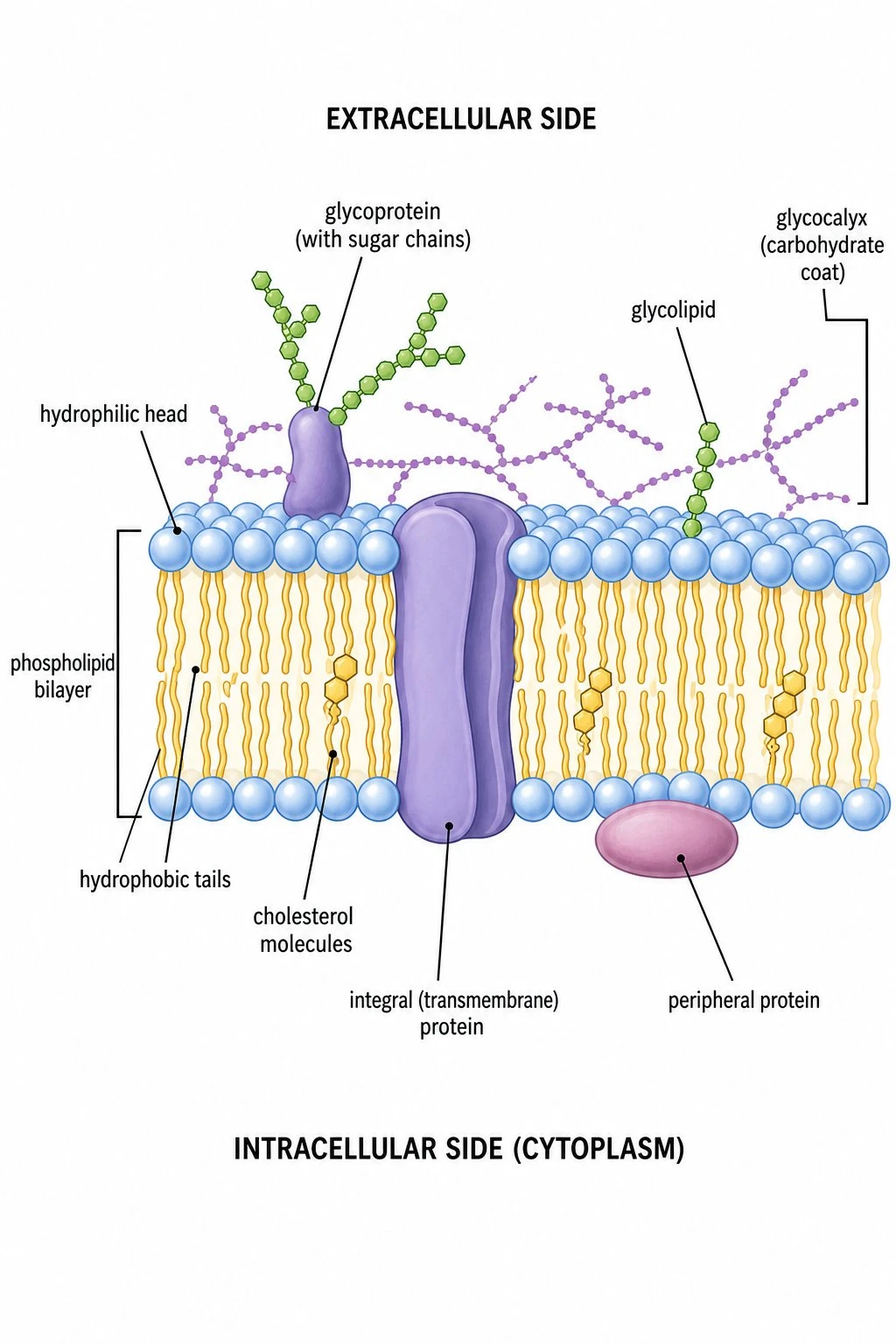
Fluid mosaic model — how membranes filter and transport at cell scale.
 Biology
Neuron Diagram
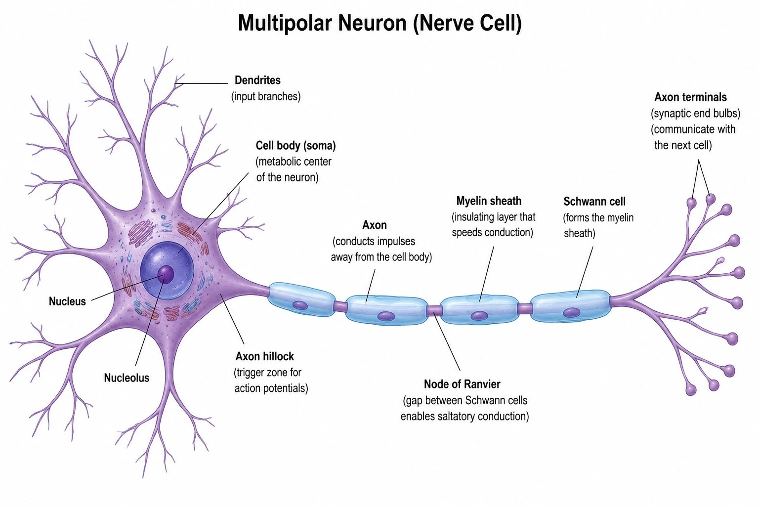
Labeled neuron structure for nervous system comparison units.