Kidney Diagram Generator Labeled Kidney Diagrams
Create labeled kidney diagrams with AI. Show the renal cortex, medulla, renal pyramids, renal pelvis, renal artery, renal vein, ureter, and a nephron close-up — cross-sections, filtration-flow diagrams, and blank worksheets. Download as PNG.
AI Kidney Diagram Generator
Free to try ·
Your kidney diagram will appear here
Describe the view and parts to label
Kidney Diagram Examples
Labeled cross-sections, nephron close-ups, filtration diagrams, and blank worksheets
Labeled Kidney Cross-Section
A full kidney cross-section with every major structure labeled — cortex, medulla, pyramids, pelvis, vessels, and ureter.
Simple Kidney Diagram
A clean, simplified kidney diagram with the key parts — great for younger students.
Nephron Close-Up
A detailed nephron diagram — glomerulus, Bowman's capsule, loop of Henle, and collecting duct.
How the Kidney Filters Blood
The filtration flow — renal artery → nephrons → collecting ducts → ureter — with labeled arrows.
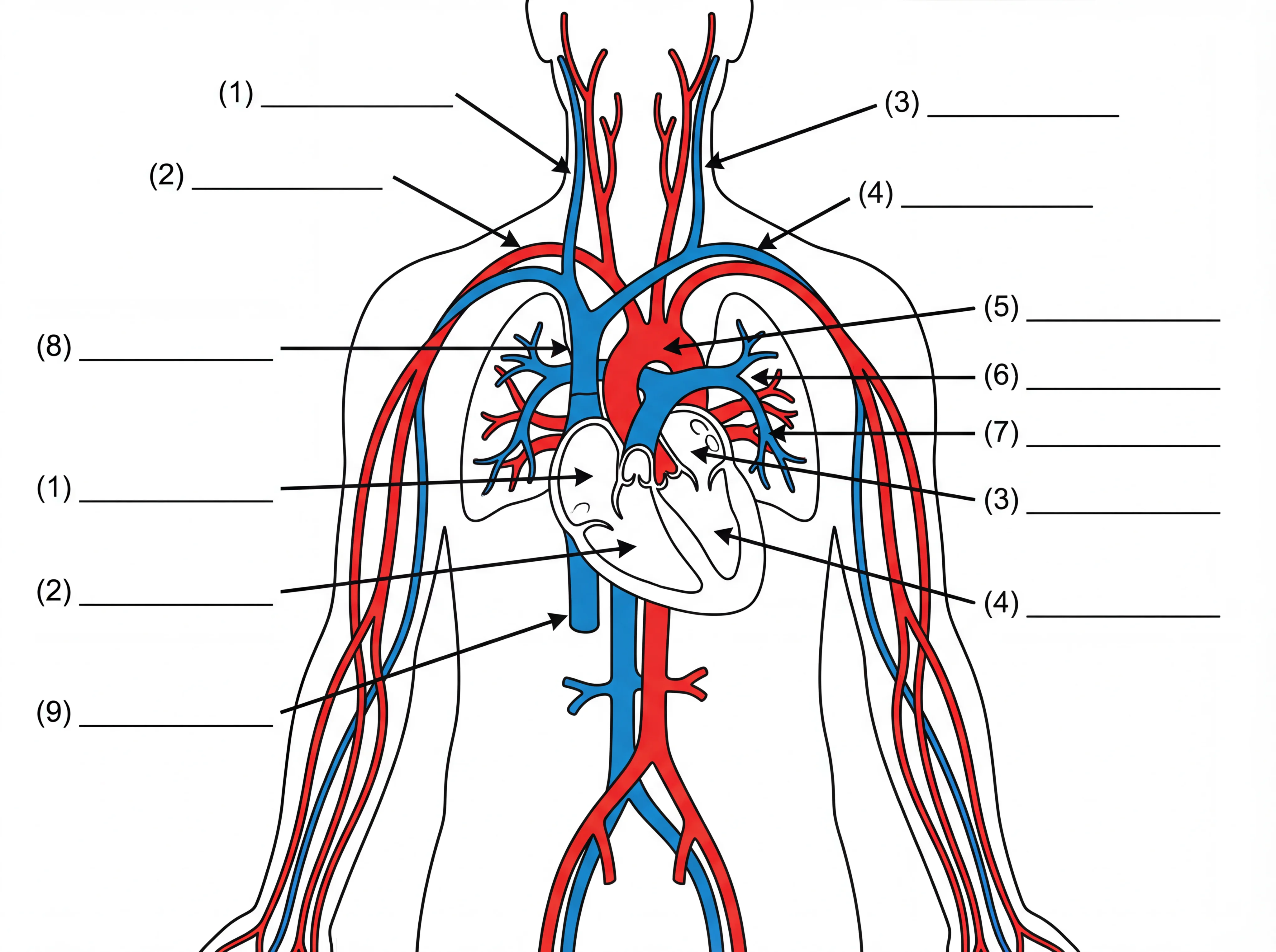
Kidney in the Urinary System
The kidney in context — its place in the full urinary system.
Blank Kidney Worksheet
An unlabeled kidney outline with numbered blanks — ready to print as a worksheet or quiz.
What does a kidney diagram show?
A kidney diagram shows the internal structure of the human kidney and the structures it contains. A labeled cross-section typically identifies the renal cortex (the outer layer), the renal medulla (the inner region), the renal pyramids (cone-shaped structures within the medulla), the renal columns, the renal pelvis (the funnel-shaped collecting space), the major and minor calyces, the renal artery, the renal vein, and the ureter. A nephron close-up adds the glomerulus, Bowman's capsule, the proximal and distal convoluted tubules, the loop of Henle, and the collecting duct. This generator creates clear, labeled kidney diagrams for biology, anatomy, and health class.
The main parts of the kidney
- Renal cortex: the outer layer of the kidney, where most of the nephrons' filtering units are located.
- Renal medulla: the inner region, organized into renal pyramids (cone-shaped sections) separated by renal columns.
- Renal pyramids and calyces: each pyramid drains filtered fluid into a minor calyx, which merges into a major calyx, feeding the renal pelvis.
- Renal pelvis: the large funnel-shaped cavity that collects urine and channels it into the ureter.
- Renal artery and vein: the renal artery brings blood in to be filtered; the renal vein carries filtered blood back to circulation.
- Ureter: the tube that carries urine from the renal pelvis down to the bladder.
The nephron — the kidney's functional unit
Each kidney contains about one million nephrons — the tiny structures that actually filter the blood. Blood enters a nephron through the glomerulus, a ball of capillaries inside the Bowman's capsule. Fluid is forced out of the blood into the Bowman's capsule (filtration). The filtrate then travels through the proximal convoluted tubule, dips into the loop of Henle (which concentrates the urine), and passes through the distal convoluted tubule before emptying into a collecting duct. Along the way, useful molecules (glucose, amino acids, water) are reabsorbed back into the bloodstream, and waste products are secreted into the filtrate, leaving concentrated urine to drain into the renal pelvis.
How the kidney filters blood
Blood arrives at the kidney through the renal artery, which branches into smaller arterioles leading to the glomeruli. Pressure inside the glomeruli forces water and small molecules (including waste products like urea and creatinine) into the Bowman's capsule, starting the filtration process. The nephron then fine-tunes the filtrate — reabsorbing water to concentrate urine, reclaiming glucose and ions, and secreting additional waste — before the urine collects in the renal pelvis and flows down the ureter to the bladder. On a good day, the kidneys filter about 180 litres of fluid but produce only 1–2 litres of urine.
Tips for a clear kidney diagram
Choose the view you need — a full labeled cross-section, a simplified version for younger learners, a nephron close-up, or a blood-filtration flow diagram with arrows. Name the structures you want labeled and specify the level of detail. For a worksheet, ask for an unlabeled version with numbered leader lines. Generate several options and download the clearest for your notes or handout.
Frequently Asked Questions
Related Biology Tools
 Biology
Human Body Systems Diagram Generator
Create labeled diagrams of the urinary, circulatory, respiratory, and other body systems.
 Biology
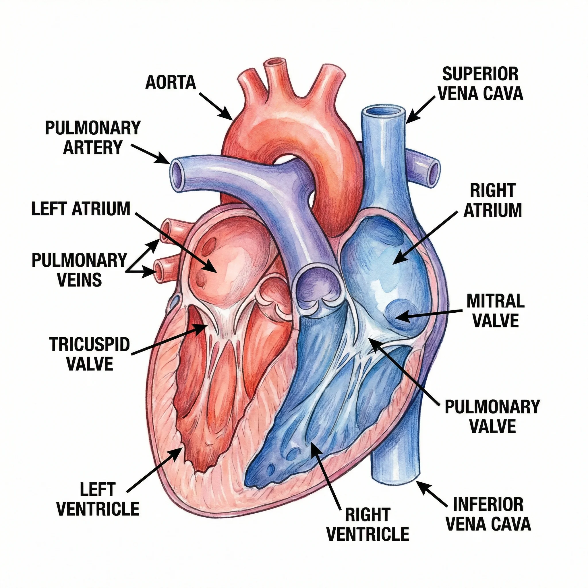
Heart Diagram Generator
Create labeled heart diagrams with chambers, valves, major vessels, and blood flow.
 Medical
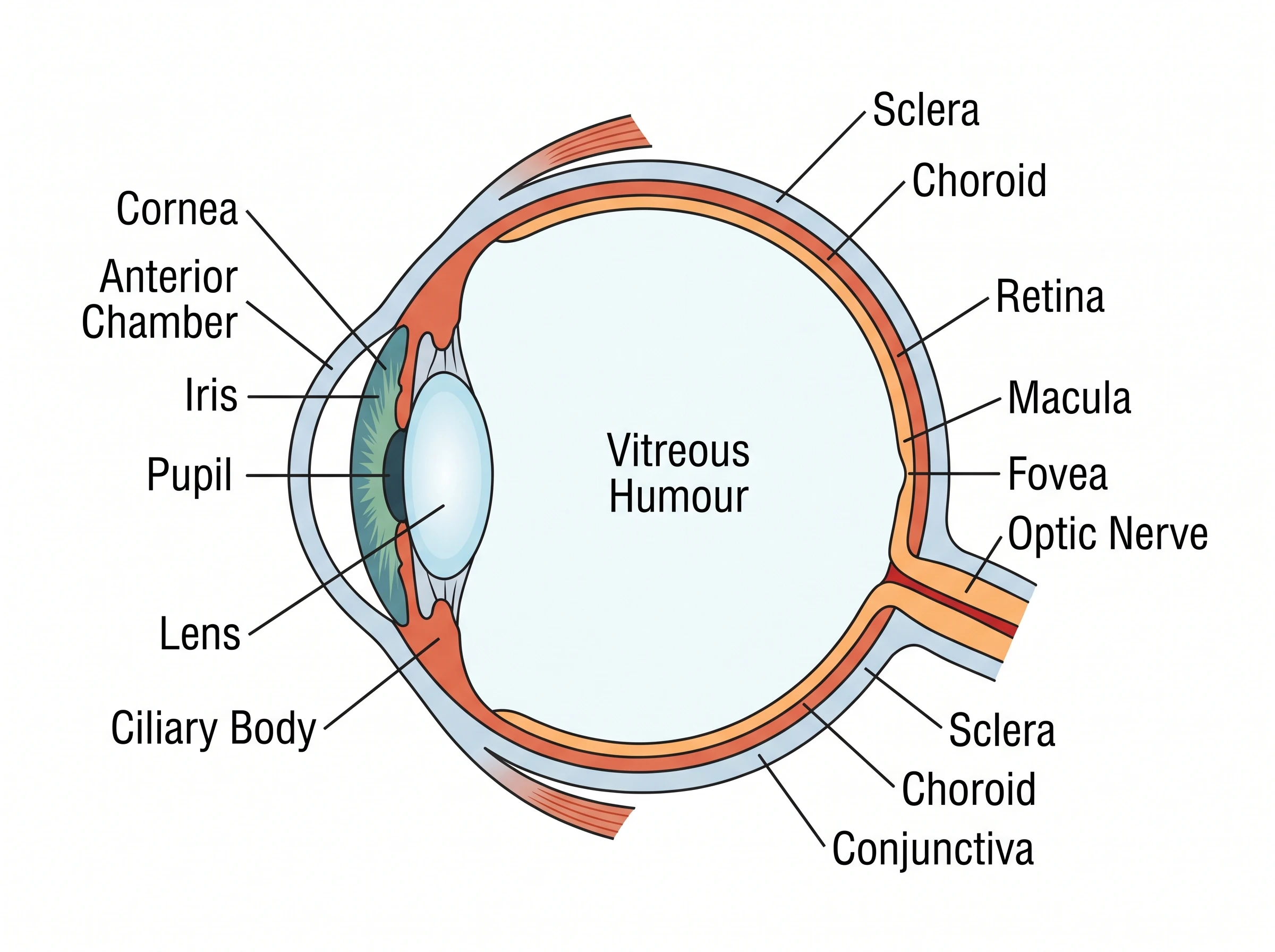
Eye Anatomy Diagram Generator
Create labeled eye diagrams — cornea, lens, retina, optic nerve — and how vision works.