ELISA Diagram Generator sandwich method figure
Free ELISA diagram generator. Draw a labeled sandwich, direct, indirect, or competitive method figure as SVG — not a western blot.
Method figure as SVG: sandwich, types, competitive, protocol, or plate map — free, no account
Figure options
Layer order is a teaching stack, not a plate-reader export. Types and competitive modes change what sits on the plate.
ELISA Diagram Generator
Preview is free on this page ·
Your AI ELISA illustration will appear here
For locked layer order, use the Precise Diagram tab instead
ELISA Diagram Examples
All six images are exact engine renders of the Precise Diagram tab.
Labeled sandwich
Exact engine render — plate, capture, antigen, detection, enzyme.
Blank worksheet
Exact engine render — same stack, names removed for revision.
Four types
Exact engine render — only sandwich keeps a capture antibody under the antigen.
Competitive
Exact engine render — more unlabeled sample means less labeled signal.
Protocol steps
Exact engine render — the sandwich built one addition at a time.
96-well plate
Exact engine render — plate map, not a gel or blot.
What an ELISA diagram is
An ELISA diagram is the method figure for an enzyme-linked immunosorbent assay: a well, what is coated on the plate, the antigen, and the antibody–enzyme stack that makes color. This page draws that lecture cartoon as an exact SVG. It is not a western blot and it is not an enzyme lock-and-key worksheet.
What the stacks lock
- Sandwich default: plate → capture antibody → antigen → detection antibody → enzyme. Capture sits under the antigen. The enzyme rides the detection antibody, not the capture antibody.
- Direct ELISA coats antigen on the plate. There is no capture antibody.
- Indirect ELISA adds a primary antibody, then an enzyme-conjugated secondary. The enzyme does not bind the antigen.
- Competitive ELISA: sample antigen and labeled antigen compete for the same capture antibody. More unlabeled sample means less color.
Labeled, blank, types, competitive, protocol, plate
Start with the labeled sandwich so the room can see the layer order. Use the blank copy for a self-test. Four types puts direct, indirect, sandwich, and competitive on one axis. Competitive isolates the competition cartoon. Protocol steps number coat, block, sample, detect, and substrate. The plate map is a top-down 96-well grid, not a gel.
Precise SVG vs AI illustration
The Precise Diagram tab is the version for class and papers: layer order is fixed in code. The AI Illustration tab is only for slide mockups. Check every capture and enzyme against the precise stacks before you project the file. Do not ask the AI tab for a western blot.
Frequently Asked Questions
Related Lab Tools
 Biology
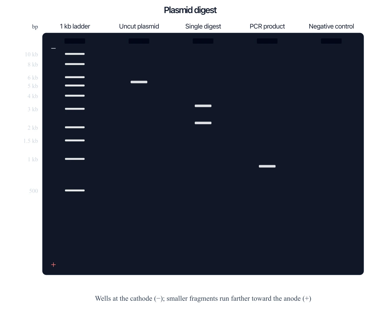
Gel Electrophoresis Diagram Generator
Lanes and fragment sizes — the blot’s gel, not the ELISA well.
 Biology
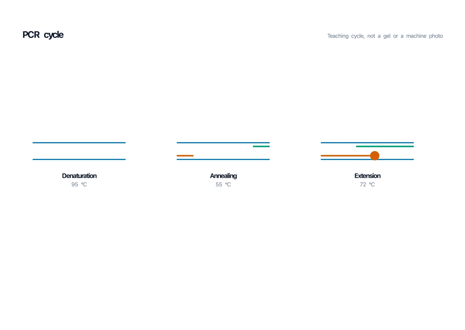
PCR Diagram Generator
Denature, anneal, extend — the DNA cycle, not an immunoassay well.
 Biology
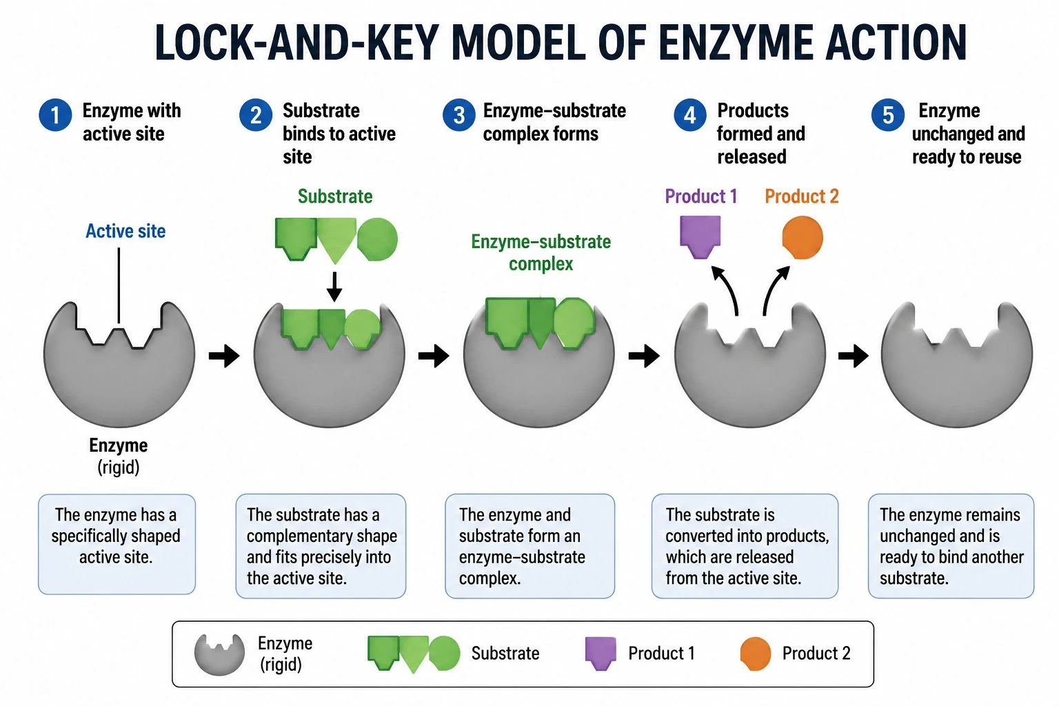
Enzyme Diagram Generator
Lock-and-key or induced fit — the enzyme as a molecule, not an ELISA conjugate.
 Medical
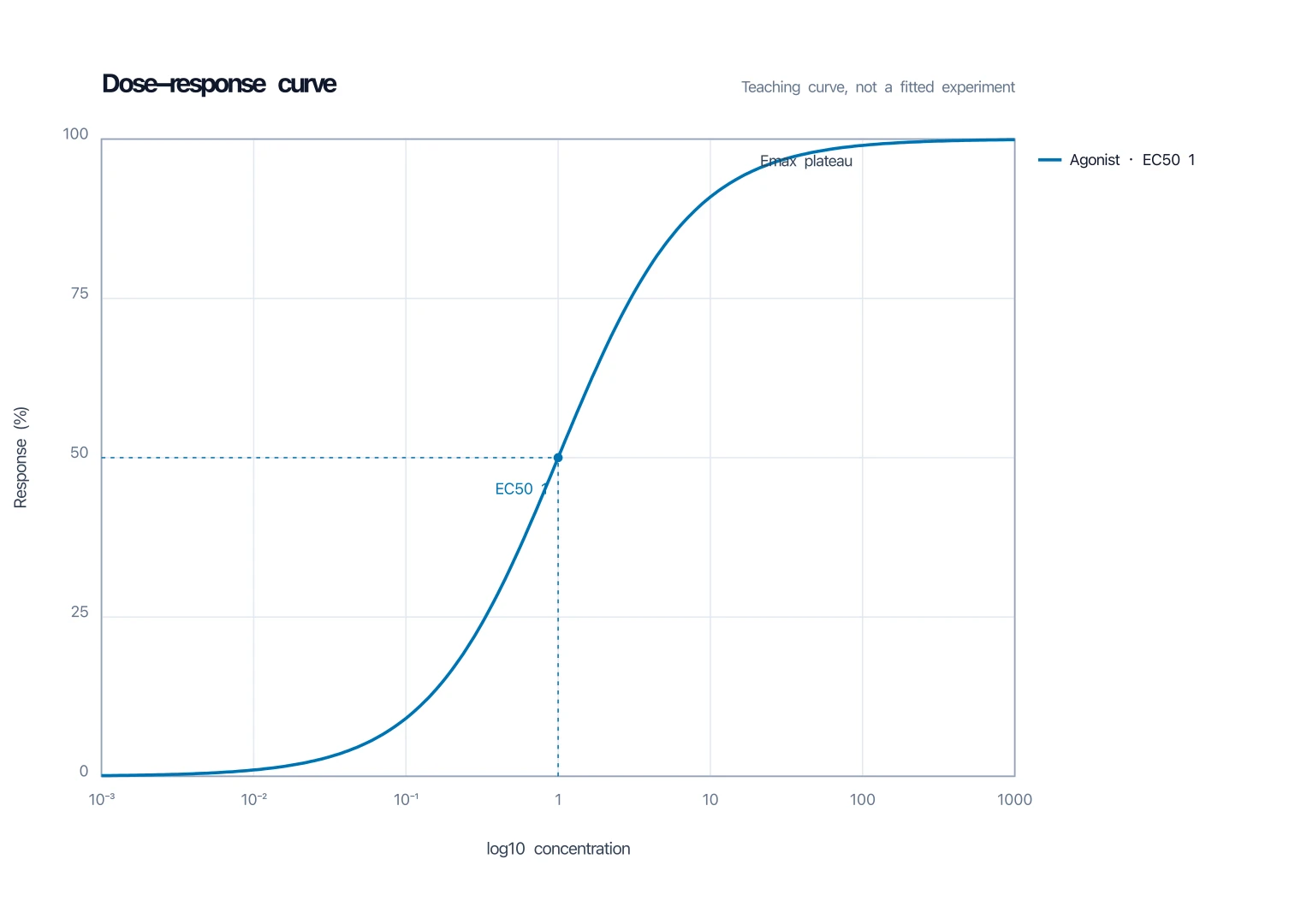
Dose-Response Curve Generator
Log-dose sigmoid when you need the quantification curve, not the method cartoon.