Flow Cytometry Diagram Generator editable schematic
Draw a flow cytometer schematic, FSC vs SSC gating plot or FACS sorting diagram. Edit lasers and channel labels, export SVG or PNG. Not for diagnosis.
Instrument schematic, illustrative gating plots, or a sorting diagram as SVG or PNG. Free, no account. Not for clinical diagnosis.
Figure options
Teaching schematic. Plots are seeded illustrations, never results.
FSC and SSC are drawn on the 488 nm laser when it is on, otherwise on the first laser.
Flow Cytometry Diagram Generator
Free preview on this page · Clean file download needs a
Your AI flow cytometry illustration will appear here
Check every label and the light path against the Precise Diagram tab. Never use an illustration as data.
Flow Cytometry Diagram Examples
Four exact diagrams from the Precise Diagram tab and two AI examples with their prompts and flaws. All are teaching schematics, not data.
Flow cytometer schematic
Exact diagram from the Precise Diagram tab. FSC sits on the beam axis behind the obscuration bar. SSC and fluorescence are collected at 90 degrees through dichroic mirrors and bandpass filters.
Three-laser schematic
Exact diagram. Each laser hits the stream at its own point and has its own detectors. BV421 sits on 405 nm, FITC and PE on 488 nm, APC on 640 nm.
FSC vs SSC gating
Exact diagram with seeded pseudo-random dots. The text "Illustrative data, not results" is inside the image. Histogram bars inside the positive gate are blue and the rest are grey, so the % positive label equals the share of plotted events inside the gate. Not a real blood sample.
FACS sorting
Exact diagram of a jet-in-air droplet sorter. A positively charged droplet moves toward the minus plate, a negatively charged droplet toward the plus plate, and uncharged droplets go to waste. "Positive" and "negative" on the tubes refer to droplet charge, not to marker staining.
AI example: workflow
AI illustration, checked by hand. Flaws: the 488 nm laser is drawn red (488 nm is blue); the three fluorescence detectors and "FSC/SSC" all sit beside the stream with no in-line FSC detector, obscuration bar or 90 degree geometry; antibodies are drawn as floating sticks on top of cells; the dot plot clusters are soft blobs, not data; no red cell lysis step is shown, and the plot axes have no values.
AI example: hydrodynamic focusing
AI illustration, checked by hand. Labels and flow arrows are correct. Flaws: the sample core is drawn nearly as wide above and below the laser, so the focusing narrowing is barely visible; the laser is drawn red although no wavelength is given, and no scatter or fluorescence detectors are shown; the beam passes through the flow cell wall as a plain line.
What a flow cytometry diagram shows
A flow cytometry diagram explains how a flow cytometer measures single cells. It usually has three parts. Fluidics: sheath fluid squeezes the sample into a narrow core so cells pass one at a time. Optics: lasers hit each cell at the interrogation point, and detectors read scattered and fluorescent light. Electronics: the detector signals become digital values that software turns into plots. This page draws all three parts with correct label placement, and you can edit the lasers, channel labels and gate names.
Hydrodynamic focusing
Sample is injected into the middle of a faster-moving sheath fluid. Laminar flow keeps the sample as a thin core, which lines cells up in single file at the interrogation point. In the flow cell panel the funnel narrows the stream, the orange core carries the cells, and each laser crosses the stream at its own height. Real instruments differ in flow cell design (cuvette or jet in air), so the drawing shows the principle, not a specific model.
FSC vs SSC: where each detector sits
- Forward scatter (FSC) is light bent a small angle from the beam. The detector is in line with the laser, usually a photodiode, behind a narrow obscuration bar (also called a shutter bar) that blocks the direct beam so only scattered light reaches it.
- Side scatter (SSC) is collected at about 90 degrees to the beam, usually by a PMT, and is separated from fluorescence by a dichroic mirror. Many instruments read FSC and SSC on the 488 nm blue laser, so the generator uses 488 nm for scatter when it is on.
- FSC is an approximate size indicator and SSC an approximate granularity or internal complexity indicator. Both also depend on refractive index, laser wavelength, collection angle and instrument settings, so they are relative measures, not microscope measurements.
Fluorescence, filters and dye-laser matching
- Fluorescence is also collected at 90 degrees. A series of long-pass dichroic mirrors sends each color range to its own detector, and a bandpass filter in front of each PMT picks the window. A label such as "FITC 530/30" means light centered at 530 nm with a 30 nm wide window.
- A dye can only be excited by a suitable laser: BV421 needs 405 nm, FITC 488 nm, APC 640 nm, and PE works with 488 or 561 nm. The generator checks known dye names against the laser you choose and shows an error when they do not match.
- Spectra of different dyes overlap, so real panels need compensation (or spectral unmixing) to remove spillover. A schematic does not show it.
Gating walk-through on lysed whole blood
- 1. Plot FSC-A against SSC-A. Debris sits near the origin and is excluded first.
- 2. Lymphocytes form a compact group with low FSC and low SSC.
- 3. Monocytes sit at higher FSC and intermediate SSC.
- 4. Granulocytes have the highest SSC because of their granules.
- 5. A gate is a region you draw around a group so later plots only show those cells. For one stained marker, a histogram on a log axis shows a negative peak and a positive peak, and a positive gate gives the percent positive.
- The dots in this tool are generated from a fixed seed. Same seed, same picture. They are labeled "Illustrative data, not results" inside the image so they cannot be mistaken for a measurement.
FACS sorting in one picture
A cell sorter adds a vibrating nozzle. The stream breaks into droplets, and a droplet that holds a wanted cell is charged as it breaks off. The droplets then fall between two high-voltage deflection plates: positively charged droplets move toward the negative plate, negatively charged droplets toward the positive plate, and uncharged droplets continue straight to waste. Collection tubes catch each group. The generator draws this jet-in-air design with editable tube labels.
Common mistakes in flow cytometry drawings
- SSC drawn in line with the beam, or FSC drawn at 90 degrees. FSC is on the beam axis, SSC at about 90 degrees.
- No obscuration bar in front of the FSC detector, which implies the full laser beam hits it.
- Dye and laser mismatch, such as APC on a 488 nm laser or BV421 without a violet laser.
- A fluorescence detector with no filter, or a PMT drawn for FSC when many instruments use a photodiode there.
- Dot plots or histograms presented as real data in a method figure. Schematic plots must say they are illustrative.
How to make a flow cytometry diagram
- 1. Pick Instrument schematic, FSC vs SSC dot plot, or FACS sorting.
- 2. For the instrument, switch on 1 to 3 lasers and set 2 to 6 fluorescence channels. Edit each label and its laser.
- 3. For the plot, rename the three gates, change the seed, and add the histogram if you need it.
- 4. Fix any red error message, then download SVG (editable) or PNG.
- Start free with 2 image credits, valid for 7 days, no card required, if you also want the AI Illustration tab. The Precise Diagram tab is free in the browser with no account.
What the AI examples got wrong
The AI examples above are useful as slide mockups, but each has flaws listed in its caption. The workflow image draws the 488 nm laser in red and puts all detectors beside the stream with no in-line FSC detector. The focusing close-up barely narrows the core. Check any AI flow cytometry image against the Precise Diagram tab before you use it, and never present an AI dot plot as data.
Not for clinical diagnosis
These diagrams are teaching schematics. They are not data, not an instrument manual, and not for clinical diagnosis or clinical decisions. Real instruments differ in laser count, filters, detector types and flow cells, so follow your instrument manual for any real configuration.
Frequently Asked Questions
Sources
- Adan et al. 2017, Flow cytometry: basic principles and applications, Crit Rev Biotechnol 37(2): overall principles, scatter, histograms and gating.
- Flow cytometry: retrospective, fundamentals and recent instrumentation (PMC3279584): FSC on the beam axis behind a shutter bar, SSC at 90 degrees, dichroic mirrors and filters, droplet charging and deflection.
- Beckman Coulter, Flow Cytometry First Principles: photodiode behind an obscuration bar and what FSC does and does not mean.
- BD FACSAria Fusion tech specs: FSC photodiode and SSC PMT on the 488 nm laser, typical dye and bandpass pairs such as FITC 530/30 and APC 670/30.
- Thermo Fisher, Electronics of a Flow Cytometer: detector, amplifier and ADC chain; FSC and SSC meaning.
Checked October 2026. Your instrument manual decides the real laser, filter and detector layout.
Related Lab Tools
 Biology
Western Blot Diagram Generator
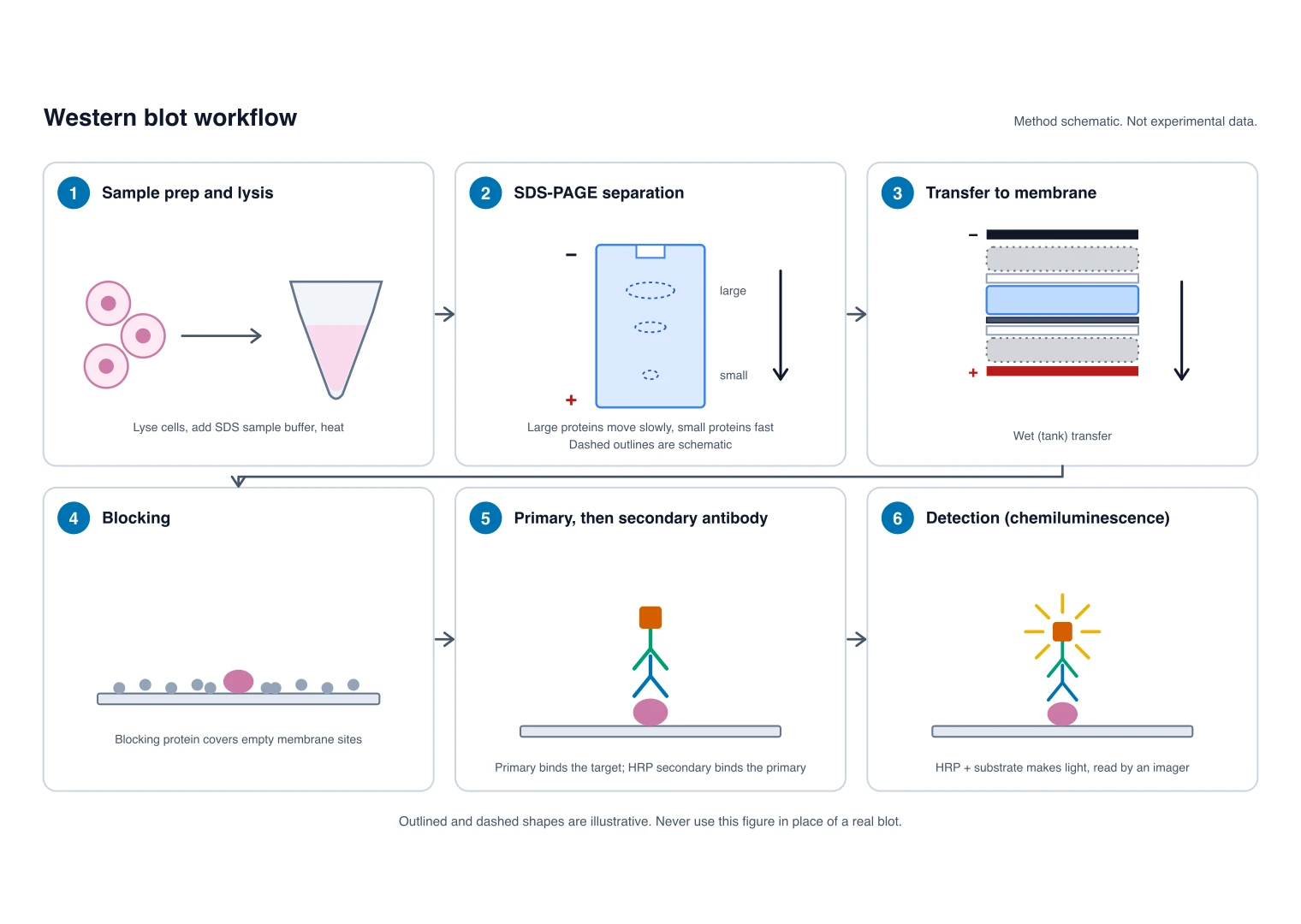
Workflow, transfer sandwich and antibody detection for protein analysis.
 Biology
ELISA Diagram Generator
Capture, antigen, detection antibody and enzyme in a well.
 Biology
PCR Diagram Generator
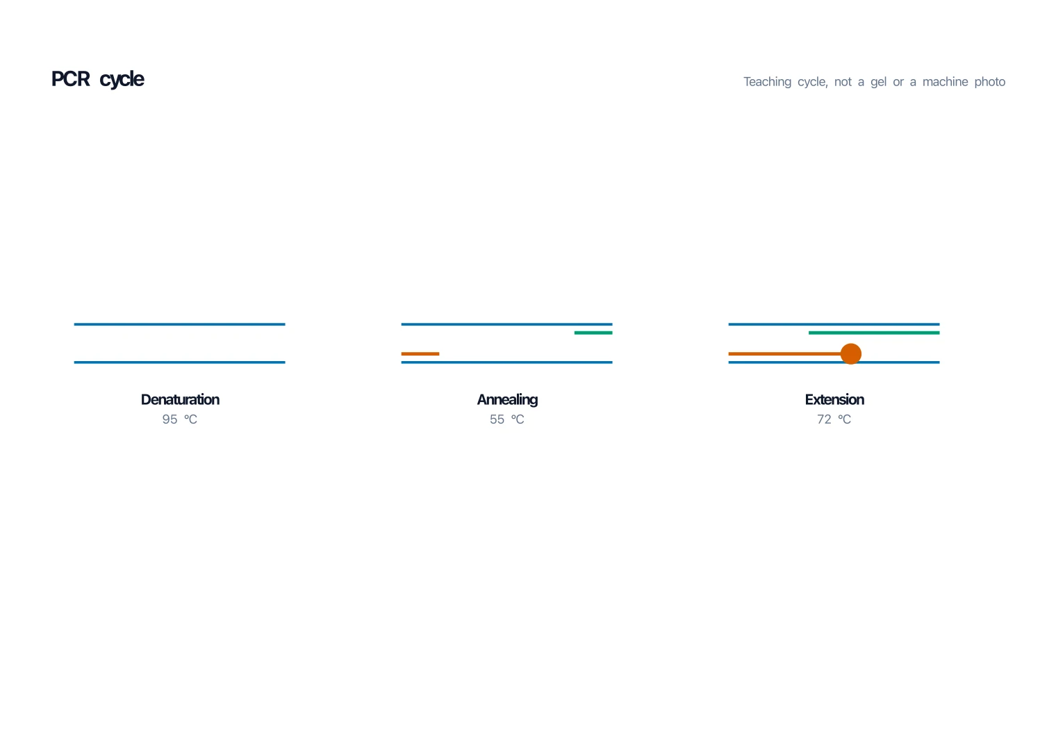
Denature, anneal, extend: the DNA cycle step by step.