Enzyme Diagram Generator for Lock-and-Key & Induced-Fit Models
Make a clearly labeled enzyme diagram in seconds. Show the lock-and-key model, induced-fit model, active site, substrate, enzyme-substrate complex, and products — plus competitive and non-competitive inhibition and the effect of temperature and pH on enzyme activity. Download labeled or blank worksheet versions, free.
Enzyme Diagram Generator
Free to try ·
Your enzyme diagram will appear here
Describe what you need and click Generate
Enzyme Diagram Examples
Lock-and-key, induced-fit, inhibition, and activity curve diagrams
Lock-and-Key Model
The classic model — a rigid active site matches only one complementary substrate, forming a complex that releases products while leaving the enzyme unchanged.
Induced-Fit Model
The induced-fit model — the active site is flexible and changes shape to better embrace the substrate, then returns to its original conformation after products are released.
Enzyme-Substrate Complex
A close-up view of the enzyme-substrate complex — active site, substrate, transition state, and products all labeled with clear leader lines.
Competitive vs Non-Competitive Inhibition
A side-by-side comparison: competitive inhibitor blocks the active site directly; non-competitive inhibitor binds the allosteric site and distorts the active site shape.
Effect of Temperature and pH
Two labeled graphs — enzyme activity versus temperature and versus pH — each showing the bell-shaped optimum curve and denaturation at extremes.
Blank Enzyme Worksheet
A printer-friendly blank worksheet based on the lock-and-key model — empty callout lines for students to label each part themselves.
What is an enzyme and how does it work?
An enzyme is a biological catalyst — almost always a protein — that speeds up a chemical reaction in a living organism without being used up in the process. After the reaction is complete, the enzyme is released unchanged and can catalyse the same reaction again. Enzymes work by lowering the activation energy needed to start a reaction, making reactions that would otherwise be too slow to sustain life happen in milliseconds. Each enzyme has a uniquely shaped active site that fits only a specific substrate (or a small group of structurally similar substrates). This specificity is what makes enzymes so precise and is the central idea behind both the lock-and-key and induced-fit models.
The lock-and-key model vs the induced-fit model
- Lock-and-key model: the active site has a rigid, fixed shape that is complementary to one specific substrate, just as a lock accepts only the matching key. The substrate slots in, forming the enzyme-substrate complex, the reaction occurs, the products are released, and the enzyme is left unchanged.
- Induced-fit model: the active site is flexible rather than rigid. When the substrate approaches, the enzyme changes shape slightly to embrace it more closely. This induced change improves the fit and strains the substrate bonds to lower activation energy. After the products leave, the enzyme returns to its original conformation.
- The induced-fit model is now considered more accurate, but both models appear in biology exams and both can be illustrated here.
The enzyme-substrate complex and the active site
The active site is the pocket or cleft on the enzyme surface where the substrate binds. It is formed by only a small number of the enzyme's amino acids, but their precise arrangement creates the three-dimensional shape that determines specificity. When the substrate enters the active site, the resulting structure is called the enzyme-substrate complex. Chemical bonds in the substrate are strained or broken, lowering the activation energy and allowing the reaction to proceed. The products then leave the active site, freeing the enzyme for another cycle. A good labeled diagram marks the enzyme, the active site, the substrate, the enzyme-substrate complex, and the products with clear callout lines.
Enzyme inhibition: competitive and non-competitive
- Competitive inhibition: a molecule with a shape similar to the substrate (the competitive inhibitor) binds to the active site and blocks the substrate from entering. The inhibitor competes directly with the substrate for the same binding site. Increasing the substrate concentration can overcome competitive inhibition by flooding the active site with substrate and out-competing the inhibitor.
- Non-competitive inhibition: the inhibitor binds to a different site on the enzyme called the allosteric site. This changes the overall shape of the enzyme and distorts the active site, reducing its ability to bind the substrate. Because the inhibitor does not bind the active site directly, increasing substrate concentration cannot reverse non-competitive inhibition.
- Both types of inhibition are important in drug design — many medicines work by inhibiting specific enzymes in pathogens or in metabolic pathways.
Effect of temperature on enzyme activity
As temperature rises, molecules move faster, collisions between enzyme and substrate become more frequent, and the rate of reaction increases — up to a point. Each enzyme has an optimum temperature at which it works fastest (around 37 °C for most human enzymes). Above this optimum, the increased heat disrupts the weak hydrogen bonds and other interactions that hold the enzyme in its three-dimensional shape. The active site loses its precise form, the enzyme can no longer bind its substrate effectively, and activity falls sharply. If the temperature is high enough, the enzyme becomes permanently denatured. A temperature-versus-activity graph shows a bell-shaped curve peaking at the optimum and dropping steeply on the right as denaturation occurs.
Effect of pH on enzyme activity
Enzymes also have an optimum pH at which their activity is highest. Most human enzymes work best around pH 7, but there are important exceptions: pepsin in the stomach has an optimum of around pH 2, and intestinal amylase works best near pH 7-8. Outside the optimum pH, the concentration of hydrogen ions changes the charges on the amino acids that form the active site, disrupting the ionic bonds and hydrogen bonds that maintain its shape. The active site distorts, substrate binding becomes less efficient, and activity drops. Extreme pH values denature the enzyme permanently. The pH-versus-activity graph is also bell-shaped, peaking at the optimum and falling on both sides.
Labeled vs blank enzyme diagrams for worksheets
For teaching, you often need two versions of the same diagram. A fully labeled diagram names every structure — enzyme, active site, substrate, enzyme-substrate complex, products, inhibitor, allosteric site — and is ideal for notes, slides, and revision. A blank version keeps the same layout but replaces the labels with empty callout lines, turning it into a fill-in-the-blank quiz. Because you describe what you want in plain English, you can generate the labeled teaching version and the blank assessment version from the same description — no redrawing needed.
Frequently Asked Questions
Related Biology Tools
 Biology
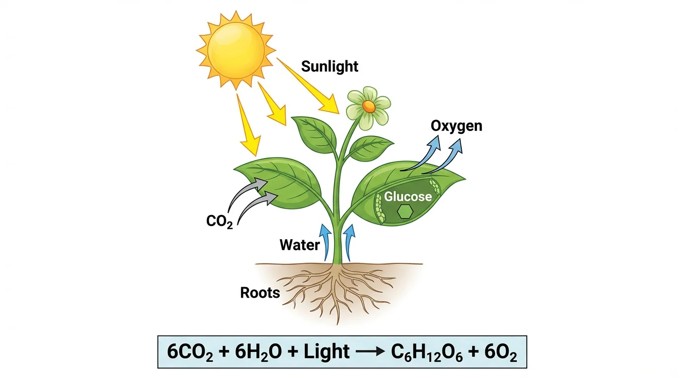
Photosynthesis Diagram Generator
Create labeled photosynthesis diagrams showing inputs, outputs, the Calvin cycle, and the light-dependent reactions.
 Biology
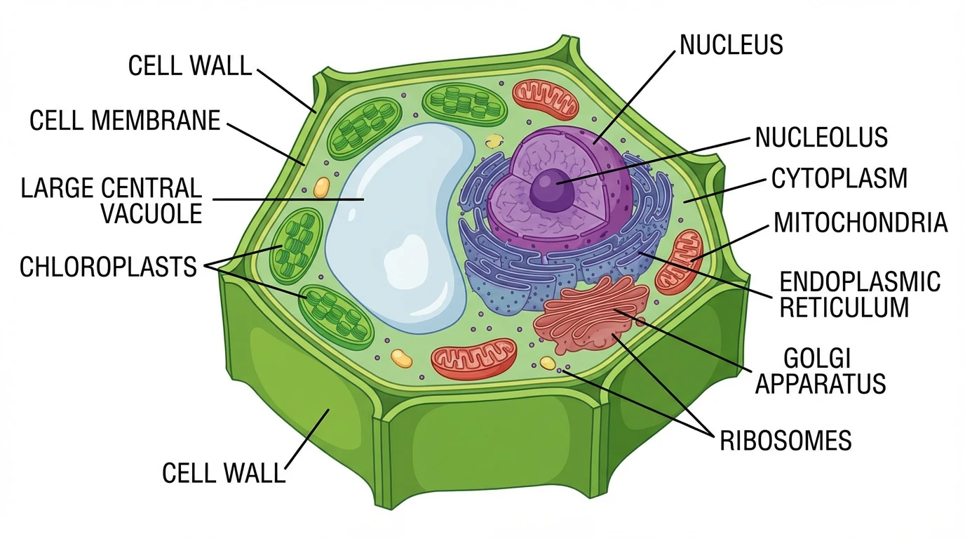
Plant Cell Diagram Generator
Make labeled plant cell diagrams with the cell wall, vacuole, chloroplasts, and other organelles.
 Biology
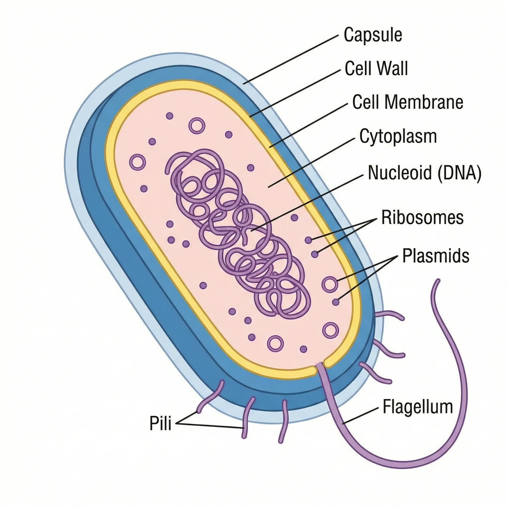
Bacteria Diagram Generator
Generate labeled bacterial cell diagrams showing the cell wall, nucleoid, plasmids, flagella, and other prokaryotic structures.