Ear Anatomy Diagram Generator Labeled Human Ear Diagrams
Create labeled ear anatomy diagrams with AI. Show the outer, middle, and inner ear — pinna, ear canal, eardrum, ossicles, cochlea, and auditory nerve — or how hearing works, for biology and health class. Download as PNG.
AI Ear Anatomy Diagram Generator
Free to try ·
Your ear anatomy diagram will appear here
Describe the view and parts to label
Ear Anatomy Examples
Labeled diagrams of the outer, middle, and inner ear, and how hearing works
Labeled Ear Cross-Section
A full cross-section with every structure of the outer, middle, and inner ear labeled.
Simple Ear Diagram
A clean, simplified version with the main parts — ideal for younger students.
Outer, Middle & Inner Ear
The three regions of the ear, with the structures grouped into each.
How Hearing Works
The path of sound — ear canal → eardrum → ossicles → cochlea → nerve.
Middle Ear Ossicles
The three tiny bones — malleus, incus, and stapes — that amplify sound.
Inner Ear & Cochlea
The inner ear — cochlea for hearing and semicircular canals for balance.
What does an ear anatomy diagram show?
An ear anatomy diagram shows the structures of the human ear, grouped into the outer, middle, and inner ear, and how they work together for hearing and balance. A typical labeled cross-section includes the pinna, ear canal, eardrum, the three ossicles (malleus, incus, stapes), the cochlea, the semicircular canals, the eustachian tube, and the auditory nerve. This generator creates clear, labeled ear diagrams for biology and health classes.
The three parts of the ear
- Outer ear: the pinna (auricle) collects sound and the ear canal channels it to the eardrum.
- Middle ear: the eardrum (tympanic membrane) vibrates and passes the vibration through three tiny bones — the malleus, incus, and stapes — to the oval window; the eustachian tube equalizes pressure.
- Inner ear: the cochlea converts vibrations into nerve signals, and the semicircular canals sense balance and head movement.
How hearing works
Sound waves are funneled by the pinna into the ear canal and make the eardrum vibrate. The three ossicles amplify and transmit this vibration to the oval window, setting fluid inside the cochlea moving. Tiny hair cells in the cochlea convert the fluid waves into electrical nerve signals, which travel along the auditory nerve to the brain, where they are interpreted as sound.
The ear and balance
Beyond hearing, the inner ear is key to balance. The three semicircular canals, set at right angles, detect rotational movements of the head, while the vestibule senses gravity and linear motion. Together they tell the brain about head position and movement, helping you stay balanced — which is why ear problems can cause dizziness.
Tips for a clear ear diagram
Choose the view you need — a full labeled cross-section, a simplified version, the three regions grouped, or a focused diagram of the ossicles or cochlea. List the parts you want labeled and the level of detail. Generate a few options and download the clearest for your worksheet or slides.
Frequently Asked Questions
Related Science Tools
 Medical
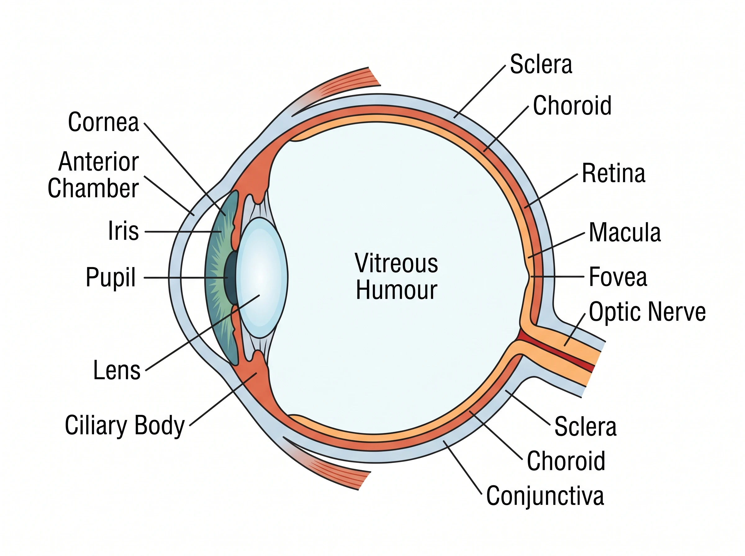
Eye Anatomy Diagram Generator
Create labeled eye diagrams — cornea, lens, retina, optic nerve — and how vision works.
 Biology
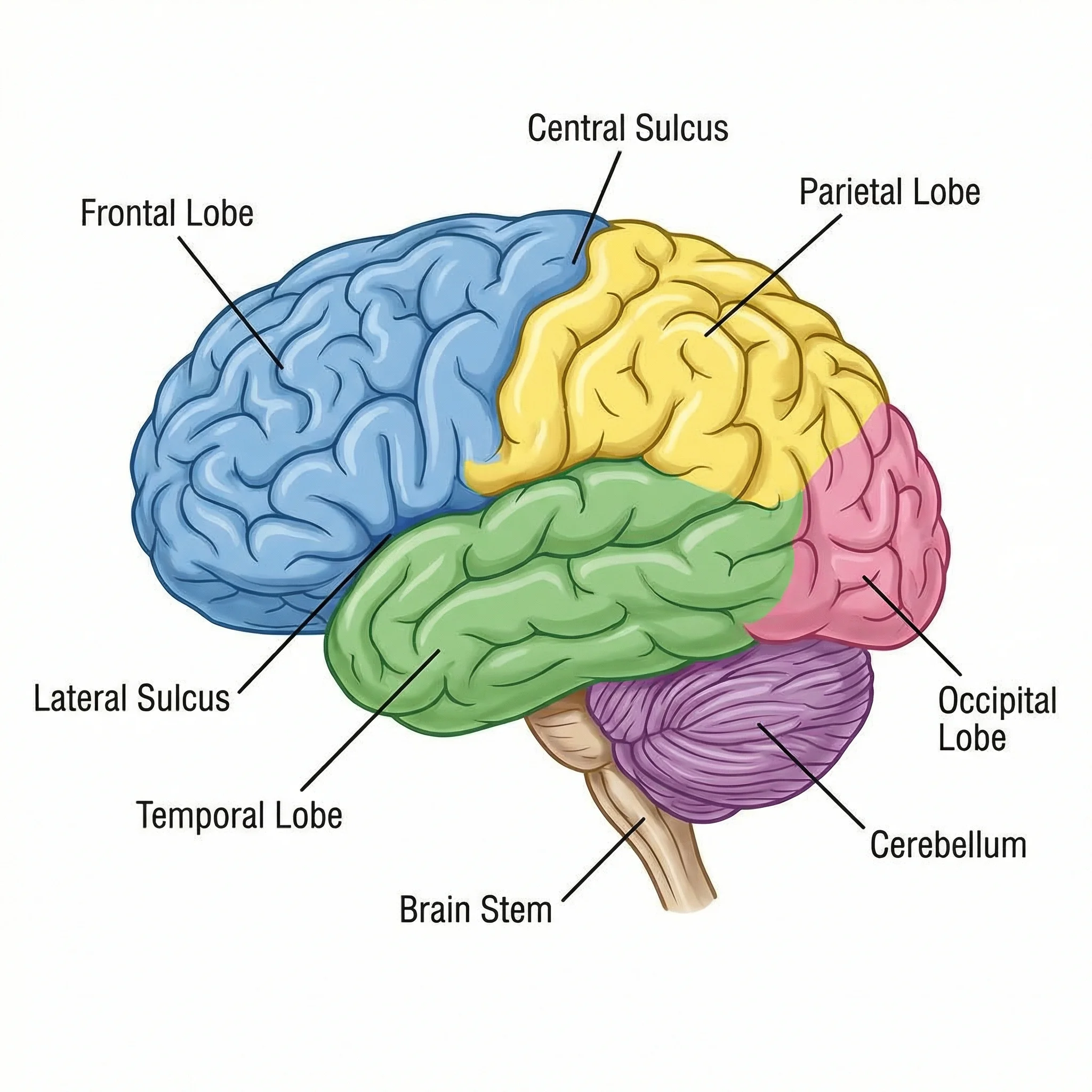
Brain Diagram Generator
Create labeled brain diagrams with lobes, regions, and structures.
 Medical
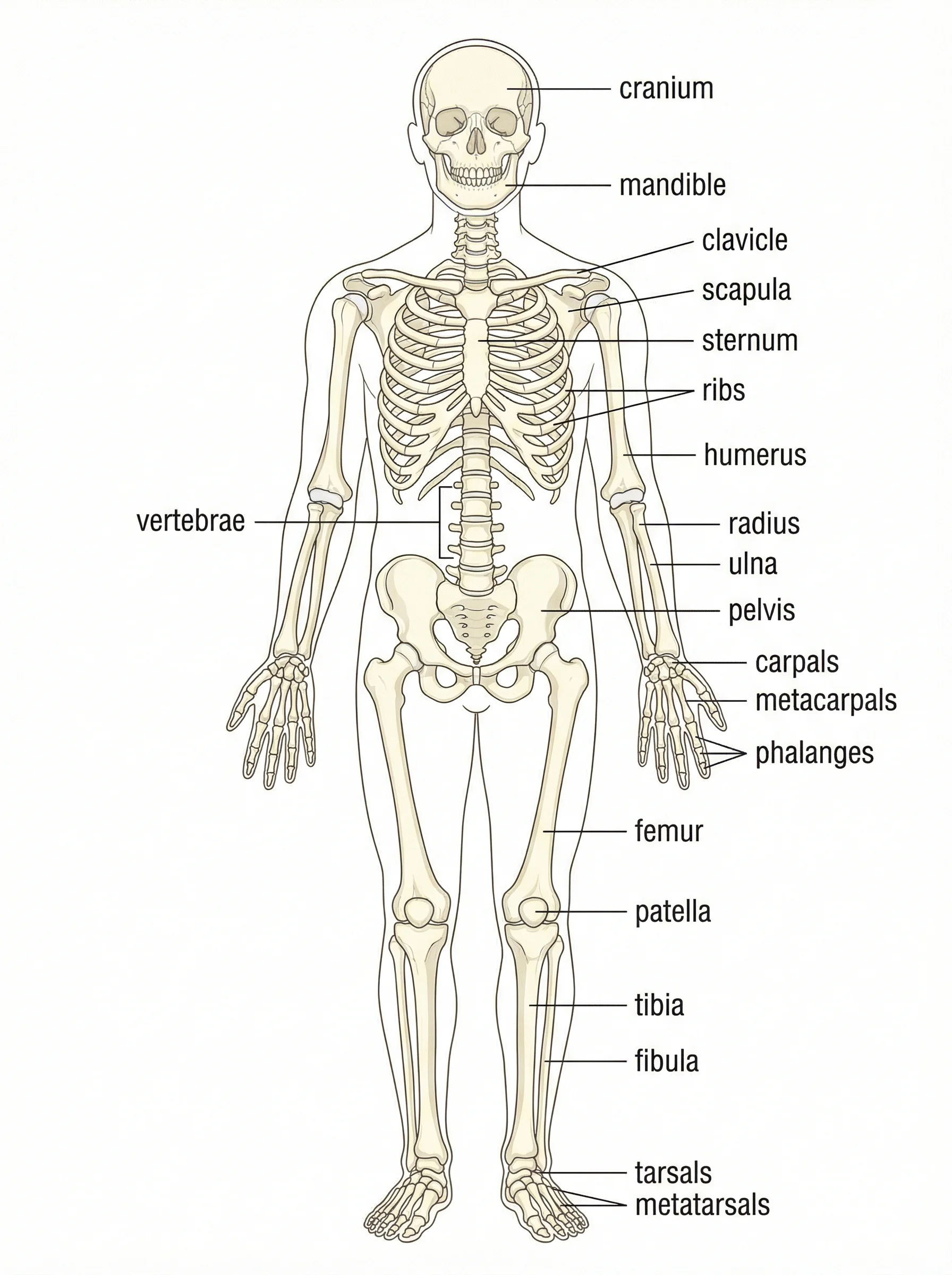
Anatomical Drawing Generator
Generate labeled anatomy illustrations of body systems and organs.