Pelvis Bones Diagram
Free labeled, blank, and printable diagrams of the bones of the pelvis: ilium, ischium, pubis, sacrum, and coccyx. Free to download.
Free Pelvis Bones Diagrams
Every version below is free to view, download, and print. No sign-up.
Labeled Bones of the Pelvis
An adult pelvis in anterior view named Ilium, Ischium, Pubis, Sacrum, Iliac crest, Acetabulum, Pubic symphysis, Obturator foramen, and Sacroiliac joint. Coccyx is visible in this anterior view but is not labeled; it is named on the posterior figure.
Core diagrams
Structure & focus
Print & practice
Test yourself: bones of the pelvis
A quick self-test. Questions reshuffle each time.
Each hip bone (coxal bone) is formed by fusion of:
Customize this diagram
Change view, grade level, or which bones are labeled. Describe it and generate your own.
Customize Your Pelvis Bones Diagram
Free to try ·
Your custom diagram will appear here
Ask for labeled overview, blank worksheet, lateral hip bone, or simple labels
How to read the labeled bones of the pelvis
Start with the labeled anterior overview. It names Ilium, Ischium, Pubis, Sacrum, Iliac crest, Acetabulum, Pubic symphysis, Obturator foramen, and Sacroiliac joint. Each hip bone (coxal bone) is three bones fused at the acetabulum: Ilium above, Ischium behind and below, Pubis in front and below. Sacrum sits in the midline between the two hip bones. Coccyx is visible in this anterior view but is not labeled here; open the posterior figure to see it named. Acetabulum is the cup-shaped hip socket on the lateral side. Obturator foramen is the large oval hole below the acetabulum. Follow every leader line to the bone it names, not to a neighbor.
Hip bone, pelvic girdle, and pelvis
Classroom diagrams treat the pelvis as four bones: the two hip bones, the sacrum, and the coccyx. Each hip bone is itself a fusion of ilium, ischium, and pubis. The pelvic girdle is the two hip bones only. It attaches the free lower limb to the axial skeleton. The bony pelvis is the girdle plus sacrum and coccyx, the complete ring. Say this out loud once when you project the figure, or students will call one hip bone “the pelvis” and miss the midline bones. Pair the anterior overview with the lateral hip-bone close-up so the three fused parts are visible on one coxal bone.
Male and female pelvis, in qualitative terms
The comparison figure places a male pelvis and a female pelvis side by side, anterior view, same scale. It names Pelvic inlet and Subpubic angle on each side. The male pelvis is narrower, taller, and more robust, with a heart-shaped inlet and a narrow V-shaped subpubic angle. The female pelvis is wider and shallower, with a round or oval inlet and a wide U-shaped subpubic angle. Those are shape words for class, not measured degrees. Use this figure after students can already name ilium, ischium, pubis, and sacrum on the labeled overview.
What the pelvis does
The pelvis carries the weight of the trunk down to the femurs. Each acetabulum receives the head of the femur, so the free lower limb hangs from the girdle. The bony ring also protects pelvic organs: bladder, rectum, and, in females, the uterus. Ischial tuberosities take the load when you sit. Iliac crests give a wide shelf for abdominal and back muscles. Sacroiliac joints lock the sacrum between the hip bones so the ring stays closed under load. The posterior figure names Sacrum, Coccyx, Ilium, Sacroiliac joint, Iliac crest, and Ischial tuberosity, which is the right view for those sitting bones.
Simple diagram, blank sheet, and coloring page
The simple diagram labels Hip bone, Sacrum, and Pubic symphysis in large type. That is the right first slide for middle school or for a warm-up before a lab. The blank worksheet matches the nine-name anterior overview: same leader-line geometry, numbers 1 to 9, no words. Project the labeled overview, hide it, and give students five minutes on the blank sheet. The coloring page is the same overview in black-and-white outline, still labeled Ilium, Ischium, Pubis, Sacrum, Iliac crest, Acetabulum, Pubic symphysis, Obturator foramen, and Sacroiliac joint. Ask the class to color ilium, ischium, pubis, and sacrum in four colors, then compare with the labeled view. All files are free to view, download, and print.
Frequently Asked Questions
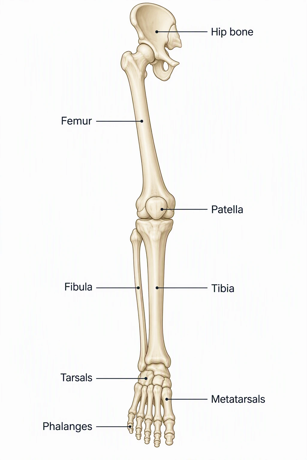
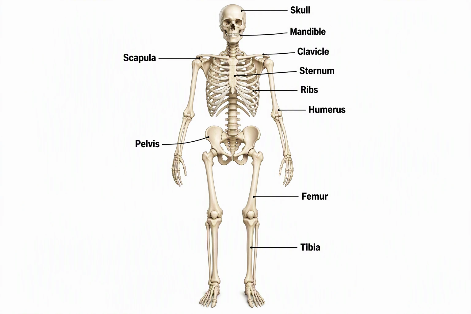
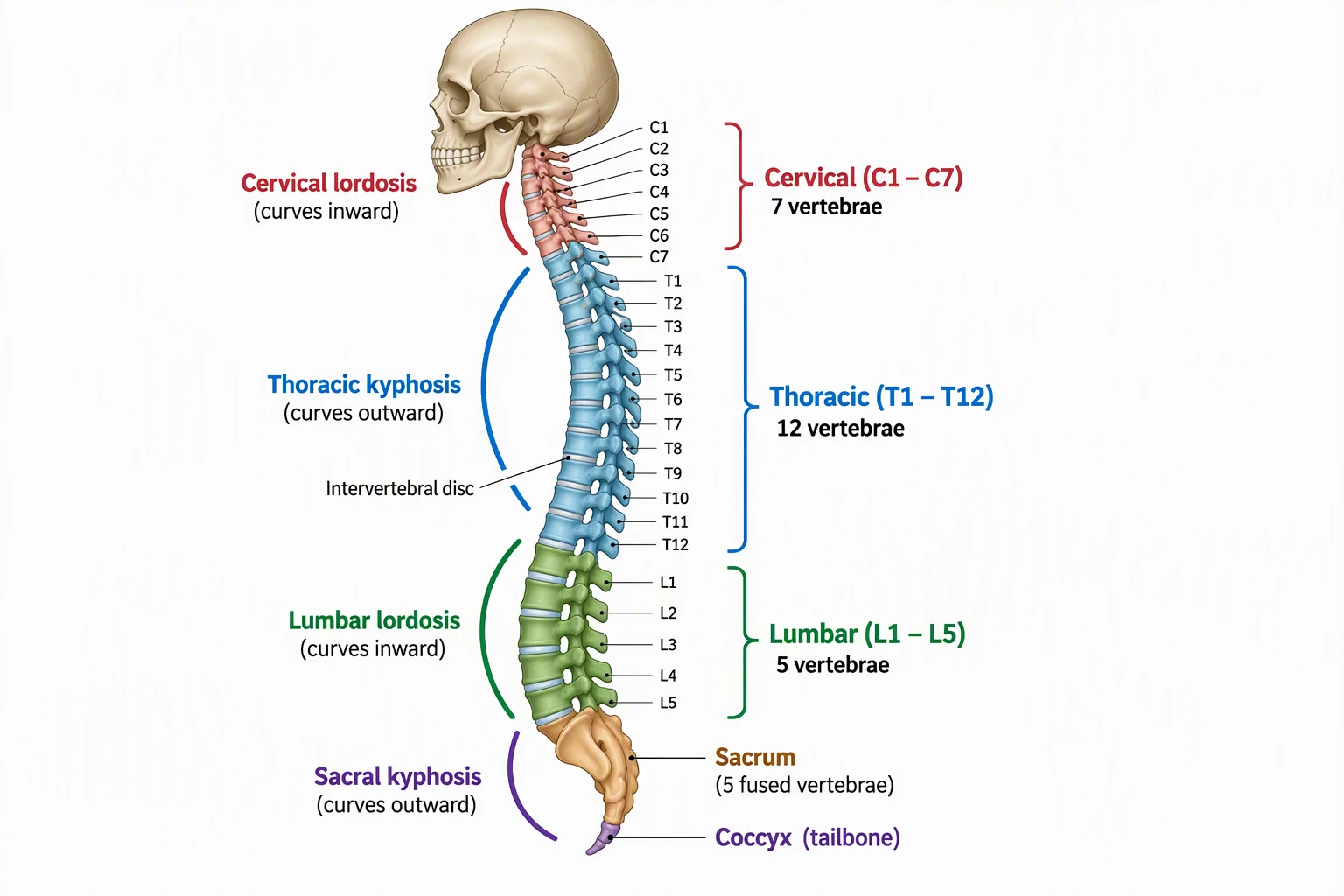
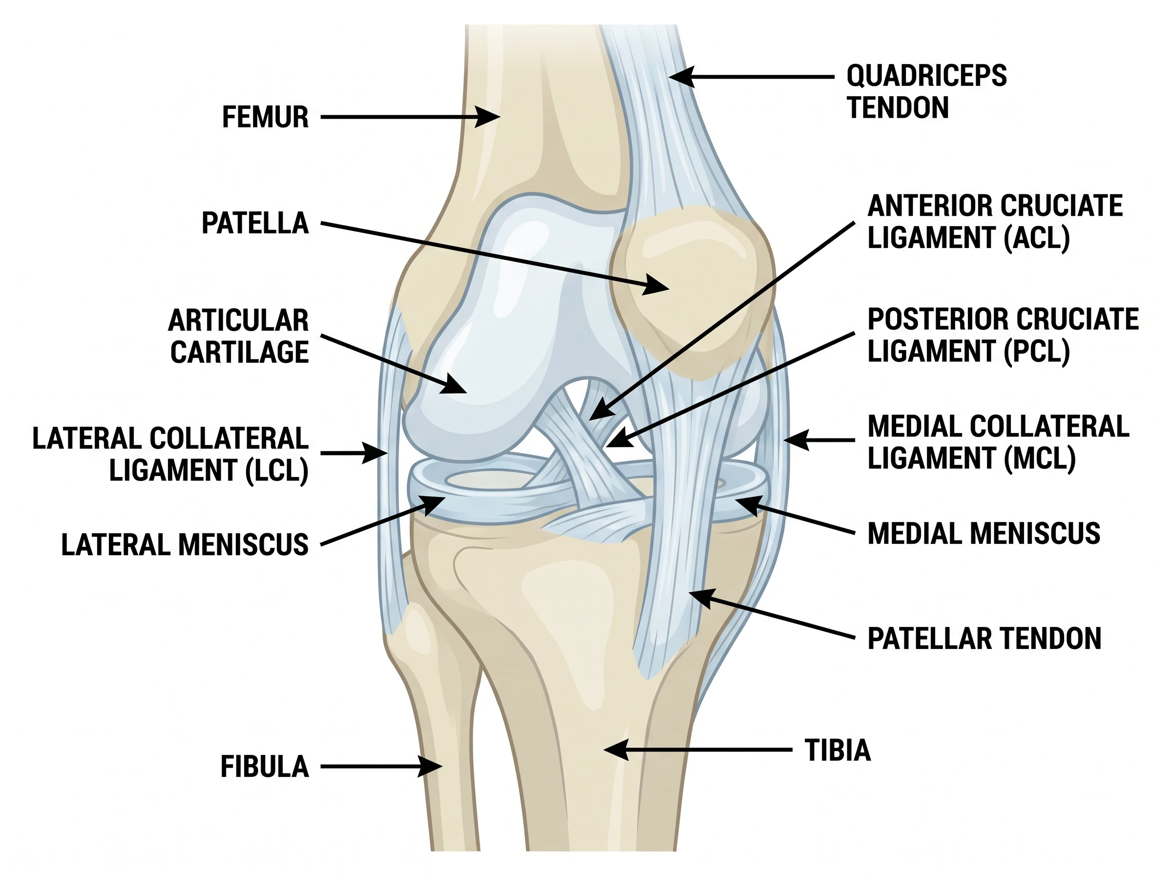
Pair this pack with the bones of the leg diagram for the free lower limb, the skeletal system diagram generator for the whole body, the spine anatomy diagram generator for sacrum and coccyx on the vertebral column, and the knee anatomy diagram generator for the joint below the hip.
Related Anatomy Tools
 Medical
Bones of the Leg Diagram
Free labeled femur, tibia, fibula, and foot groups.
 Biology
Skeletal System Diagram Generator
Full-body skeleton maps; zoom here for the pelvis.
 Medical
Spine Anatomy Diagram Generator
Spinal regions, sacrum, and coccyx on the same axis.
 Medical
Knee Anatomy Diagram Generator
Joint close-up on the same limb as the pelvis.
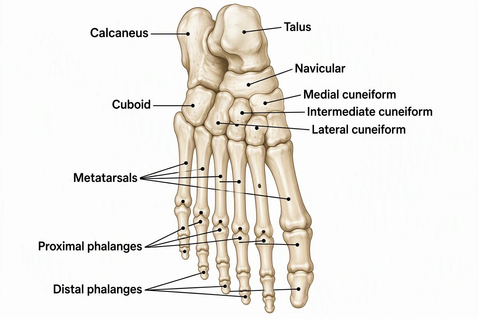 Medical
Foot Bones Diagram
Free labeled tarsals, metatarsals, and phalanges.
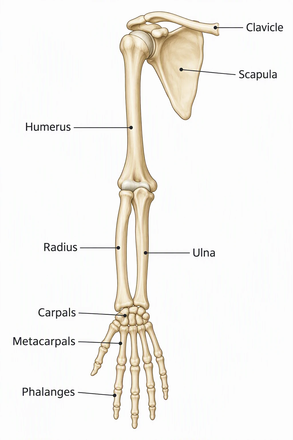 Medical
Bones of the Arm Diagram
Free labeled humerus, radius, ulna, and hand groups.