Bones of the Leg Diagram
Free bones of the leg diagrams: labeled femur, tibia, and fibula, blank worksheet, simple version, and coloring page. Printable; free download.
Free Bones of the Leg Diagrams
Every version below is free to view, download, and print. No sign-up.
Labeled Bones of the Leg
A right lower limb in anterior view with the bones named: Hip bone, Femur, Patella, Fibula, Tibia, Tarsals, Metatarsals, and Phalanges. Fibula is lateral, on the left of the figure.
Core diagrams
Structure & focus
Print & practice
Test yourself: bones of the leg
A quick self-test. Questions reshuffle each time.
One adult lower limb, not counting the girdle, has:
Customize this diagram
Change view, grade level, or which bones are labeled. Describe it and generate your own.
Customize Your Leg Bones Diagram
Free to try ·
Your custom diagram will appear here
Ask for labeled overview, blank worksheet, femur landmarks, or simple labels
How to read the labeled bones of the leg
Start with the labeled overview. It names Hip bone, Femur, Patella, Fibula, Tibia, Tarsals, Metatarsals, and Phalanges on a right lower limb in anterior view, with Fibula on the lateral side (left of the figure). Hip bone is a pelvic girdle bone at the top of the figure. Distal to the ankle, Tarsals, Metatarsals, and Phalanges are labeled as groups so the map stays at eight names. Follow every leader line to the bone it names, not to a neighbor. Keep this print on the desk while the class opens the close-ups.
What “bones of the leg” means in class
In anatomy class, “bones of the leg” usually means the whole lower limb, not only the femur. Classroom count for one side is 30 free bones: femur, patella, tibia, fibula, 7 tarsals, 5 metatarsals, and 14 phalanges. Hip bone belongs to the pelvic girdle; it appears on the labeled overview so students can see where the limb hangs, but it is not added into that 30. Say this out loud once when you project the figure, or students will mix girdle bones with free limb bones. Pair the overview with the foot bones page when you need tarsals named one by one.
Femur landmarks and the tibia-fibula pair
The anterior femur figure names Head of femur, Neck of femur, Greater trochanter, Lesser trochanter, Shaft of femur, Lateral condyle, Medial condyle, and Patellar surface. Head and neck are proximal; the condyles and Patellar surface are distal at the knee. The tibia-fibula figure then names Head of fibula, Tibial tuberosity, Fibula, Tibia, Interosseous membrane, Lateral malleolus, and Medial malleolus. Tibia is medial and weight-bearing; Fibula is lateral. Medial malleolus is distal tibia; Lateral malleolus is distal fibula. Print the two close-ups on one sheet so students can match the distal femur with the proximal tibia.
Lateral view of the limb
The lateral figure is a side-on limb, toes pointing right, not a front map. It names Femur, Patella, Fibula, Tibia, Talus, and Calcaneus. Patella sits in front of the distal femur. Talus sits at the ankle; Calcaneus is the heel. Use this view after the two close-ups, so students already know the long bones before they look at the ankle stack. Cuboid and the cuneiforms are not on this side view; they belong on the foot bones page.
Simple diagram, blank sheet, and coloring page
The simple diagram labels Femur, Patella, Fibula, Tibia, and Foot bones in large type. That is the right first slide for middle school or for a warm-up before a lab. The blank worksheet matches the eight-name overview: same leader-line geometry, numbers 1 to 8, no words. Project the labeled overview, hide it, and give students five minutes on the blank sheet. The coloring page is the same overview in black-and-white outline, still labeled Hip bone, Femur, Patella, Fibula, Tibia, Tarsals, Metatarsals, and Phalanges. Ask the class to color girdle, thigh, leg, and foot in four colors, then compare with the labeled view.
How to use these files in class
A reliable sequence is simple five-name diagram, labeled overview, femur landmarks, tibia-fibula, lateral view, then the blank sheet. Homework can be the coloring page plus the blank sheet on the reverse. All files are free to view, download, and print. Drop the labeled overview into slides, or print letter or A4. This page is a lower-limb close-up. Use the foot bones page for named tarsals, the bones of the arm page for the matching upper limb, the knee anatomy generator for the joint, and the skeletal system generator for the whole body. Keep each download set on its own page so labels stay short and readable.
Frequently Asked Questions
Pair this pack with the foot bones diagram for named tarsals, the bones of the arm diagram for the matching upper limb, the knee anatomy diagram generator for the joint, and the skeletal system diagram generator for the whole body.
Related Anatomy Tools
 Medical
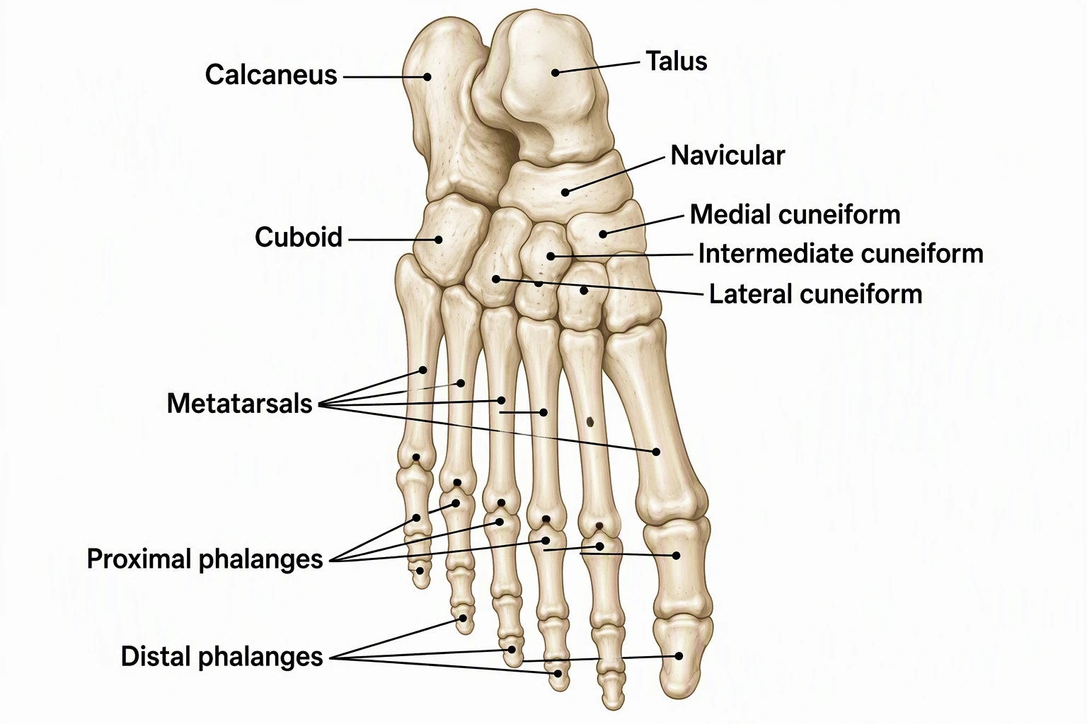
Foot Bones Diagram
Free labeled tarsals, metatarsals, and phalanges.
 Medical
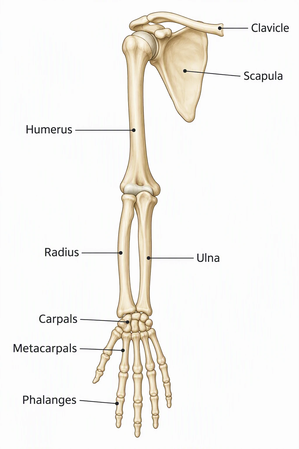
Bones of the Arm Diagram
Free labeled humerus, radius, ulna, and hand groups.
 Medical
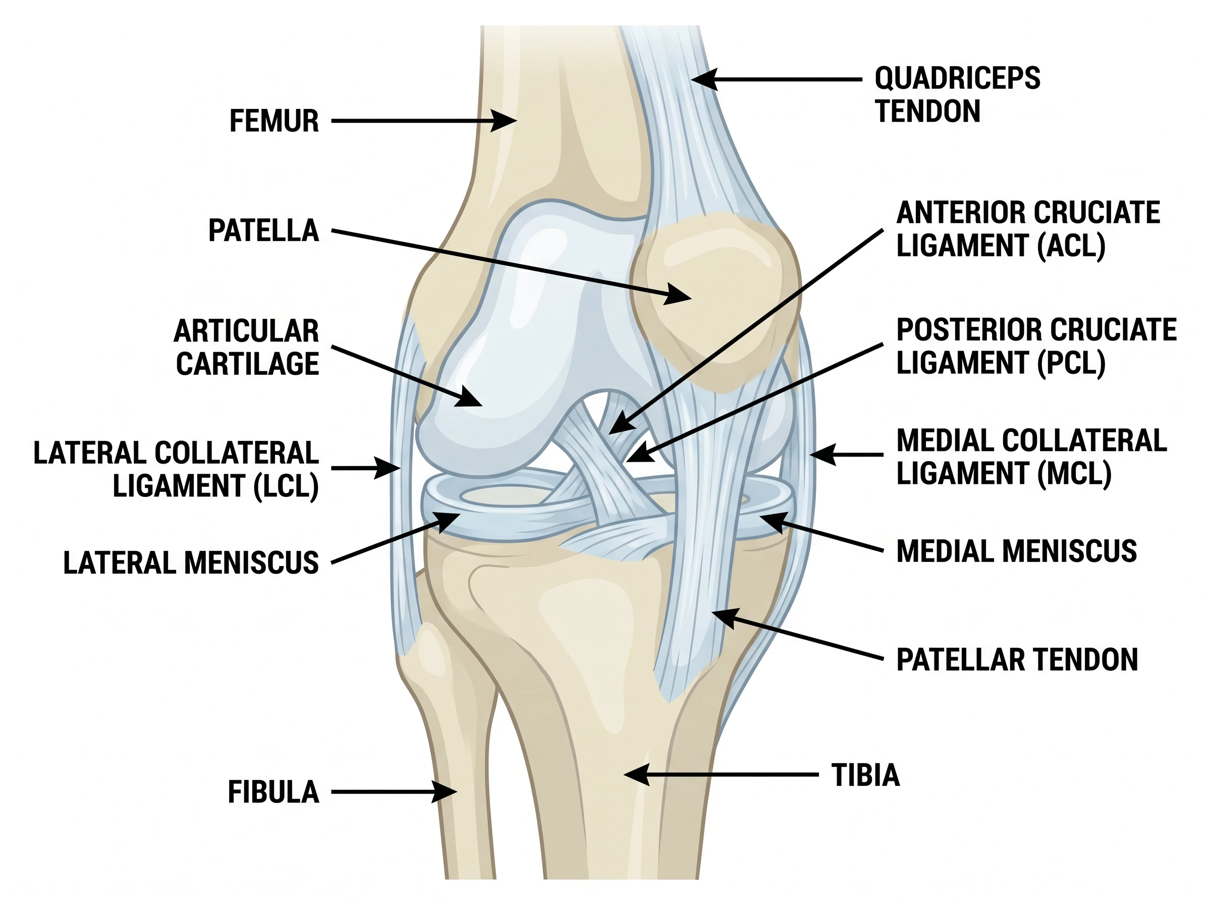
Knee Anatomy Diagram Generator
Joint close-up on the same limb as the leg.
 Biology
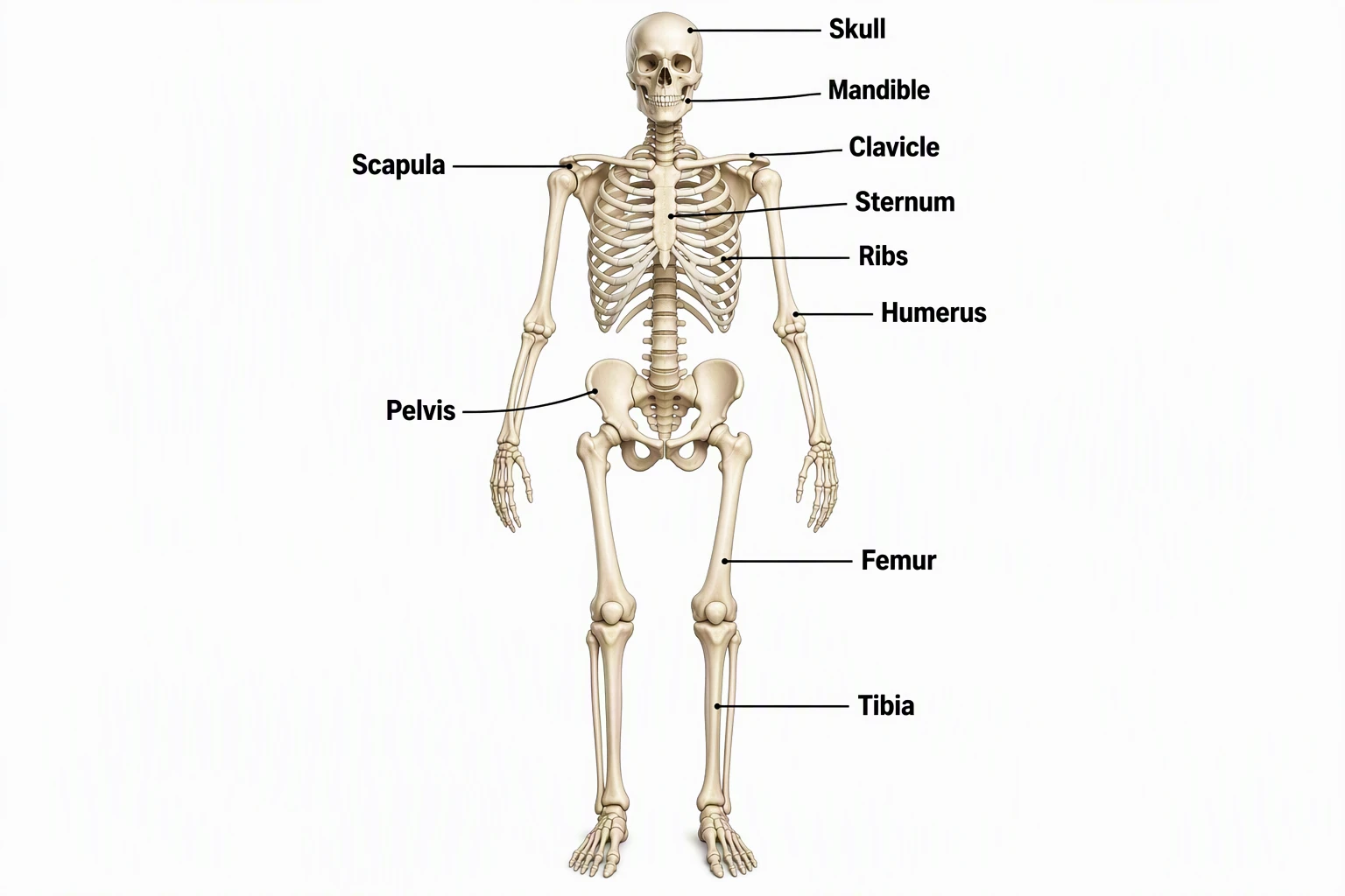
Skeletal System Diagram Generator
Full-body skeleton maps; zoom here for the leg.
 Medical
Hand Bones Diagram
Free labeled carpals, metacarpals, and phalanges.
 Medical
Skull Diagram
Free labeled anterior and lateral skull views.