Protein Synthesis Diagram
Free protein synthesis diagrams: labeled transcription and translation, blank worksheet, simple version, and coloring page. Printable; free download.
Free Protein Synthesis Diagrams
Every version below is free to view, download, and print. No sign-up.
Labeled Protein Synthesis Overview
The whole path from gene to protein in one picture: Nucleus, DNA, RNA polymerase, mRNA, Nuclear pore, Ribosome, tRNA, Codon, Anticodon, and Polypeptide chain.
Core diagrams
Structure & focus
Print & practice
Test yourself: protein synthesis
A quick self-test. Questions reshuffle each time.
Transcription happens mainly in the…
Customize this diagram
Change grade level, which stages are labeled, or a nucleus versus ribosome focus. Describe it and generate your own.
Customize Your Protein Synthesis Diagram
Free to try ·
Your custom diagram will appear here
Ask for a labeled overview, blank worksheet, or translation stages
How to read the labeled overview
Start with the fully labeled overview. It names Nucleus, DNA, RNA polymerase, mRNA, Nuclear pore, Ribosome, tRNA, Anticodon, Codon, and Polypeptide chain. Transcription sits in the nucleus: RNA polymerase copies DNA into mRNA. The mRNA strand then leaves through a nuclear pore. Translation sits on a ribosome: tRNA brings amino acids, each anticodon matching one codon, and the polypeptide chain grows. Ten labels keep the picture printable. If a name is not on this overview (for example a specific amino acid), it belongs on the codon-anticodon close-up or on a codon chart, not as extra text on the overview.
Transcription focus, then translation stages
After the overview, open the nucleus close-up. It labels Nucleus, DNA, RNA polymerase, mRNA, and Nuclear pore. That is the whole transcription story this pack needs. Then open the three-stage translation figure. Headings are 1. Initiation, 2. Elongation, and 3. Termination. Each panel labels Ribosome, tRNA, and Amino acid. Initiation shows the ribosome on mRNA. Elongation shows tRNA delivering amino acids as the chain grows. Termination shows the ribosome subunits separating and the tRNA and finished chain released. Teach those three words as a sequence, not as three separate organelles.
Codon and anticodon at one site
The codon-anticodon close-up is a zoom, not a second pathway. It labels Amino acid, tRNA, Anticodon, mRNA, and Codon. One mRNA codon (three bases) lines up with one tRNA anticodon. The amino acid rides on that tRNA. Print this figure next to the overview so students can find mRNA, tRNA, and Codon at two scales. If the lesson then asks “which amino acid is that?”, switch to the codon chart rather than writing amino-acid names onto this zoom. Keeping the zoom at five labels is what makes the letters large enough to read from the back of a room.
Simple diagram, blank sheet, and coloring page
The simple diagram labels DNA, mRNA, Ribosome, tRNA, and Protein. Use it as a middle-school poster or as a warm-up slide. The blank worksheet matches the ten-label overview: same layout, numbers 1 to 10 with blank lines, no words. Project the labeled version, hide it, and give students five minutes on the blank sheet. The coloring page is an outline of nucleus, mRNA, and a ribosome with tRNAs and chain, with no text. Ask the class to color transcription one color and translation another so the two stages stay visually separate on the page.
How to use these files in a lesson
A reliable sequence is simple diagram (two minutes), labeled overview (ten minutes), nucleus focus plus three-stage translation (ten minutes), then the blank worksheet. Homework can be the coloring page plus the blank sheet on the reverse. All files are free to view, download, and print. Drop the labeled overview into slides at 16:9, or print letter or A4. Do not treat this pack as a sequence decoder: it does not list every codon. For a typed DNA string that becomes mRNA and amino acids, use the transcription and translation generator linked below.
Where this pack sits next to replication
Protein synthesis uses the information that DNA replication copied. Keep the replication pack for the fork (helicase, polymerase, Okazaki fragments). Keep this pack for DNA to RNA to protein. Linking the two pages is enough; do not redraw a replication fork on these protein-synthesis figures. That split keeps each download set small enough to print and each label set at or under ten names.
Frequently Asked Questions
Related Biology Tools
 Biology
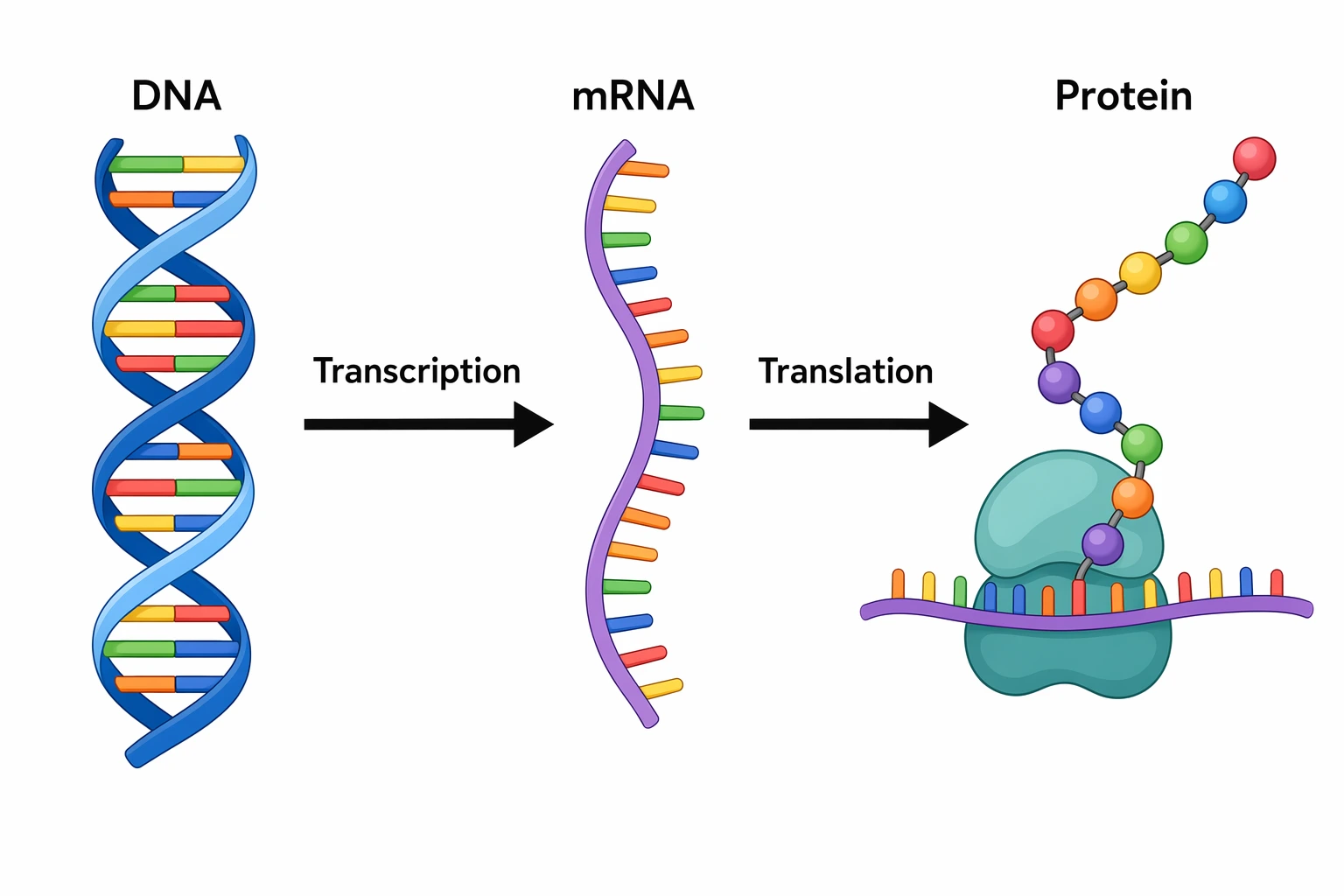
Transcription and Translation Diagram Generator
Type a DNA sequence and get mRNA, codons, and amino acids as a labeled diagram.
 Biology
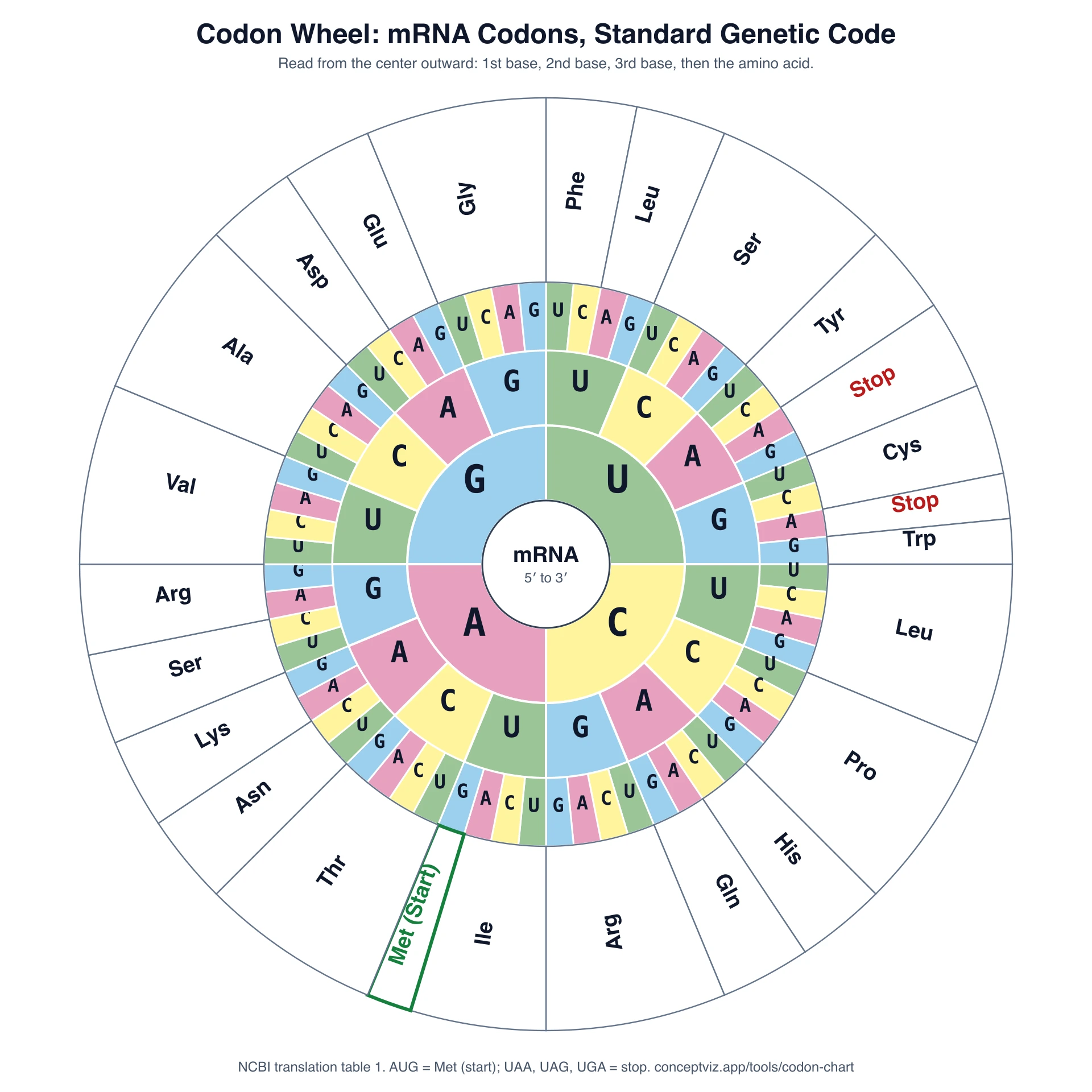
Codon Chart
mRNA codon table and wheel for the amino-acid lookup step.
 Biology
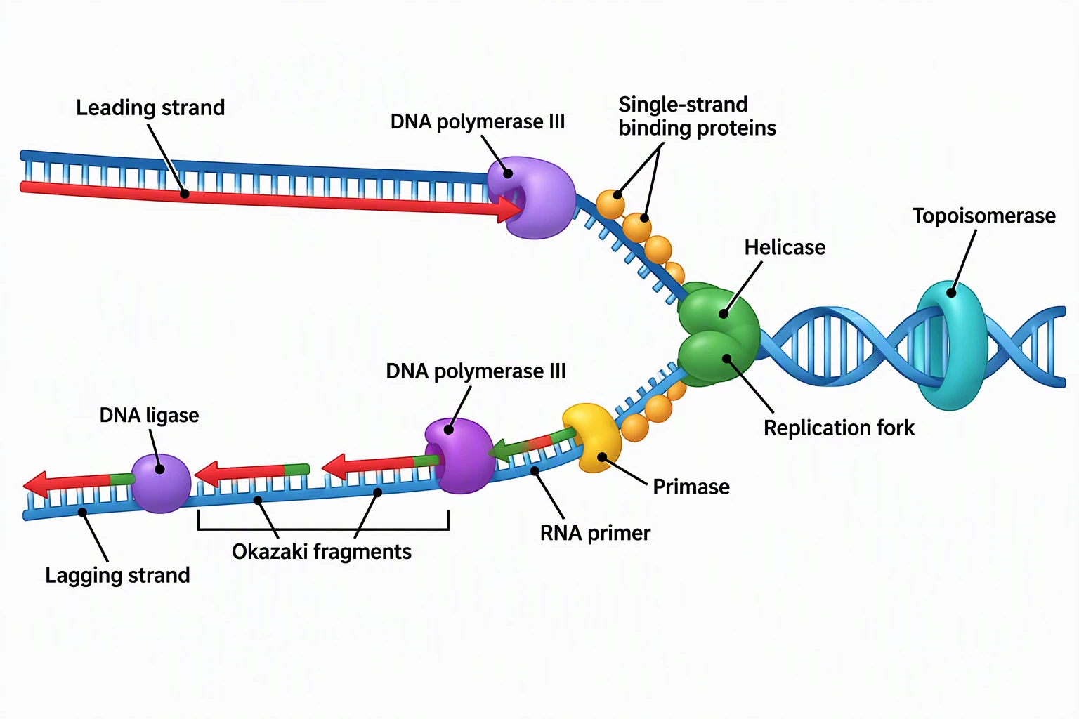
DNA Replication Diagram
Free replication-fork pack: helicase, polymerase, Okazaki fragments.
 Biology
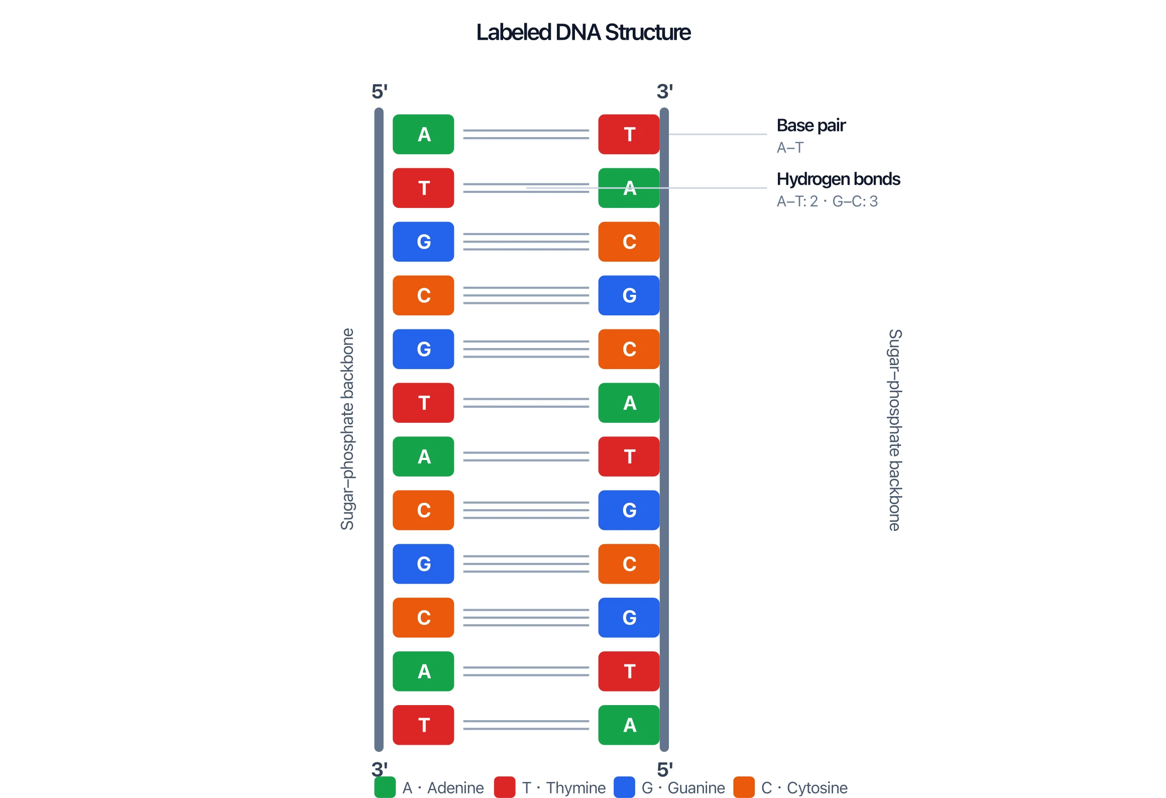
DNA Structure Diagram Generator
Double helix and base pairs that transcription reads.
 Biology
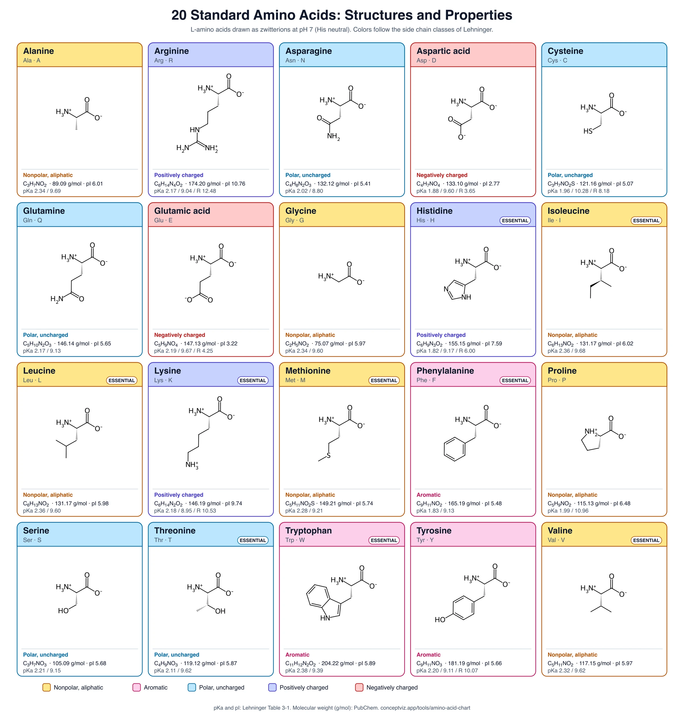
Amino Acid Chart
Structures and properties of the twenty amino acids.
 Biology
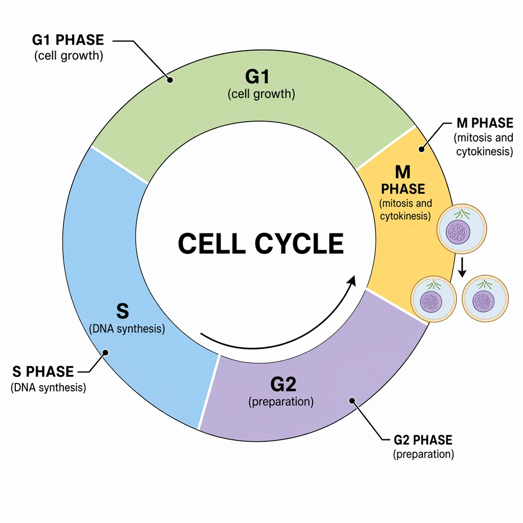
Cell Cycle Diagram Generator
When DNA is copied and when protein synthesis is busy.