Codon Chart Table, Wheel and Lookup
Free codon chart for the standard genetic code: mRNA and DNA codon table, codon wheel, one-codon lookup and print or PNG and SVG export. No sign-up.
View
Sequence type
Amino acid labels
Type a codon, or click a codon in the chart.
Swipe sideways inside the chart to see every column.
Amino acid index: pick one to highlight every codon that encodes it
Codon Chart Images
Ready-made codon tables and wheels, free to view and download with no sign-up. They come from the same chart engine as the tool above.
mRNA Codon Chart Table
The classic 4 by 4 mRNA codon table with three-letter amino acid codes. AUG start and UAA, UAG, UGA stop codons are marked.
Codon tables
Codon wheels
Print and practice
What is a codon chart?
A codon chart, also called a genetic code table or codon table, lists which amino acid each three-letter RNA or DNA codon specifies during protein synthesis. There are four possible bases, so there are 4 x 4 x 4 = 64 codons. Sixty-one of them code for one of the 20 standard amino acids, and three (UAA, UAG and UGA) tell the ribosome to stop. The chart on this page uses the standard genetic code, NCBI translation table 1, which is shared by nearly all nuclear genes in eukaryotes, bacteria and archaea. A few organelles and organisms use small variants, such as vertebrate mitochondria, so check the right table before applying this one to unusual genomes. When you color amino acids by property, note that textbooks do not fully agree on Cys, Tyr, Gly and His; this chart uses the common four-class grouping (nonpolar, polar, acidic, basic).
How to read the codon table
- Write the codon 5′ to 3′, for example AUG. Find the first base (A) in the left column, the second base (U) in the top row, and then the third base (G) in the right column. The row where they meet is the amino acid: Met.
- The first base picks one of four large blocks, the second base picks a column inside it, and the third base picks one of four lines in that cell. Type any codon in the lookup box to see the same path highlighted.
- Switch the sequence type to DNA to show T in place of U. The DNA table is the coding (sense) strand, which reads the same as the mRNA, so the codon ATG on the coding strand matches AUG on the mRNA.
How to read the codon wheel
The codon wheel packs the same information into rings. Start in the center with the first base, move outward through the second and third bases, and stop at the outer ring for the amino acid. Where neighboring codons share an amino acid, one outer wedge spans them. Leucine, serine and arginine each appear as two separate wedges because their six codons sit in two different blocks, while methionine and tryptophan each get a single narrow wedge. The wheel also makes the pattern in the third base easy to see: for many amino acids the third base barely matters.
Start codon, stop codons and degeneracy
- AUG is the start codon. It codes for methionine and sets the reading frame, so the first amino acid of a new protein is usually Met.
- UAA, UAG and UGA are stop codons. In the standard code they have no matching tRNA, and release factors end translation when the ribosome reaches one.
- The code is degenerate, meaning most amino acids have more than one codon. Leucine, serine and arginine have six, while methionine and tryptophan have one. Codons for the same amino acid usually differ only at the third base, which is why a third-base change is often a silent mutation.
- Wobble pairing explains this: the third codon position pairs more loosely with the first position of the tRNA anticodon, so fewer tRNAs are needed than there are codons.
mRNA codons, DNA codons and anticodons
People mix these up often, so keep the strands straight. An mRNA codon is copied from the DNA template strand and matches the coding strand with U in place of T. A tRNA anticodon is complementary and antiparallel to the codon: for the codon 5′-AUG-3′ the anticodon is 3′-UAC-5′, which is written 5′-CAU-3′ when read in the usual direction. When a question gives you the template strand, first write the mRNA as the complement of the template strand (the template is antiparallel, so reverse it if it is given 3′ to 5′), then read codons from the mRNA 5′ end. The lookup box shows the mRNA codon, the DNA coding-strand codon and the anticodon together. To translate a whole sequence in one go, use the transcription and translation diagram tool linked below.
Printing and exporting the chart
Use PNG for slides and documents, SVG when you need a vector that stays sharp at any size, and Print for a paper copy; your browser print dialog can also save the page as a PDF. Practice mode hides every amino acid so students can fill the blanks and then check against the filled chart. Everything is drawn in your browser and is free to use without an account.
Frequently Asked Questions
Related Biology Tools
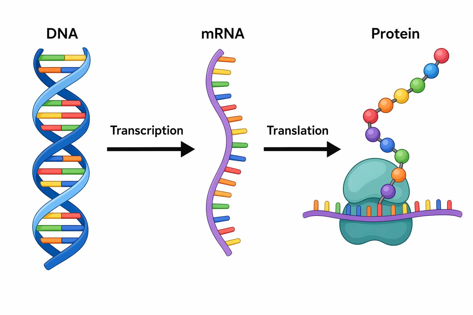 Biology
Transcription and Translation Diagram
Paste a DNA sequence and see the mRNA, codons and amino acid chain step by step.
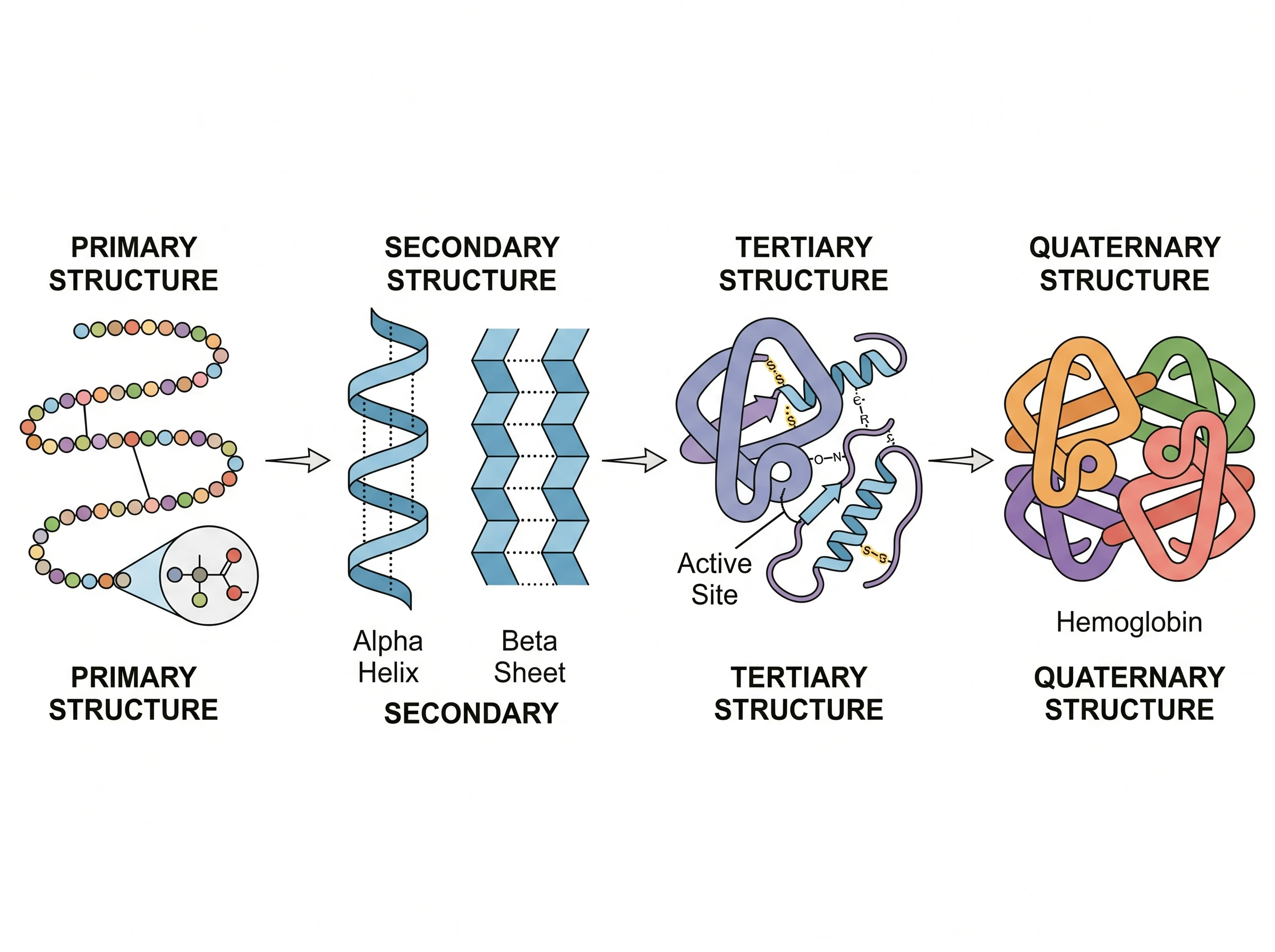 Biology
Protein Structure Diagram Generator
Illustrate the four levels of protein structure, the product of translation.
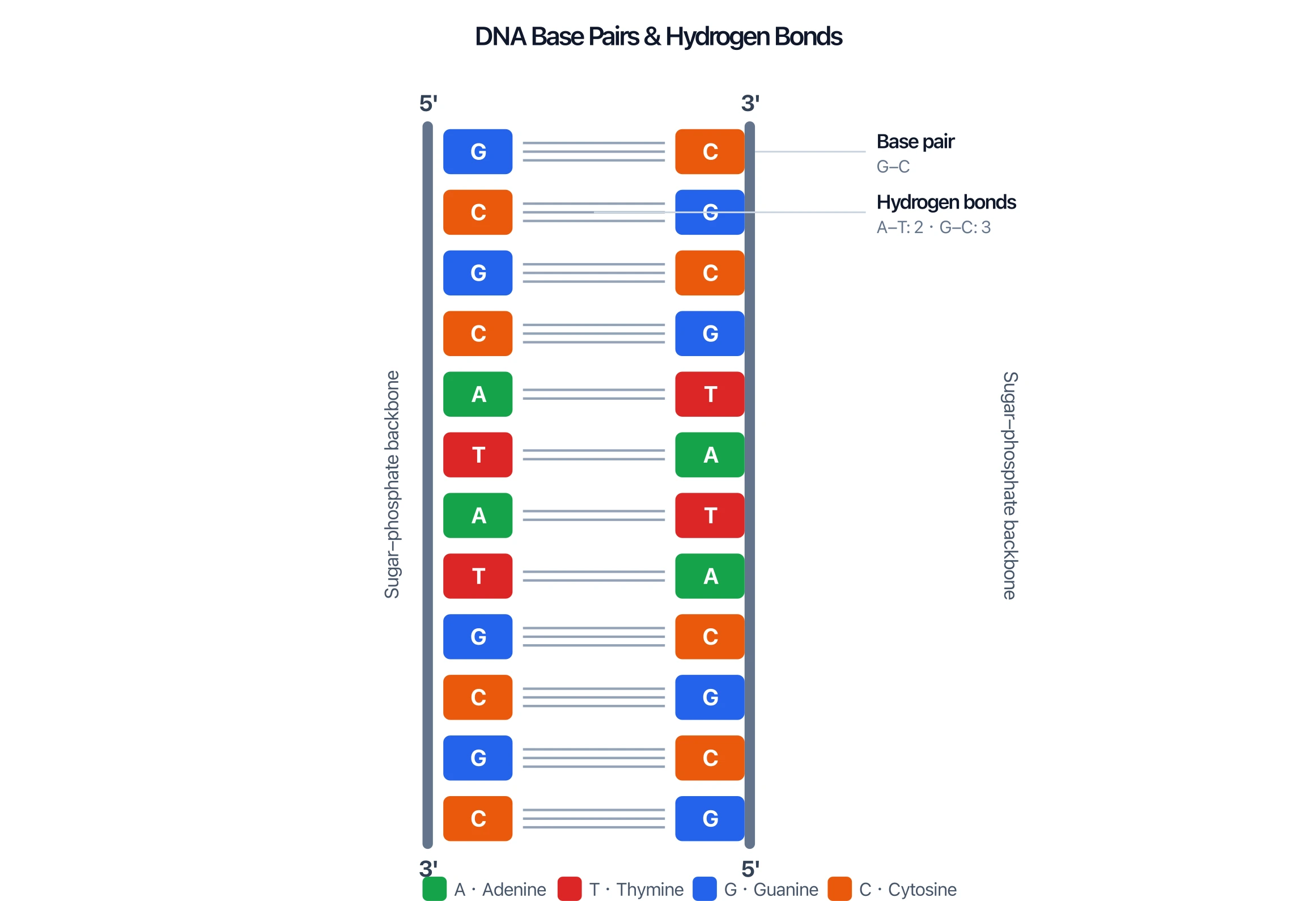 Biology
DNA Structure Diagram Generator
Draw labeled DNA double-helix and base-pair diagrams for biology class.