DNA Replication Diagram
Free DNA replication diagrams: labeled fork, blank worksheet, simple version, and coloring page. Printable; free download, no sign-up.
Free DNA Replication Diagrams
Every version below is free to view, download, and print. No sign-up.
Labeled Replication Fork
The full replication fork with every enzyme and strand named: Helicase, Topoisomerase, Single-strand binding proteins, Primase, RNA primer, DNA polymerase III on both strands, Leading strand, Lagging strand, Okazaki fragments, DNA ligase, and Replication fork.
Core diagrams
Structure & focus
Print & practice
Test yourself: DNA replication
A quick self-test. Questions reshuffle each time.
At the replication fork, helicase…
Customize this diagram
Change grade level, which enzymes are labeled, or leading versus lagging focus. Describe it and generate your own.
Customize Your DNA Replication Diagram
Free to try ·
Your custom diagram will appear here
Ask for a labeled fork, blank worksheet, or Okazaki close-up
How to read the labeled replication fork
Start with the fully labeled fork. The parental helix is on the right and the fork moves right. The figure has 11 distinct names and 12 callouts: Leading strand, DNA polymerase III (two callouts, one on the leading strand and one on the lagging strand), Single-strand binding proteins, Helicase, Topoisomerase, Replication fork, Primase, RNA primer, DNA ligase, Okazaki fragments, and Lagging strand. Helicase separates the two strands. Topoisomerase sits on the still-twisted helix ahead of the opening, where supercoils would otherwise pile up. Single-strand binding proteins coat the exposed templates so they do not snap shut. Follow every leader line to its tip: the line should land on the protein or strand it names.
Leading strand, lagging strand, and Okazaki fragments
The two new strands are not copied the same way. Open the leading-versus-lagging figure next to the fork overview. That focus figure labels Leading strand, DNA polymerase III, Replication fork, Okazaki fragments, RNA primer, and Lagging strand. The leading strand is synthesized continuously toward the fork after one RNA primer. The lagging strand is copied in short bursts away from the fork; each burst starts with an RNA primer from primase and becomes an Okazaki fragment. The close-up then labels Parental strand, RNA primer, DNA polymerase I, DNA ligase, Lagging strand, and Okazaki fragments. DNA polymerase I removes primers and fills the gaps; DNA ligase seals the remaining nicks. Print those two focus figures at the same scale so students can match labels across pages.
Simple diagram for a first pass
The simple diagram labels DNA, Helicase, DNA polymerase (two callouts), Leading strand, and Lagging strand. Use it as a middle-school poster or as a one-minute warm-up before a high-school lesson. Point to the Y shape first, then to the two new strands, then to the enzyme at the opening. Once that picture is stable, switch to the 12-callout fork. Do not start with Okazaki fragments if the class has not yet seen the simple version; the extra names land better on a second pass.
Blank worksheet, coloring page, and a classroom routine
The blank worksheet matches the labeled fork: same proteins, same leader-line geometry, numbers 1 to 12 with blank lines instead of words. Project the labeled version, hide it, and give students five minutes on the blank sheet. You can also print labeled on the front and blank on the back of one page for homework. The coloring page is an outline fork with no text. Ask students to color parental DNA one color and newly made DNA another; that color split is a quiet preview of the semiconservative figure. All of these files are free to view, download, and print.
The semiconservative replication figure
The last labeled figure is not a fork. It labels Parental DNA, Original strand, New strand, Generation 1, and Generation 2. Generation 1 shows two hybrid helices (one original strand and one new strand each). Generation 2 shows two hybrid helices plus two fully new helices. Show this figure after the fork diagrams so students do not mix up “how the fork copies DNA” with “what the helices look like after one or two rounds.” If you need the double-helix chemistry (bases, backbone, 5-prime and 3-prime ends), switch to the DNA structure pack linked below rather than crowding those names onto the fork.
Where this pack sits in a unit
Replication is one step in a larger story. The cell-cycle pack shows when DNA is copied (S phase) relative to mitosis. The protein-synthesis pack shows what happens when those copies are later transcribed and translated. Keep this page for the fork itself: enzymes, strands, fragments, and the semiconservative product cartoon. That split keeps each download set small enough to print and large enough to teach from.
Frequently Asked Questions
Related Biology Tools
 Biology
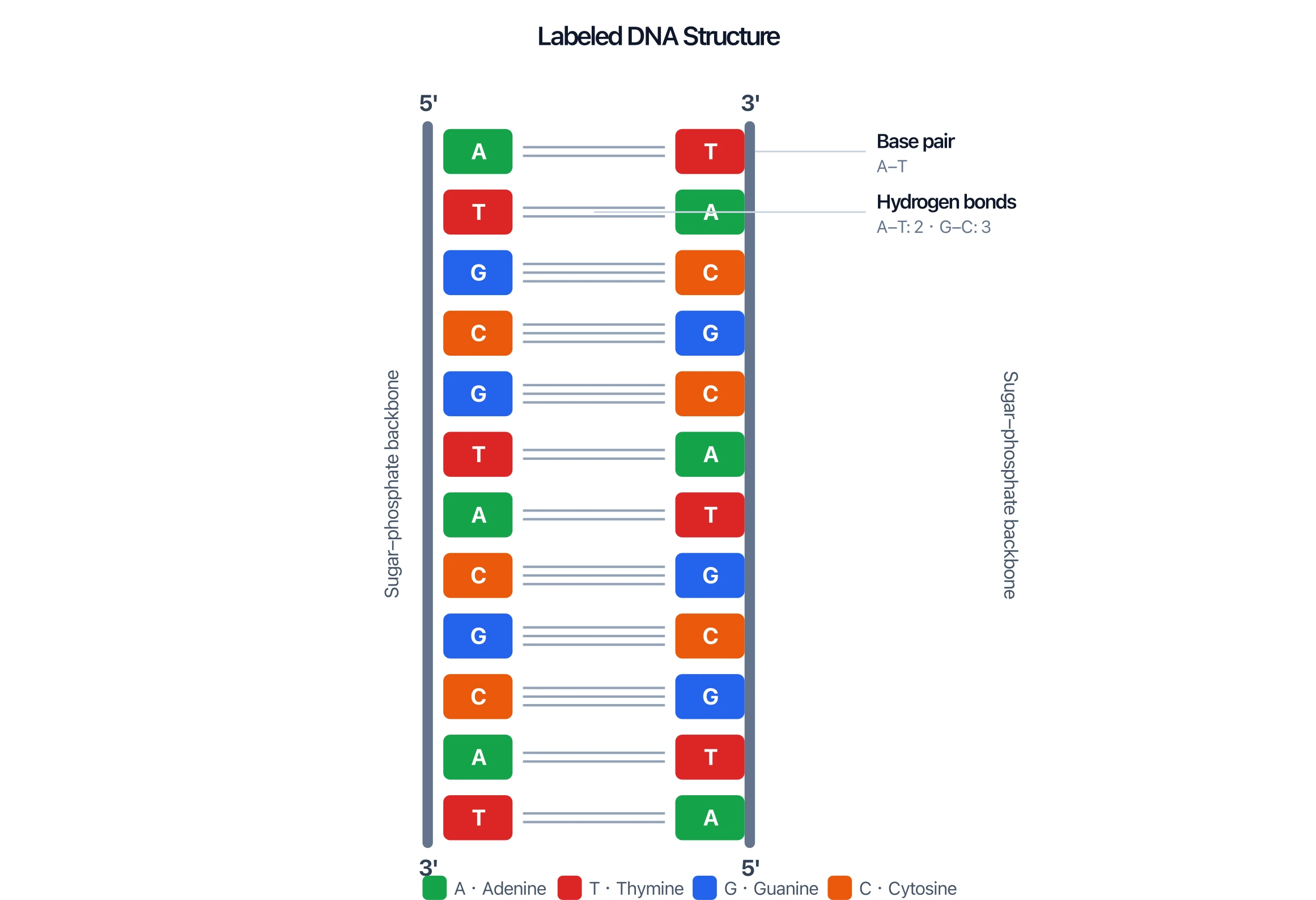
DNA Structure Diagram Generator
Double helix, base pairs, and antiparallel ends for the chemistry behind the fork.
 Biology
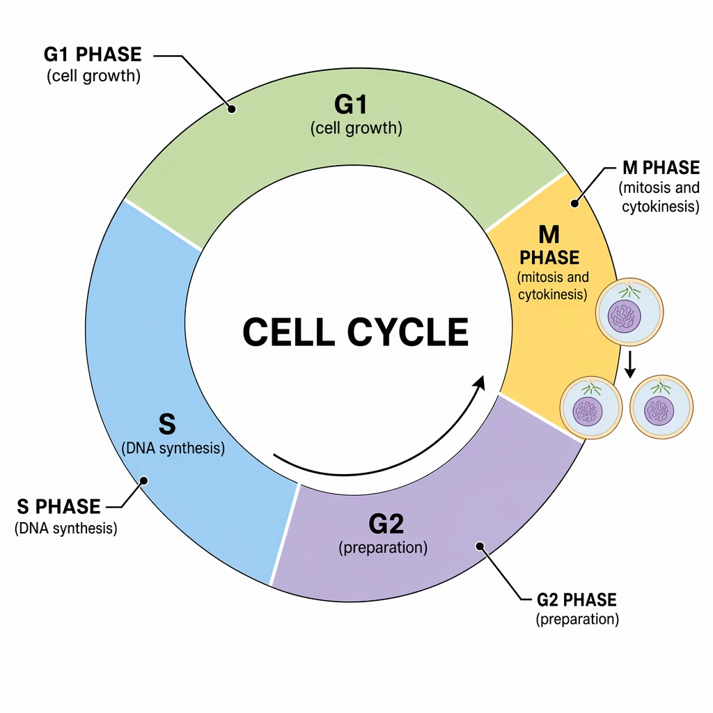
Cell Cycle Diagram Generator
S phase in context: when DNA is copied relative to mitosis.
 Biology
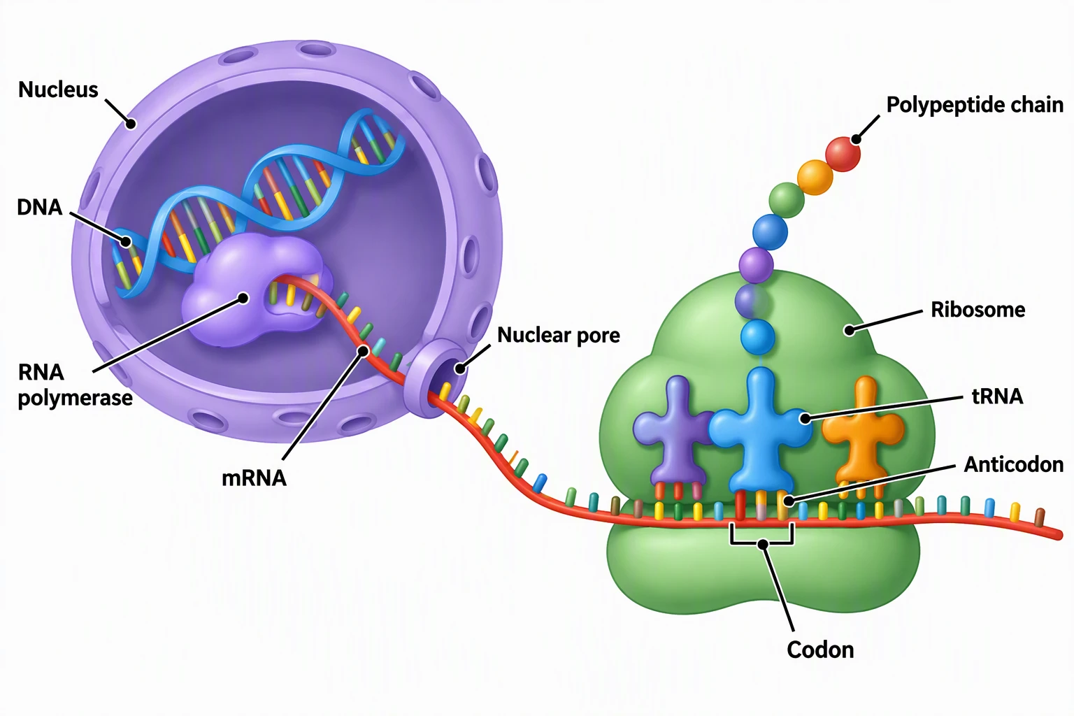
Protein Synthesis Diagram
Free transcription and translation pack that follows a copied gene.
 Biology
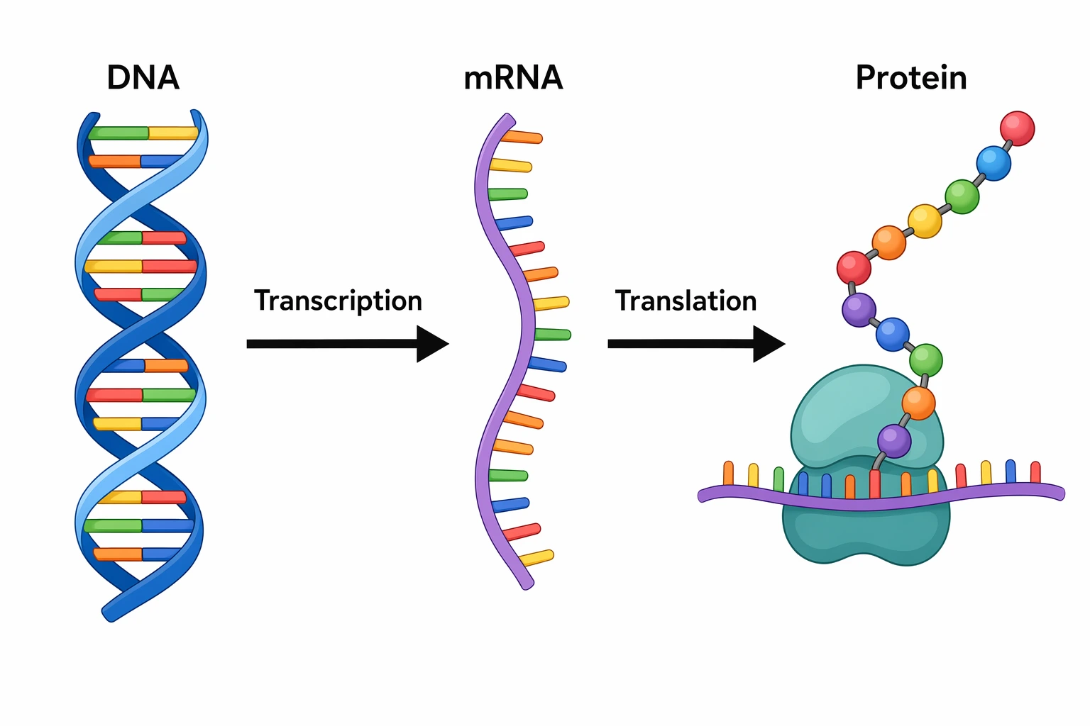
Transcription and Translation Diagram Generator
Custom sequence-to-protein diagrams for a chosen DNA string.
 Biology
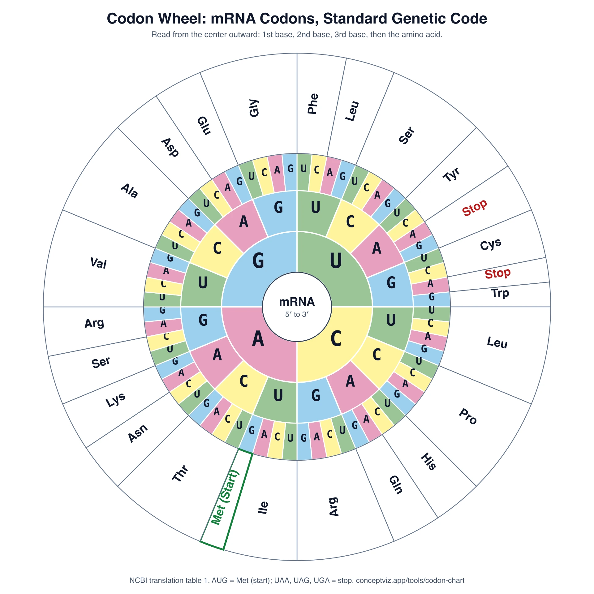
Codon Chart
mRNA codon table and wheel for decoding the new strand later.
 Biology
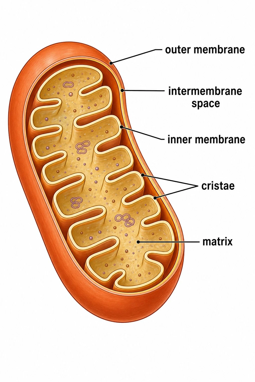
Mitochondria Diagram
Free organelle pack for linking replication to ATP context.