Coagulation Cascade Diagram Generator for Lecture & Review Figures
Generate a labeled, blank, or simplified coagulation cascade for a lecture or review figure. Intrinsic, extrinsic, and common pathways — then download as PNG.
Coagulation Cascade Diagram Generator
Free preview on this page · Clean file download needs a
Used by 40,000+ teachers, students & researchers
Your coagulation cascade diagram will appear here
Ask for labeled, blank, simpler, or a drug / disease annotation
Coagulation Cascade Examples
Labeled, blank, simpler, and annotated lecture figures — not a what-is page
Labeled Classical Cascade
The lecture original: intrinsic, extrinsic, and common paths, factors named, fibrin at the end.
Blank Self-Test
Same topology, labels removed — the second artifact in a teaching set.
Simplified Lecture Slide
Fewer boxes, larger type — the simpler PPT for the same hour.
Anticoagulant Action Sites
Where heparin, warfarin, and a DOAC sit on the same cascade.
Cell-Based vs Classical
Classical Y-shape next to initiation / amplification / propagation.
Hemophilia A Break
VIII missing, aPTT path broken, PT path intact — a figure you can stand in front of.
What this page makes (and what it does not)
This page makes a coagulation cascade figure you can take to a lecture or drop into a review: labeled, blank for self-test, simpler for a first slide, or annotated with a drug or a disease break. It does not replace the NIH or Osmosis “what is the clotting cascade” explainer. Those pages already own the canonical picture. The job here is a custom variant of that topology — the set a tutor or lecturer actually pays to finish.
Classical pathways the diagram should lock
- Intrinsic (contact) path: XII → XI → IX, with VIII as a cofactor, measured by aPTT.
- Extrinsic (tissue-factor) path: TF + VII, measured by PT / INR.
- Common path: X (with V) → II (prothrombin → thrombin) → I (fibrinogen → fibrin).
- Cofactors often worth labeling: Ca2+, phospholipid / platelet surface, and vitamin K for II, VII, IX, and X.
Labeled, blank, and simpler — one topic, three artifacts
A paying teaching workflow is usually a pack, not a single download. Start with a fully labeled classical cascade so the room can see every factor. Make a blank copy of the same layout for a self-test. Then make a simpler slide that keeps TF → VII → X → thrombin → fibrin if the audience is nursing or first-year. Ask for those three outputs in three prompts; do not expect one image to be both the atlas and the quiz.
Drug sites and disease breaks
The cascade becomes a clinical figure when you mark where a drug or a missing factor sits. Unfractionated heparin works through antithrombin on Xa and IIa. Warfarin hits the vitamin-K–dependent factors II, VII, IX, and X. Direct oral anticoagulants typically block Xa (or thrombin, for dabigatran). Hemophilia A removes VIII and lengthens aPTT while PT stays normal. Describe the annotation you need; the generator should keep the same three-path layout underneath so the class can still read it.
Classical Y-shape vs the cell-based model
Most exam slides still use the Y-shaped classical cascade. Many hematology courses also teach the cell-based model: initiation on tissue-factor–bearing cells, amplification on platelets, then propagation of a thrombin burst. If you need both, ask for a two-panel figure with the same factor names in each panel. Do not mix the two models into one unlabeled tangle.
Honesty note on AI factor labels
These are AI illustrations. The model can swap Roman numerals, drop a cofactor, or put heparin on the wrong node. That is why this page is a lecture-pack generator and not a Precise SVG engine: the topology branches, and a boxy “correct” renderer would look like a stacked org chart. Check every factor against your textbook, Osmosis, or a hematology atlas before you project the slide or put it in a paper. Treat the file as a draft you verify.
Frequently Asked Questions
Related Medical Tools
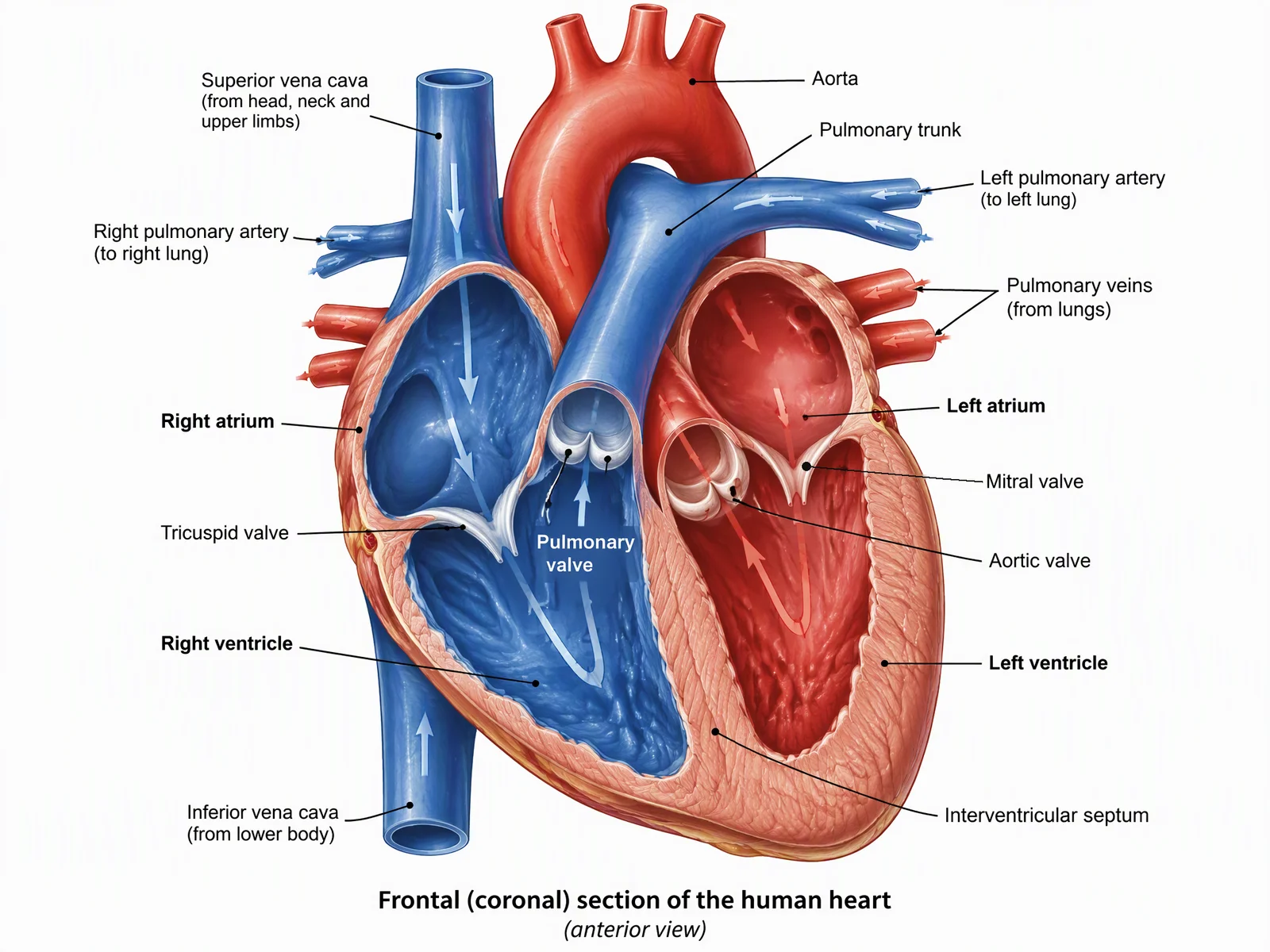 Medical
Medical Illustration Generator
Broader clinical and anatomy figures when the cascade is only one panel in a larger slide.
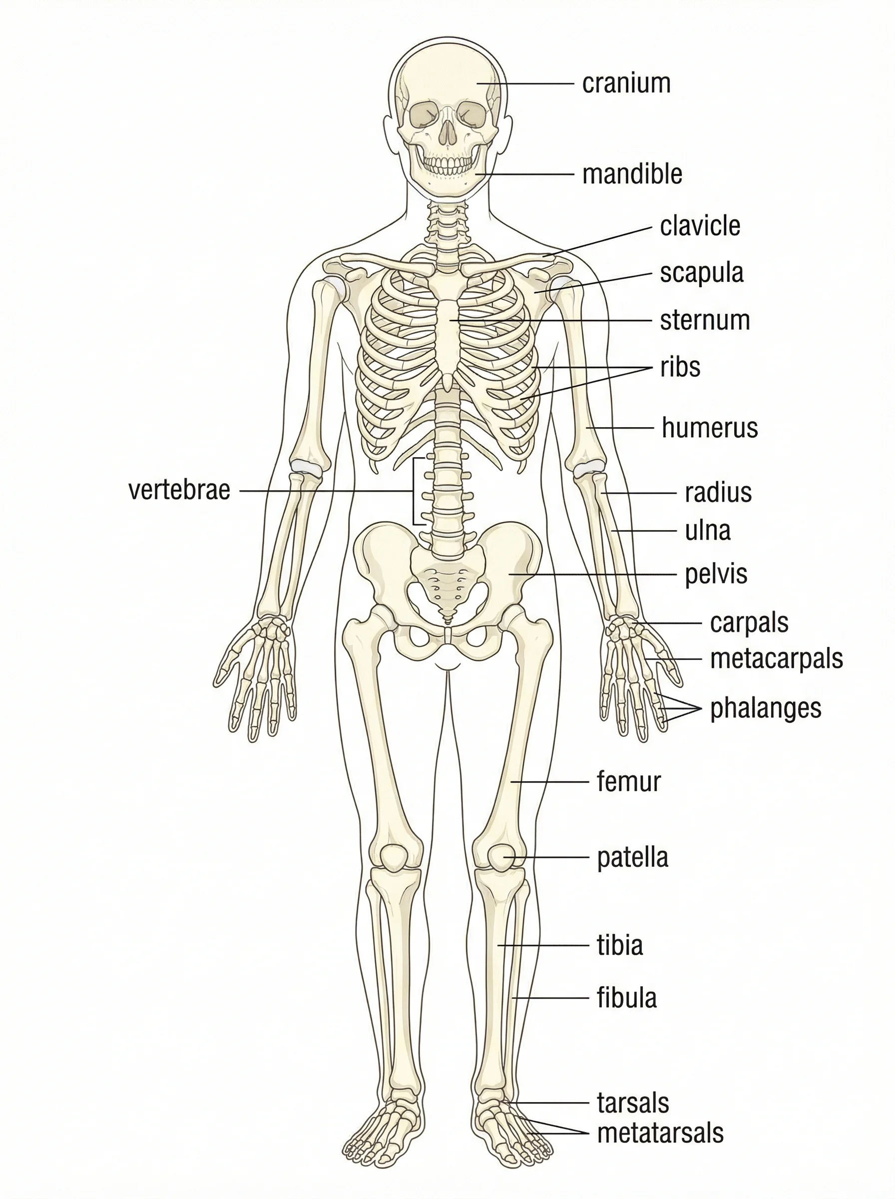 Medical
Anatomical Drawing Generator
Anatomy drawings for the same lecture deck when you also need vessels or organs.
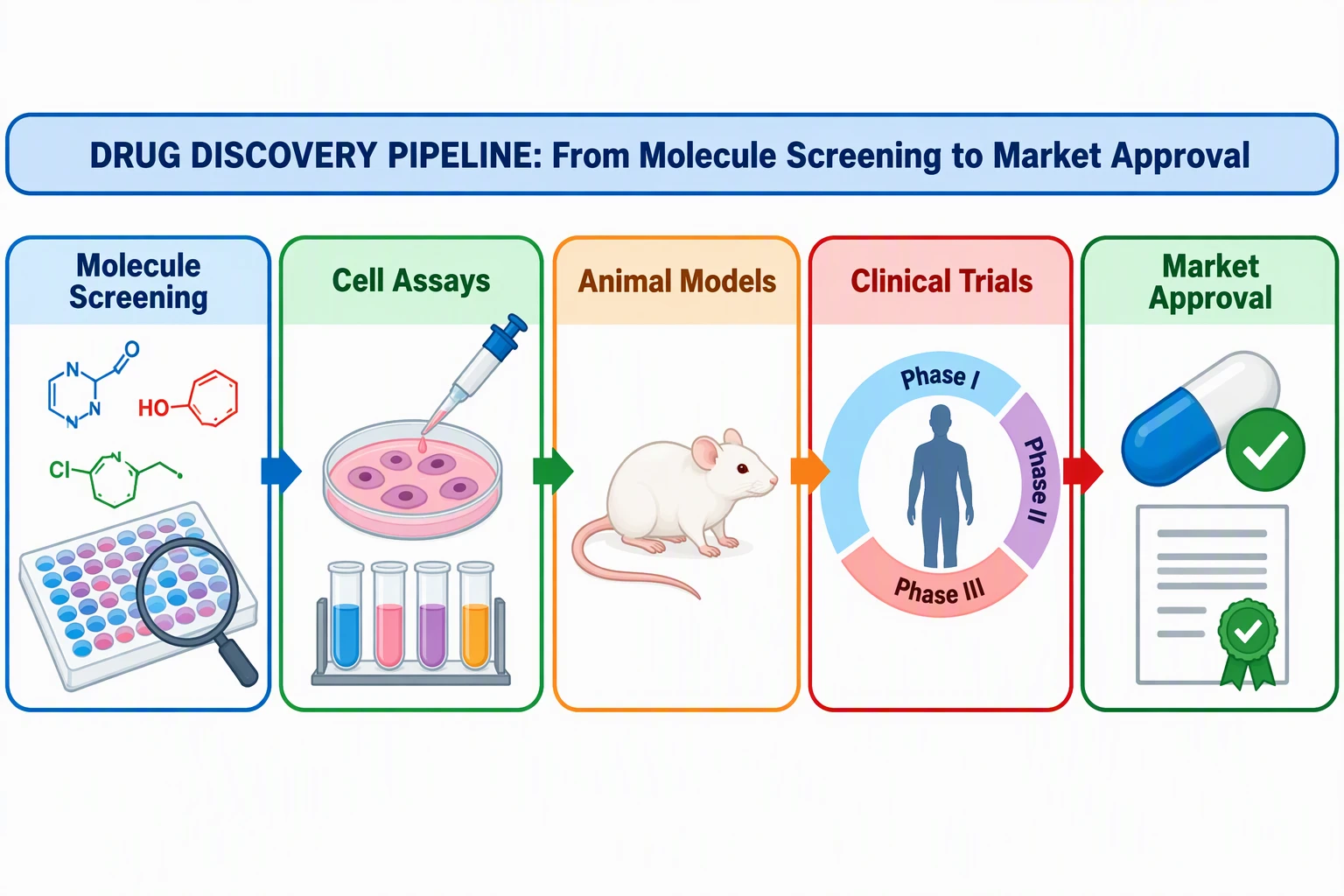 Research
Graphical Abstract Maker
Journal-style visual abstracts if the cascade is going into a paper, not a slide.