Phospholipid Diagram Generator for Structure & the Bilayer
Create a clearly labeled phospholipid diagram from a description: the hydrophilic phosphate head, two hydrophobic fatty acid tails, and how the molecules line up into the phospholipid bilayer of the cell membrane.
AI Phospholipid Diagram
Free to try ·
Your phospholipid diagram will appear here
Illustrative art — review the labels and structure against your textbook
Phospholipid Diagram Examples
Labeled structures, bilayers, and cell membrane illustrations
Single Phospholipid Structure
A single phospholipid with the parts labeled — phosphate head, glycerol, and the two fatty acid tails.
Phospholipid Bilayer
Two layers of phospholipids — heads out toward the water, tails tucked inside, away from it.
Fluid Mosaic Cell Membrane
The bilayer as the basis of the cell membrane, with proteins, cholesterol, and glycoproteins added.
Transport Across the Membrane
How molecules cross the bilayer — simple diffusion, facilitated diffusion, and active transport.
Liposome Structure
A phospholipid bilayer curved into a sphere — the same heads-out, tails-in rule, in 3D.
Membrane Fluidity
Why unsaturated, kinked tails and cholesterol keep the membrane fluid rather than rigid.
What is a phospholipid?
A phospholipid is the main building-block molecule of every cell membrane. It belongs to the lipid family, but unlike a fat it has a phosphate group attached, which gives it a special split personality: one end loves water and the other end avoids it. That single feature is what lets phospholipids spontaneously arrange themselves into the thin, flexible sheet that surrounds every cell. This generator draws a phospholipid and its bilayer with every part labeled, so the link between the molecule and the membrane is easy to see.
The structure of a phospholipid: head and two tails
- Hydrophilic head: a phosphate group attached to a glycerol backbone. "Hydrophilic" means water-loving — this end is polar and mixes happily with water.
- Glycerol backbone: the small three-carbon molecule that links the phosphate head to the fatty acid tails.
- Two fatty acid tails: long hydrocarbon chains that are hydrophobic (water-fearing) and non-polar, so they turn away from water.
- One tail is often unsaturated, meaning it has a double bond that puts a kink in the chain; the other is usually saturated and straight.
- A common shorthand draws the phospholipid as a circle (the head) with two wavy lines (the tails) — the "lollipop" or "balloon" shape you see in textbooks.
Why a phospholipid is amphipathic
Because one end is hydrophilic and the other is hydrophobic, a phospholipid is described as amphipathic (or amphiphilic) — it has both properties in one molecule. This is the key idea that explains everything else. The water-loving phosphate head wants to face water, while the water-fearing fatty acid tails want to hide from it. A single molecule cannot satisfy both ends at once, but a crowd of them can, by organizing into a structure where every head touches water and every tail is shielded. Labeling the head as hydrophilic and the tails as hydrophobic is the most important annotation on any phospholipid diagram.
How phospholipids form a bilayer in water
Drop phospholipids into water and they arrange themselves automatically into a phospholipid bilayer — a double layer two molecules thick. The hydrophilic heads point outward on both surfaces, facing the watery environment inside and outside the cell, while the hydrophobic tails point inward toward each other, away from water. This creates a stable sheet with a water-friendly outside and an oily, water-repelling core. No energy is needed to assemble it; it forms because it is the lowest-energy arrangement. The membrane is also self-sealing, which is why small tears close on their own. This tool can draw both a single labeled phospholipid and the full bilayer so you can show the molecule and the structure it builds.
The phospholipid bilayer and the cell membrane
The phospholipid bilayer is the foundation of the cell membrane, but a real membrane is more than just lipids. The accepted picture is the fluid mosaic model: "fluid" because the phospholipids drift sideways and the membrane behaves like a 2D liquid, and "mosaic" because many other molecules are embedded in it. Proteins float in the bilayer — integral (transmembrane) proteins span it to act as channels and carriers, while peripheral proteins sit on the surface. Cholesterol slots between the phospholipids to control how fluid the membrane is, and glycolipids and glycoproteins carry carbohydrate chains on the outer surface for cell recognition. The hydrophobic core is what makes the membrane selectively permeable, letting small non-polar molecules pass while blocking ions and large polar molecules.
Labeled vs blank diagrams, and how to generate one
For learning, a fully labeled diagram shows you where every part goes; for revision and quizzes, a blank version lets you test yourself by filling in the labels. You can generate either one here by describing what you want in plain English — for example, "a labeled phospholipid bilayer with hydrophilic heads and hydrophobic tails marked" or "a blank cell membrane diagram with empty label lines." The tool turns your description into a clean, presentation-ready illustration you can drop into notes, slides, or a worksheet. One honest note: this is an AI image generator, so the diagrams are illustrative and you should always check the labels and structure against your textbook before relying on them for an assignment.
Frequently Asked Questions
Related Biology Tools
 Biology
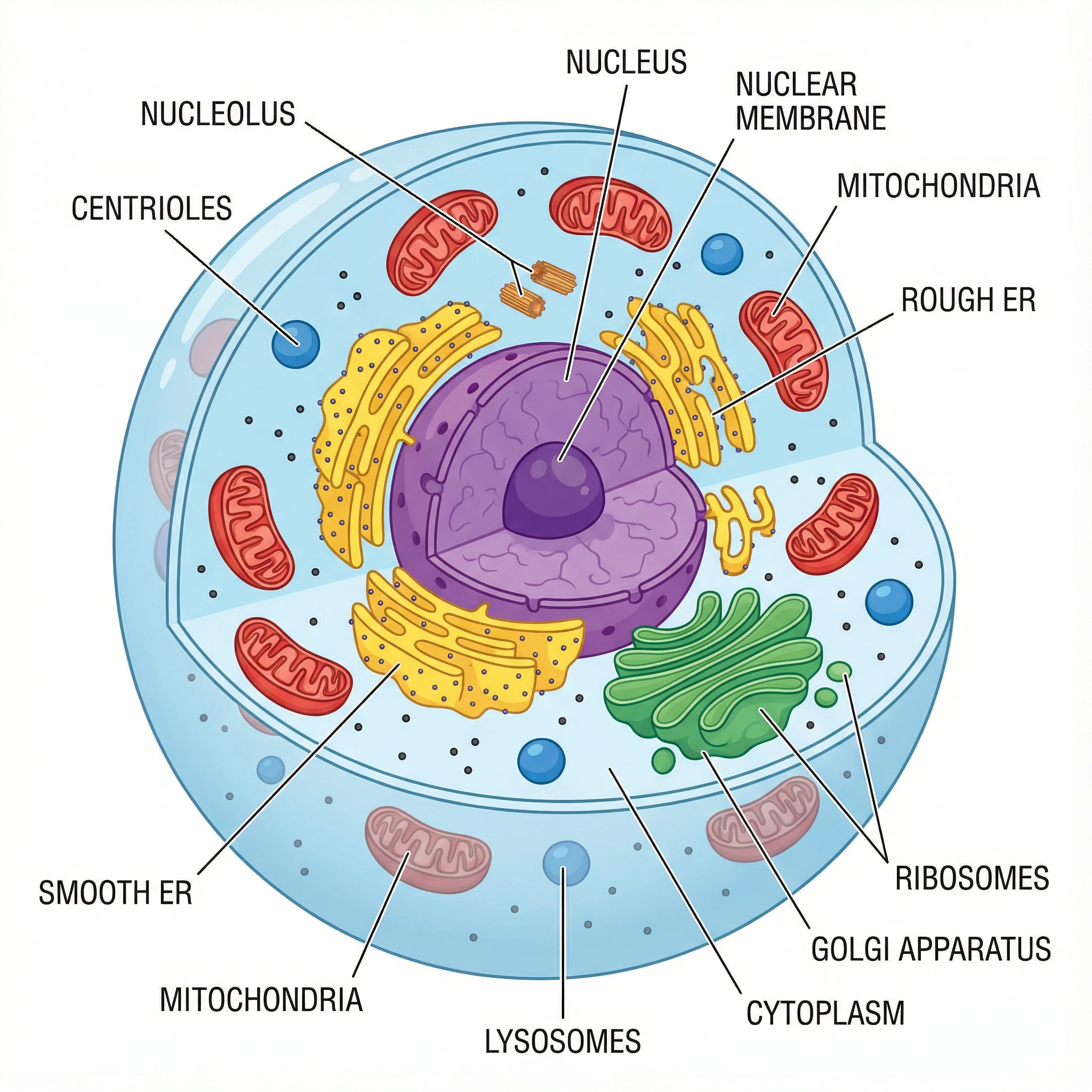
Animal Cell Diagram Generator
Create labeled animal cell diagrams with the cell membrane, nucleus, mitochondria, and other organelles.
 Research
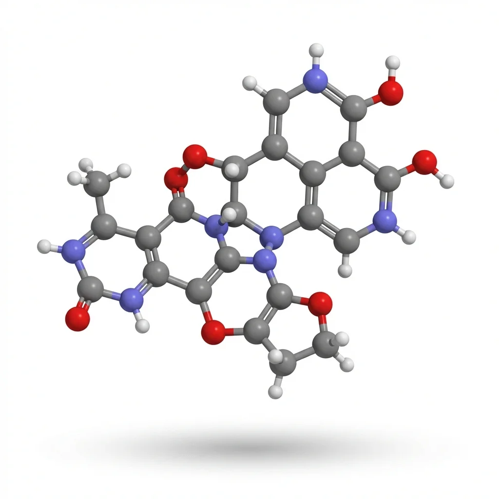
AI Scientific Image Generator
Generate accurate scientific illustrations of molecules, structures, and biological processes.
 Biology
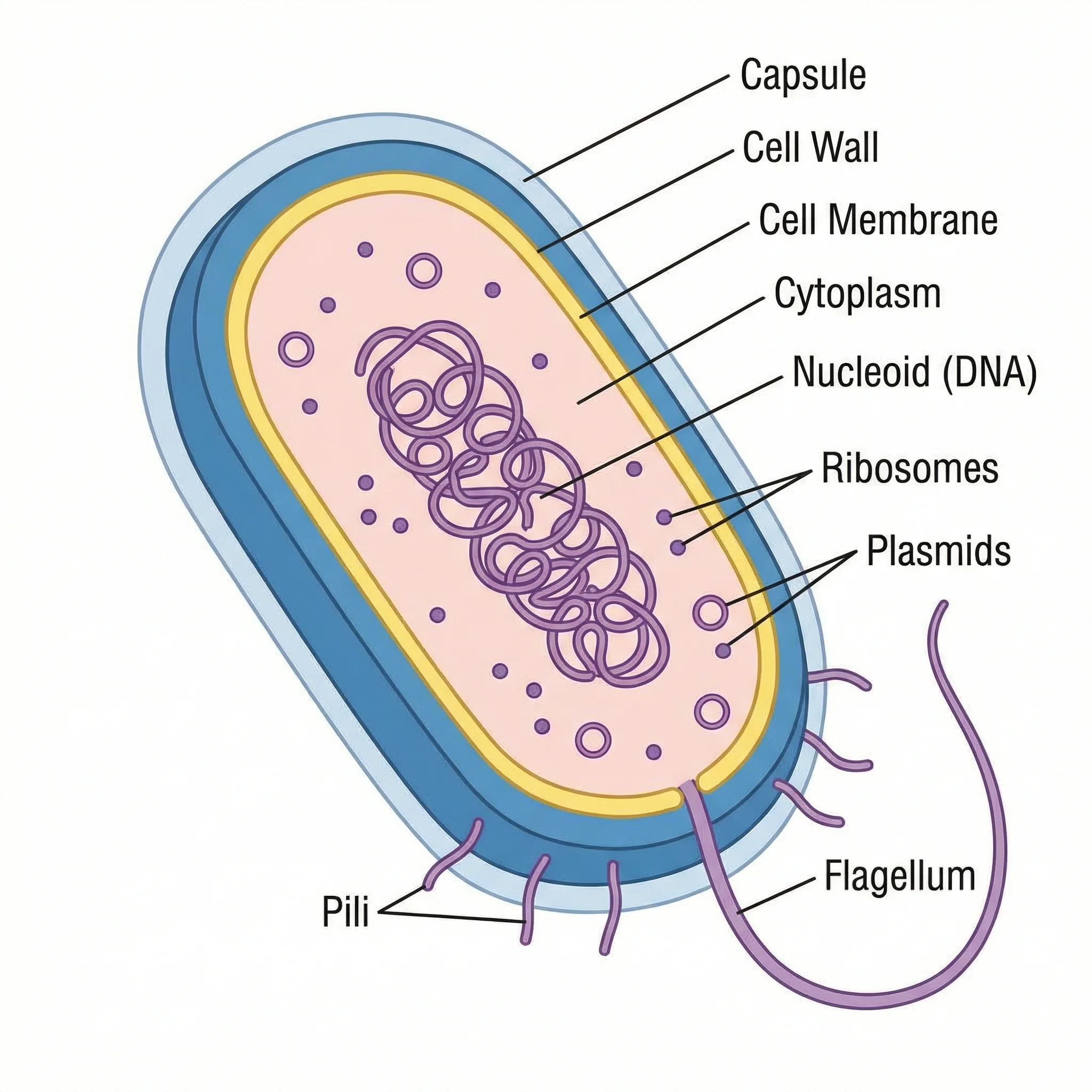
Bacteria Diagram Generator
Draw labeled bacterial cell diagrams showing the cell wall, membrane, flagella, and other structures.