Mitochondria Diagram Generator — Labeled & Blank
Generate a clearly labeled mitochondria diagram in seconds. Show the outer membrane, inner membrane, cristae, matrix, and intermembrane space — plus blank, unlabeled versions for worksheets. Describe what you need and download a classroom-ready diagram, free.
AI Mitochondria Diagram
Free to try ·
Your mitochondria diagram will appear here
AI-generated — review the parts and labels for accuracy before using in class
Mitochondria Diagram Examples
Labeled, unlabeled, and cellular-respiration diagrams for biology class
Labeled Mitochondria Diagram
Every part labeled — outer membrane, inner membrane, cristae, matrix, intermembrane space, ribosomes, and mitochondrial DNA.
Unlabeled (Blank) Mitochondria
A blank version for worksheets and quizzes — students fill in each part themselves.
Cellular Respiration Diagram
Shows where respiration happens: the Krebs cycle in the matrix and the electron transport chain on the cristae.
Simple Mitochondria Diagram
A clean, simplified version for younger students learning the basic shape and parts.
Cristae Close-Up
A close-up of the cristae — the folds that hold ATP synthase and the electron transport chain.
ATP Production Diagram
Traces how the cell makes ATP, from the matrix to the electron transport chain.
What is a mitochondrion?
A mitochondrion is the organelle often called the "powerhouse of the cell" — it is where most of a cell's usable energy, ATP, is made through cellular respiration. (The plural is mitochondria; a single one is a mitochondrion.) Most animal and plant cells contain many of them, and cells that need a lot of energy, such as muscle and nerve cells, hold the most. On a biology diagram, a mitochondrion is usually drawn as a bean-shaped organelle with a smooth outer layer and a folded inner layer. This tool generates that diagram with every part labeled, so students can see what a mitochondrion looks like and where each structure sits.
The labeled parts of a mitochondria diagram
- Outer membrane: the smooth outer boundary that surrounds the whole organelle and controls what enters and leaves.
- Intermembrane space: the narrow gap between the outer and inner membranes, where protons build up to drive ATP synthesis.
- Inner membrane: the membrane just inside the outer one, folded into cristae and packed with the proteins of the electron transport chain.
- Cristae: the folds of the inner membrane — they increase surface area so more energy reactions can happen at once.
- Matrix: the fluid-filled space enclosed by the inner membrane, where the Krebs cycle takes place.
- Mitochondrial DNA and ribosomes: mitochondria carry their own small circular DNA and ribosomes, so they can make some of their own proteins.
What each part does
Each structure has a job, and a good labeled diagram makes the relationships clear. The outer membrane is the protective boundary; the inner membrane is the active workspace, folded into cristae to maximize surface area; the intermembrane space stores the protons that power ATP production; and the matrix is the reaction chamber that holds enzymes, mitochondrial DNA, and ribosomes. The cristae are the key feature to understand — the more folds a mitochondrion has, the more electron transport chain proteins it can hold, and the more ATP it can make. When students can point to each labeled part and say what it does, the structure starts to make sense rather than being a shape to memorize.
Mitochondria and cellular respiration: where ATP is made
The mitochondrion is the site of aerobic cellular respiration, the process that releases energy from glucose and stores it as ATP. It happens in two main stages inside the organelle. The Krebs cycle (also called the citric acid cycle) runs in the matrix, breaking down molecules and releasing electron carriers such as NADH and FADH2. Those carriers feed the electron transport chain, which sits on the inner membrane and cristae and uses oxygen to pump protons into the intermembrane space; ATP synthase then uses that proton gradient to produce most of the cell's ATP. (The first stage, glycolysis, happens in the cytoplasm outside the mitochondrion.) Knowing where each stage happens — matrix versus cristae — is one of the most common things biology exams test, which is why labeling the diagram by location is so useful.
Labeled vs. unlabeled (blank) diagrams for worksheets
For teaching, you usually want two versions of the same diagram. A labeled mitochondria diagram is for instruction and study — every part is named so students learn the structure. A blank, unlabeled diagram is for practice and assessment — the same diagram with the labels removed so students can fill in the outer membrane, inner membrane, cristae, matrix, and intermembrane space themselves. This generator makes both: describe a labeled version for your notes or slides, then describe an unlabeled version for a quiz or worksheet, and download each as an image. Having a matching pair makes it easy to build a study sheet and the answer key together.
How to generate a labeled mitochondria diagram
- Describe the diagram you want in plain English — for example, "a labeled mitochondria diagram showing the outer membrane, inner membrane, cristae, matrix, and intermembrane space."
- Say who it is for and how much detail you need: a simple version for elementary students, a full labeled diagram for high school, or a cellular-respiration version showing the Krebs cycle and electron transport chain.
- For worksheets, ask for an unlabeled or blank version so students can fill in the parts themselves.
- Generate the diagram, then download the image to drop into slides, notes, a study guide, or a printed worksheet.
Frequently Asked Questions
Related Biology Tools
 Biology
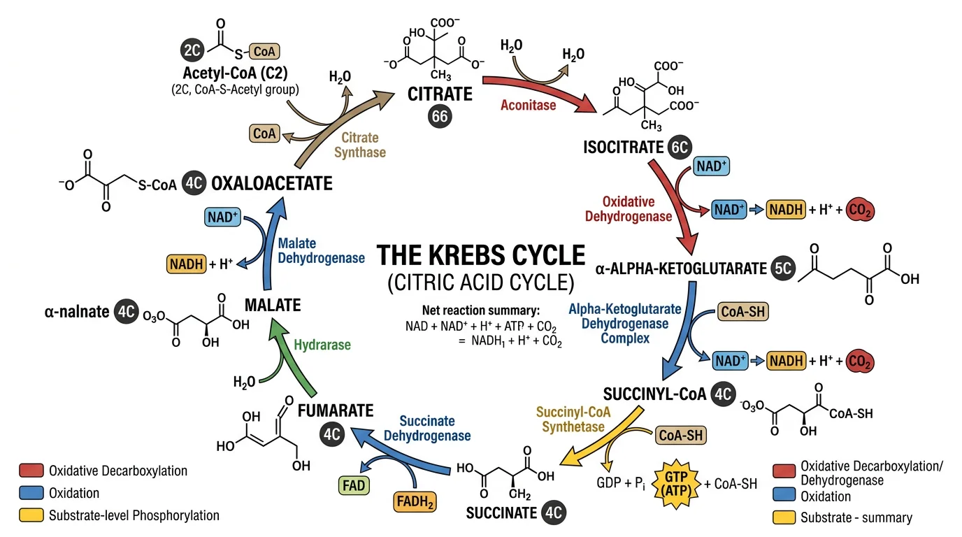
Krebs Cycle Diagram Generator
Draw the citric acid cycle that runs in the mitochondrial matrix, with labeled steps and energy carriers.
 Biology
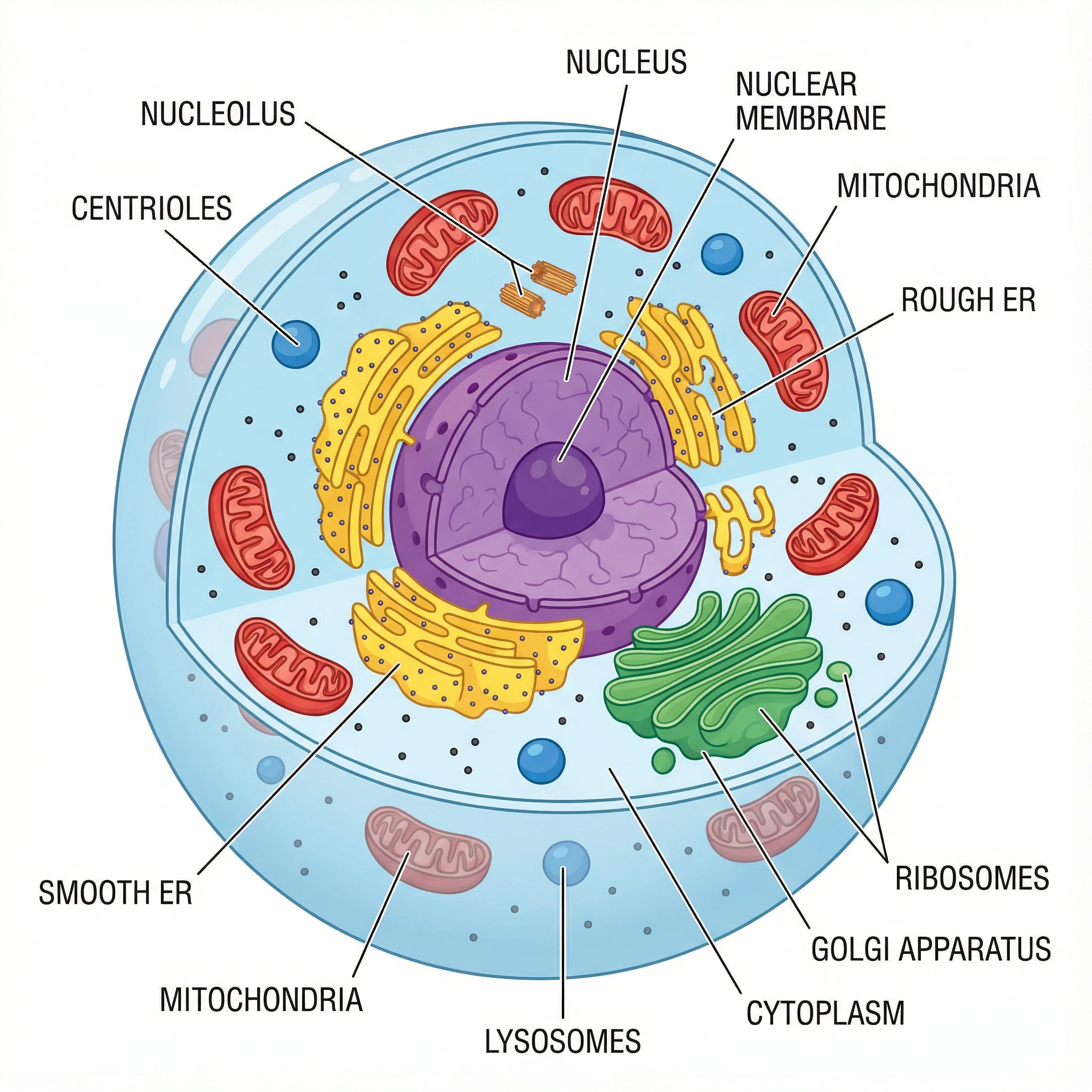
Animal Cell Diagram Generator
Create labeled animal cell diagrams with the nucleus, mitochondria, and other organelles.
 Biology
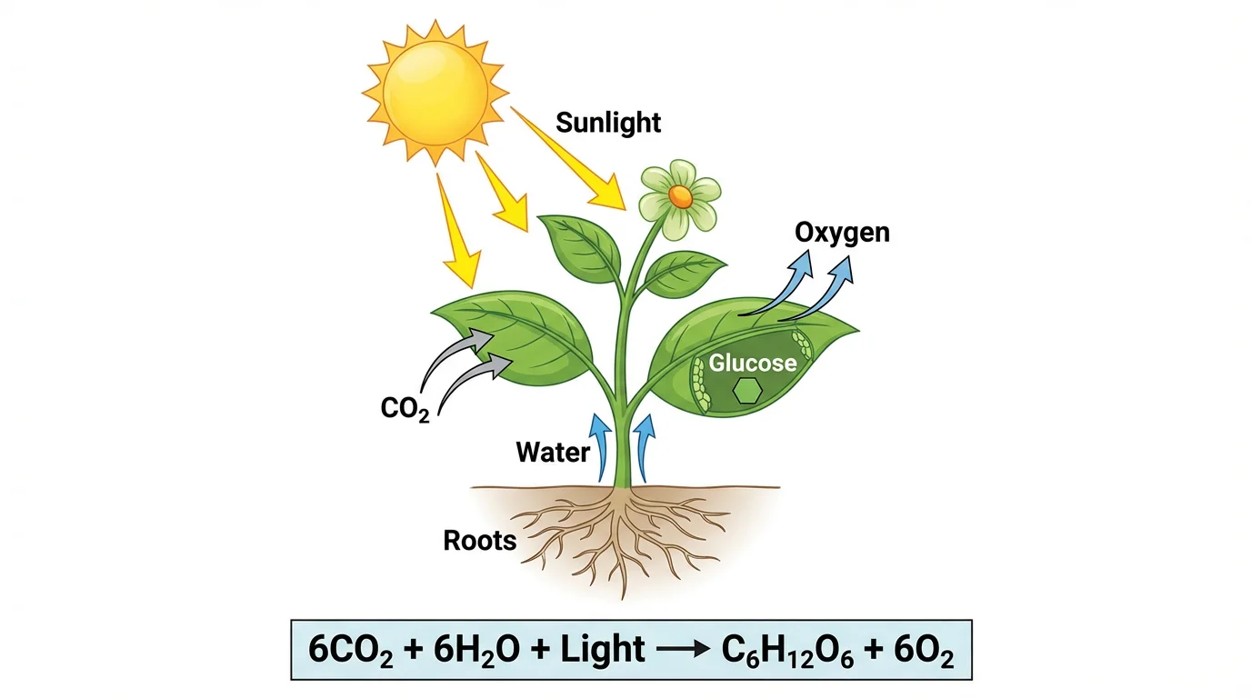
Photosynthesis Diagram Generator
Make labeled photosynthesis diagrams showing inputs, outputs, and the chloroplast.