Animal and Plant Cell Diagram Labeled, Blank & Venn
Free animal and plant cell comparison diagrams — labeled, blank, and Venn-diagram versions. View, download, or print any format, or make your own.
Free Animal vs Plant Cell Diagrams
Every version below is free to view and download — no sign-up.
Labeled Side-by-Side Comparison
Both cells fully labeled, with arrows on the three plant-only parts — cell wall, chloroplasts, and the large central vacuole.
Diagrams & tables
Print & practice
Venn diagrams
Test yourself: animal vs plant cell
A quick self-test — questions and answers reshuffle each time, with a review of anything you miss at the end.
What are animal and plant cells both classified as?
Need a custom version?
Change the layout, grade level, or which parts are highlighted — describe it and generate your own.
Customize Your Cell Comparison
免费试用 ·
Your animal and plant cell comparison will appear here
For a study-ready diagram, ask for labels and a side-by-side or Venn layout
Animal and plant cells side by side
Animal cells and plant cells are both eukaryotic, so they share most of the same internal machinery — but a plant cell has a few extra structures that an animal cell lacks. The clearest way to study this is a side-by-side diagram: the animal cell on one side, the plant cell on the other, with the same organelles labeled on both so the differences jump out. A Venn-diagram version groups the parts into shared, plant-only, and animal-only.
Organelles that both cells share
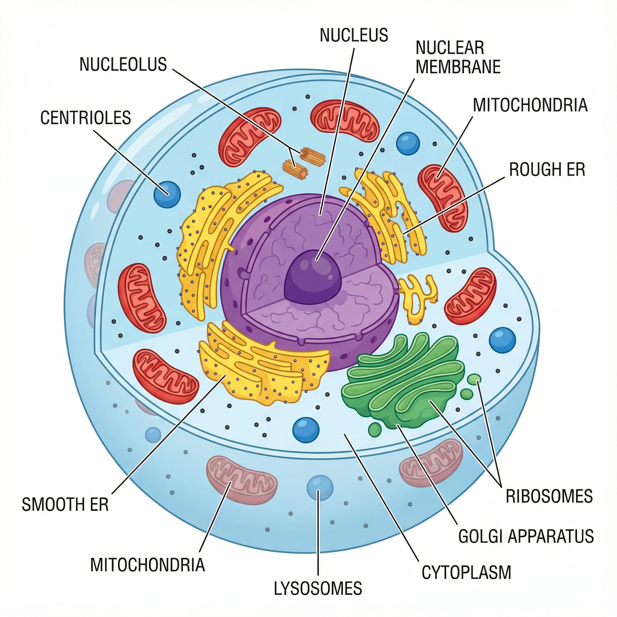
- Nucleus: the control center holding the cell’s DNA — present in both.
- Cell membrane (plasma membrane): the flexible outer boundary that controls what enters and leaves — both cells have one.
- Cytoplasm: the jelly-like fluid that fills the cell and surrounds the organelles.
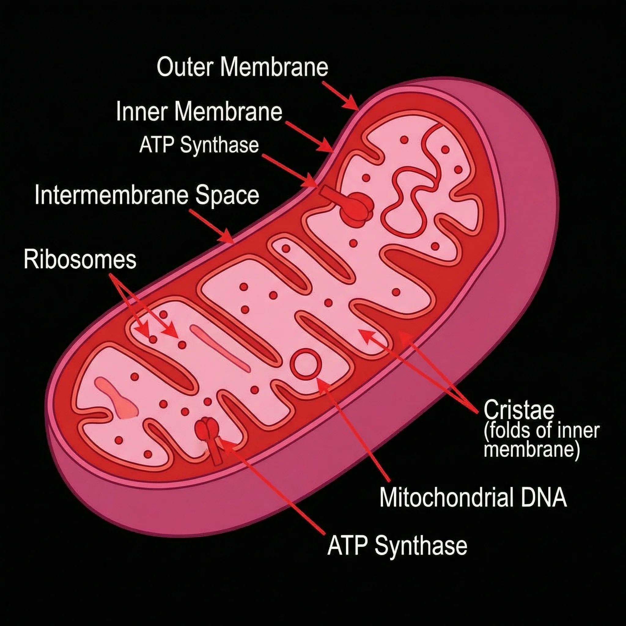
- Mitochondria: the sites of cellular respiration that release energy — yes, plant cells have them too.
- Ribosomes: the structures that build proteins, found in both cells.
- Endoplasmic reticulum (rough and smooth): the membrane network for making and transporting proteins and lipids.
- Golgi apparatus: packages and ships proteins, present in both.
Plant-only parts: cell wall, chloroplasts, and a large vacuole
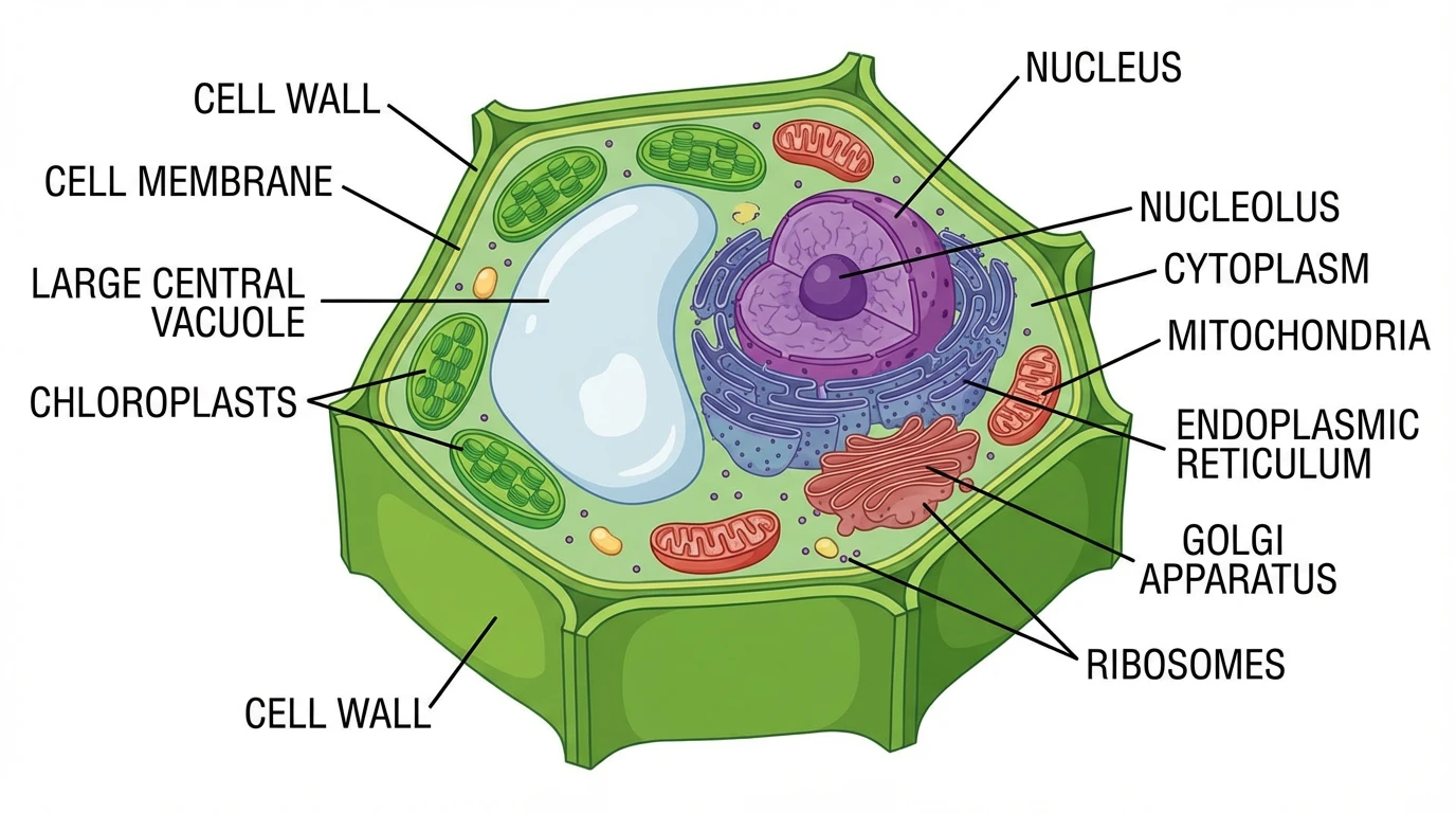
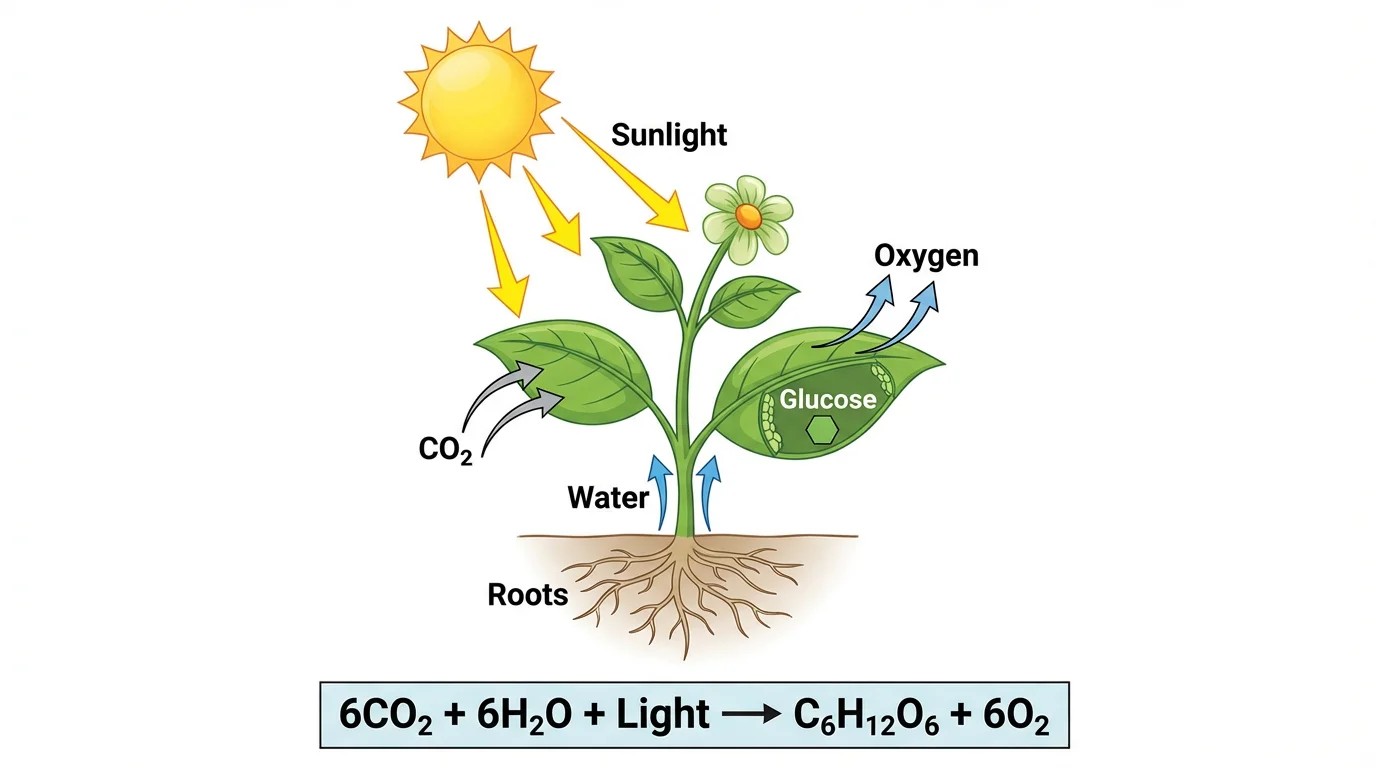
Three structures appear in plant cells but not in animal cells. The cell wall is a rigid outer layer of cellulose that surrounds the membrane and gives the plant cell its fixed, box-like shape and support. Chloroplasts contain chlorophyll and carry out photosynthesis, turning light into food — this is why plants are green and why animal cells, which cannot make their own food, have none. The large central vacuole is a single big fluid-filled sac that stores water and keeps the cell firm (turgid); animal cells may have small vacuoles, but never one dominant central vacuole. These three parts are the differences most diagrams highlight.
Animal-only structures: centrioles and lysosomes
Animal cells contain a couple of structures usually described as animal-only in textbooks. Centrioles are small cylindrical structures that help organize the spindle fibers during cell division; plant cells generally divide without them. Lysosomes are sacs of digestive enzymes that break down waste and worn-out parts and are routinely shown only in animal cells, though similar functions in plants are handled by the vacuole. Animal cells also lack a rigid wall, so they take on more rounded, irregular shapes rather than the fixed rectangular outline of a plant cell.
Plant vs animal cell Venn diagram
A Venn diagram is a popular way to compare the two cell types because it sorts every part into one of three regions: the overlapping center for shared organelles (nucleus, mitochondria, ribosomes, endoplasmic reticulum, Golgi apparatus, cell membrane, and cytoplasm), one circle for plant-only parts (cell wall, chloroplasts, large central vacuole), and the other circle for animal-only structures (centrioles, lysosomes). The Venn-diagram sheet above uses exactly this layout, which is ideal for compare-and-contrast study notes and homework that asks for the similarities and differences in one picture.
Labeled or blank — for teaching and for quizzes
Use a fully labeled diagram when you want a reference or a slide, with every organelle named on both cells. Use a blank version with empty callout lines when you want a worksheet — students fill in the labels themselves and list the differences, which is a common biology assessment. The detail level ranges from a simplified, cartoon-style comparison for elementary students to a complete, color-coded diagram for high-school and beyond.
常见问题
Related Biology Tools
 Biology
Animal Cell Diagram
Free labeled, blank, and printable animal cell diagrams — nucleus, mitochondria, lysosomes, and more.
 Biology
Plant Cell Diagram
Free labeled, blank, and printable plant cell diagrams with the cell wall, chloroplasts, and vacuole.
 Biology
Mitochondria Diagram
Labeled mitochondria diagrams — the powerhouse organelle shared by animal and plant cells.
 Biology
Photosynthesis Diagram
See how chloroplasts turn light, water, and CO₂ into glucose — the process only plant cells can do.
 Biology
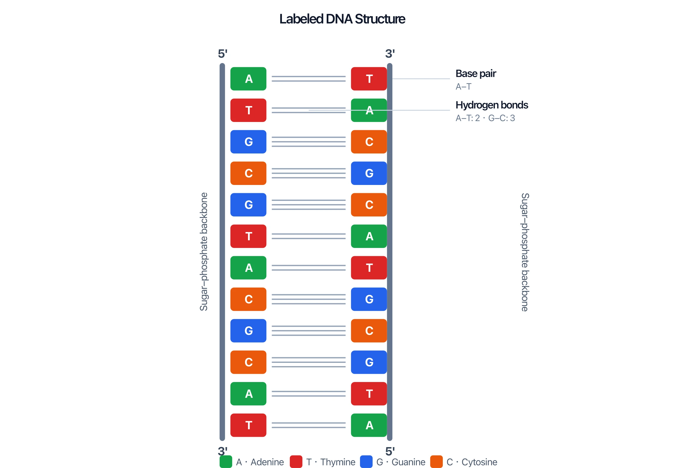
DNA Structure Diagram
Labeled DNA double-helix diagrams — the genetic material stored in the nucleus of both cells.
 Biology
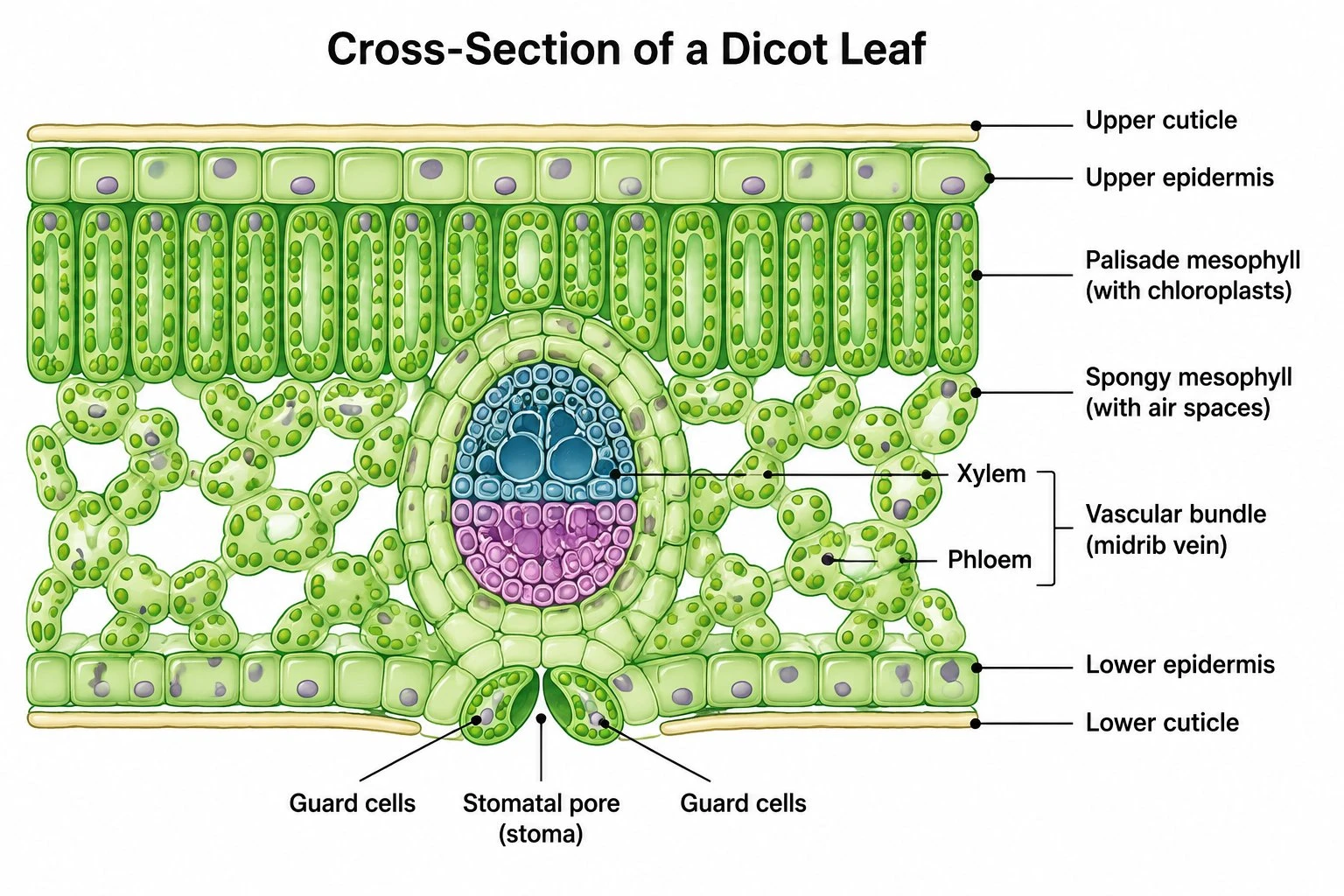
Leaf Structure Diagram
Labeled leaf cross-sections showing the layers where plant cells carry out photosynthesis.