Leaf Structure Diagram Generator Labeled Leaf Diagrams
Create labeled leaf structure diagrams with AI. Show the internal cross-section — cuticle, epidermis, palisade mesophyll, spongy mesophyll, stomata, guard cells, vascular bundle — or the external parts — blade, petiole, midrib, veins, margin — for biology class. Download as PNG.
AI Leaf Structure Diagram Generator
Free to try ·
Your leaf structure diagram will appear here
Describe the tissue layers or external parts to label
Leaf Structure Diagram Examples
Internal cross-sections, external parts, and stomata diagrams for biology class
Leaf Cross-Section (Labeled)
A full internal cross-section with every tissue layer labeled — ideal for GCSE and high-school biology.
External Leaf Parts
External leaf morphology — blade, petiole, midrib, veins, margin, apex, and base, all labeled.
Stomata & Guard Cells
Zoom in on the stoma — guard cells, pore, and gas exchange labeled in detail.
Leaf Tissue Layers
Each tissue layer color-coded and labeled — a clear visual for learning the leaf's internal organisation.
Simple Leaf Diagram for Kids
A friendly, simple leaf with just the main parts labeled — perfect for younger students.
Blank Leaf Structure Worksheet
An unlabeled cross-section with empty leader lines — a ready-to-print worksheet or quiz.
What does a leaf structure diagram show?
A leaf structure diagram shows either the internal cross-section of a leaf — its tissue layers from cuticle to stomata — or the external morphology of the whole leaf, naming parts like the blade, petiole, midrib, and veins. Internal cross-section diagrams label the upper cuticle, upper epidermis, palisade mesophyll, spongy mesophyll, lower epidermis, lower cuticle, stomata, guard cells, and the vascular bundle (containing xylem and phloem). External diagrams label the leaf blade (lamina), petiole, midrib, lateral veins, leaf margin, apex, and base. This generator creates clear, labeled leaf diagrams for GCSE, middle-school, and high-school biology.
The layers inside a leaf (cross-section)
- Cuticle: a thin, waxy layer on the upper and lower surface that reduces water loss.
- Upper epidermis: a single layer of transparent cells that lets light through to the mesophyll below.
- Palisade mesophyll: tightly packed, column-shaped cells just below the upper epidermis, packed with chloroplasts — this is where most photosynthesis happens.
- Spongy mesophyll: loosely arranged cells with large air spaces between them, allowing CO₂, O₂, and water vapour to move through the leaf.
- Lower epidermis: the bottom cell layer, containing stomata — tiny pores flanked by guard cells.
- Stomata and guard cells: the stomata open and close (controlled by the guard cells) to regulate gas exchange and water loss.
- Vascular bundle (midrib/vein): contains xylem (carries water and minerals up) and phloem (carries sugars made by photosynthesis down).
External parts of a leaf
- Blade (lamina): the flat, broad part of the leaf — the main surface for capturing light and carrying out photosynthesis.
- Petiole: the stalk connecting the blade to the stem.
- Midrib: the large central vein running the length of the blade from the petiole.
- Veins: a network of smaller veins branching from the midrib that carry water and nutrients and support the blade.
- Leaf margin: the edge of the leaf blade, which can be smooth, serrated, or lobed depending on species.
- Leaf apex: the tip of the leaf.
- Leaf base: the bottom of the leaf blade where it meets the petiole.
Why leaves are shaped for photosynthesis
The flat, broad shape of the leaf blade maximises the surface area for capturing light. The upper epidermis is transparent so light passes straight through to the palisade cells below. The palisade mesophyll is packed with chloroplasts and positioned at the top of the leaf where light intensity is highest. The spongy mesophyll's air spaces create a large internal surface for CO₂ to dissolve into cell surfaces before entering the chloroplasts. The stomata on the lower surface open during the day to let CO₂ in and O₂ out, while the cuticle slows water loss from the rest of the surface.
How to use this leaf structure diagram generator
- Choose the view you need: a full labeled internal cross-section, an external leaf parts diagram, a close-up of stomata and guard cells, or a simplified version for younger students.
- Describe the diagram: the tissue layers or external parts to label, the level of detail (elementary, middle school, or GCSE/high school), and any style preferences (color-coded, black-and-white, worksheet).
- Generate the image — the tool draws a clean, scientific-style diagram on a white background.
- Download as PNG and drop it into your notes, slides, worksheet, or handout.
Frequently Asked Questions
Related Biology Tools
 Biology
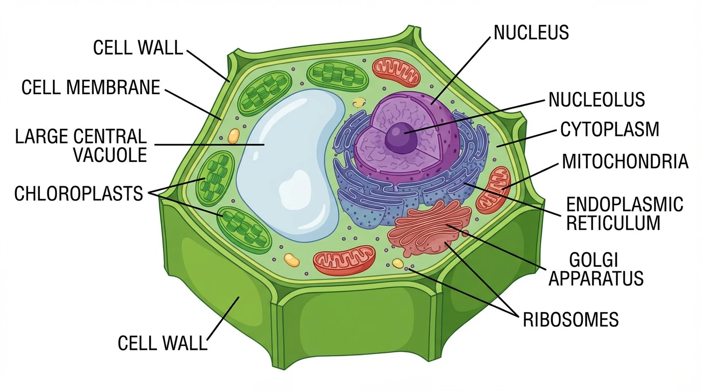
Plant Cell Diagram Generator
Create labeled plant cell diagrams showing the cell wall, chloroplasts, large central vacuole, and all organelles.
 Biology
Parts of a Flower Diagram Generator
Create labeled flower diagrams showing petals, sepals, stamen, pistil, and all reproductive parts.
 Biology
Photosynthesis Diagram Generator
Create labeled photosynthesis diagrams showing light reactions, the Calvin cycle, and glucose production.