Animal Cell Diagram Labeled, Blank & Printable
Free animal cell diagrams — labeled, blank, coloring, and exam-review sheets. View, download, or print any version, or customize your own.
Free Animal Cell Diagrams
Every version below is free to view and download — no sign-up.
Labeled Animal Cell Diagram
Every major organelle labeled — nucleus, mitochondria, ER, Golgi, lysosomes, ribosomes, and cell membrane. Ready for study, notes, or slides.
Core diagrams
Structure & focus
Print & study
Test yourself: animal cell
A quick self-test — questions and answers reshuffle each time, with a review of anything you miss at the end.
Which organelle is known as the "powerhouse" of the cell?
Need a custom version?
Change the grade level, colors, or which organelles are labeled — describe it and generate your own.
Customize Your Animal Cell Diagram
免费试用 ·
Your custom animal cell diagram will appear here
Ask for a labeled, blank, or printable version of the cell
What is an animal cell diagram?
An animal cell diagram is a cross-section drawing that shows the parts of a typical animal cell — the organelles — and where they sit inside the cell. Animal cells are eukaryotic, which means their genetic material is stored inside a membrane-bound nucleus, and the diagram makes that internal structure easy to see. Use a labeled version for studying, or a clean unlabeled version as a worksheet or quiz.
Labeled vs. blank (unlabeled) diagrams
- Labeled diagram: every organelle is named — ideal for studying, presentations, and answer keys.
- Blank / unlabeled diagram: the structures are drawn but the labels are left empty so students can fill them in — perfect for worksheets, quizzes, and homework.
- Printable coloring page: black-and-white line art with blank label lines, ready to print and hand out.
- Use a matching pair — a labeled key and a blank version of the same cell — so the quiz and the answer key always line up.
The organelles in an animal cell
- Nucleus: the control center, holding the DNA and surrounded by the nuclear membrane; the nucleolus inside makes ribosomes.
- Mitochondria: the "powerhouse" organelles that release energy through respiration.
- Ribosomes: tiny structures that build proteins, found free in the cytoplasm or attached to the ER.
- Endoplasmic reticulum (ER): a network of membranes — rough ER (with ribosomes) makes proteins, smooth ER makes lipids.
- Golgi apparatus: packages and ships proteins and other molecules around the cell.
- Lysosomes: contain enzymes that break down waste and worn-out parts.
- Centrioles: paired structures that help organize the spindle during cell division.
- Cell membrane and cytoplasm: the flexible outer boundary and the jelly-like fluid that holds the organelles.
Animal cell vs. plant cell
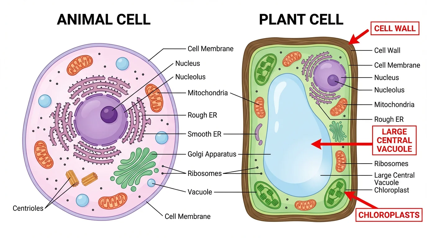
Animal and plant cells share most organelles — both have a nucleus, mitochondria, ribosomes, ER, and a Golgi apparatus. The key differences are structures plant cells have that animal cells do not: a rigid cell wall outside the membrane, chloroplasts for photosynthesis, and one large central vacuole. Animal cells, by contrast, have only a flexible cell membrane (no wall), no chloroplasts, and small or no vacuoles — plus centrioles, which most plant cells lack. Knowing these differences is one of the most common exam questions, so the comparison sheet places the two side by side.
Using animal cell diagrams in the classroom
Teachers use labeled diagrams to introduce cell structure, then hand out blank or unlabeled versions for students to complete from memory — a quick, reliable way to check understanding. Printable coloring pages work well for younger grades, while detailed labeled cross-sections suit high school and introductory biology. Because a labeled key and a matching blank diagram of the same cell come together, building a worksheet and its answer key takes seconds instead of hunting for two images that happen to match.
常见问题
Related Biology Tools
 Biology
Animal vs. Plant Cell Comparison
Compare animal and plant cells side by side, with shared and unique organelles labeled.
 Biology
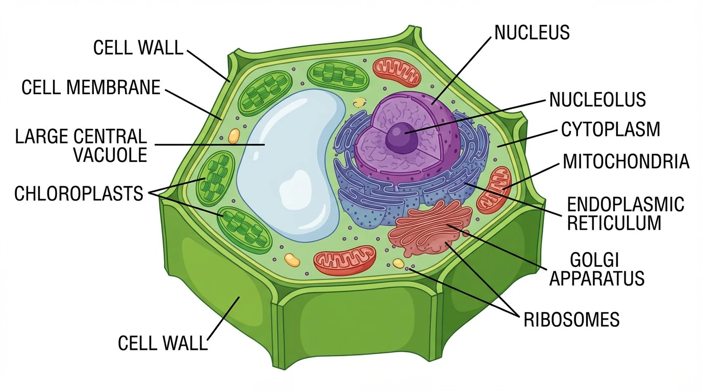
Plant Cell Diagram
Free labeled, blank, and printable plant cell diagrams with the cell wall, chloroplasts, and vacuole.
 Biology
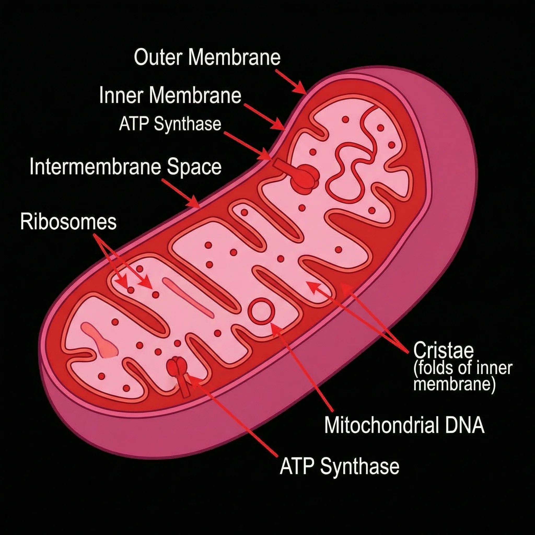
Mitochondria Diagram
Labeled mitochondria diagrams — the powerhouse organelle inside animal and plant cells.
 Biology
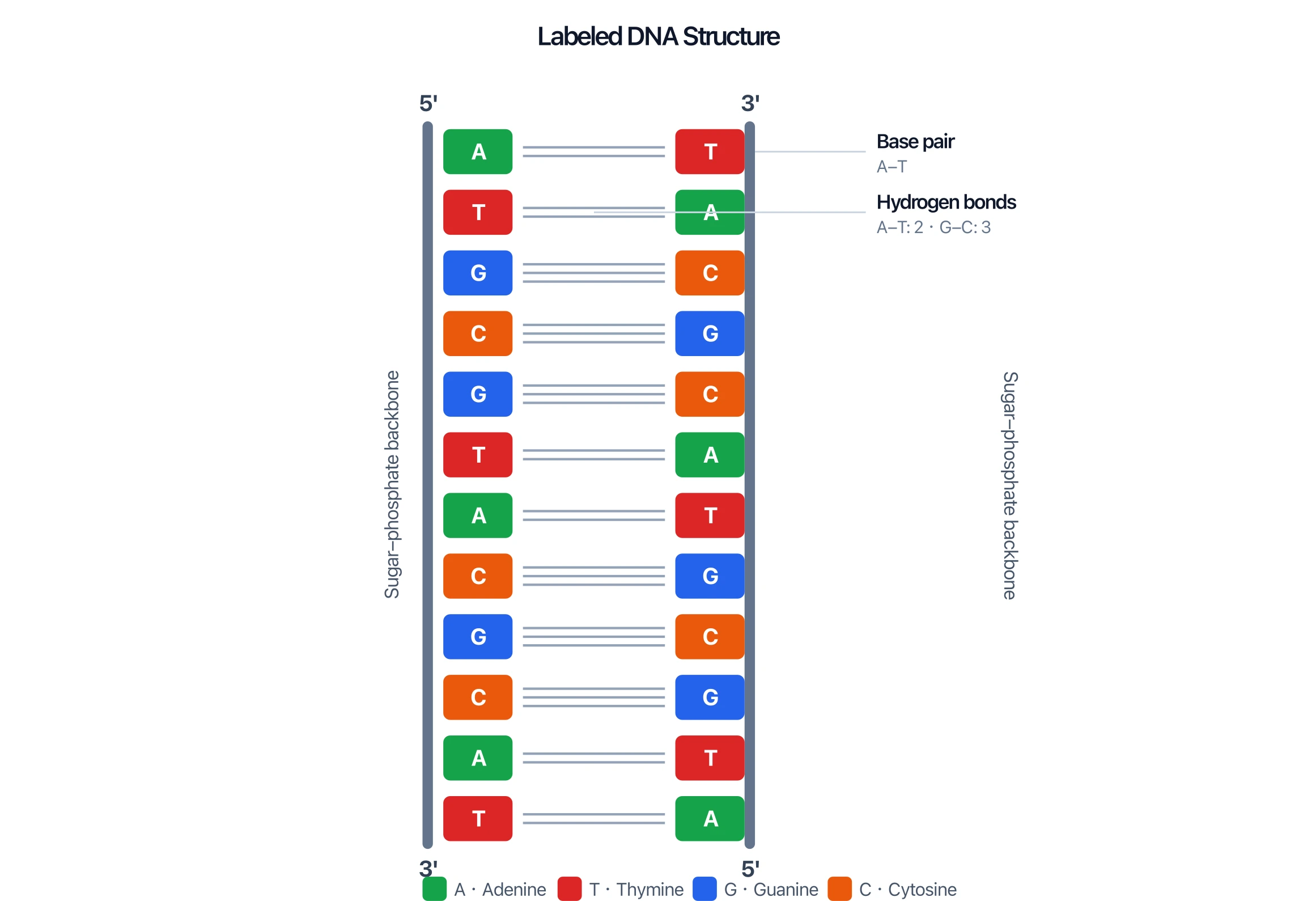
DNA Structure Diagram
Labeled DNA double-helix diagrams — the genetic material stored in the nucleus.
 Biology
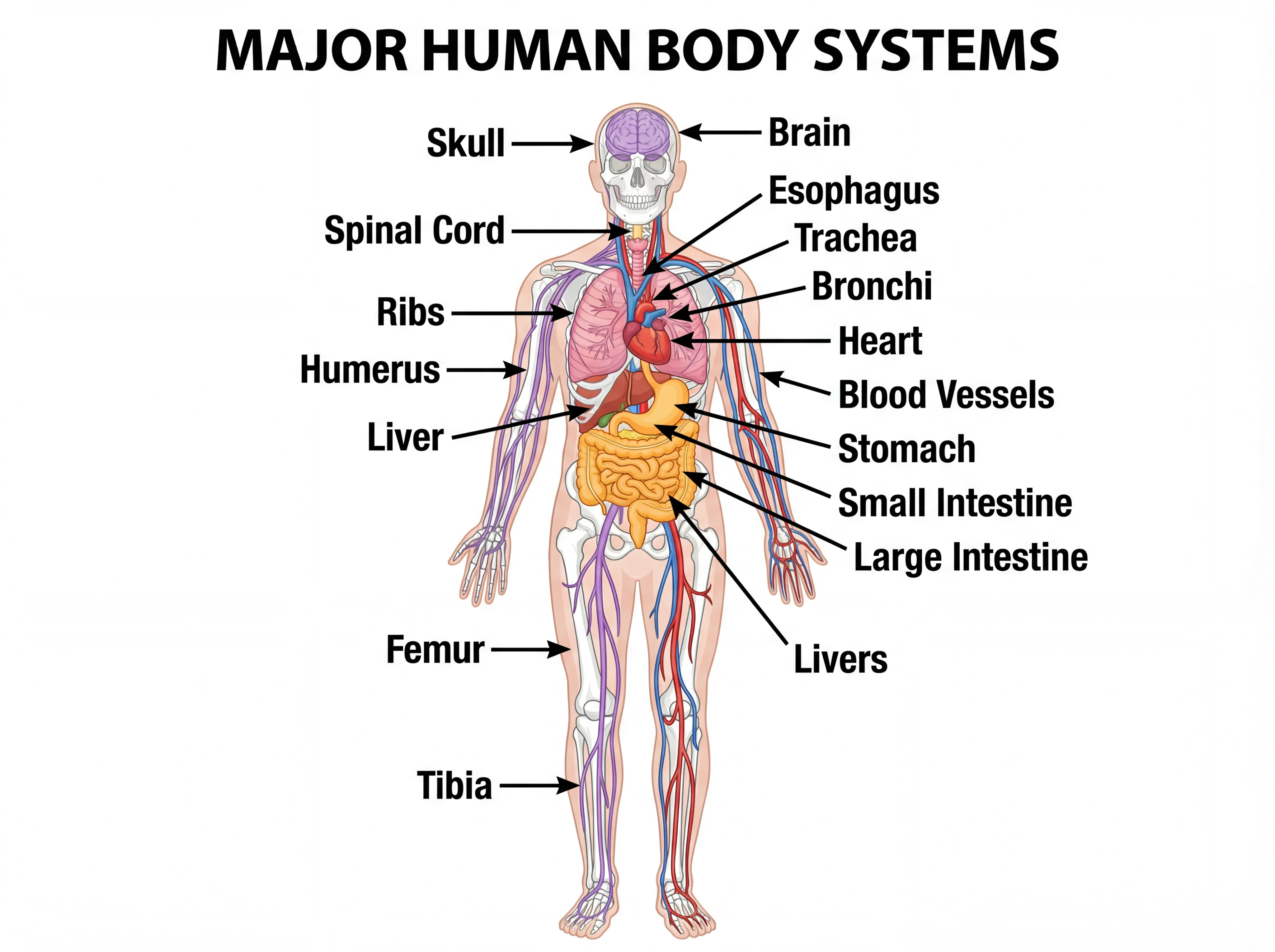
Human Body Systems Diagram
Labeled diagrams of the body systems built from cells, tissues, and organs.
 Biology
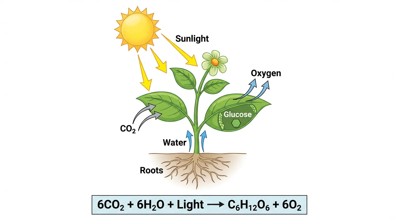
Photosynthesis Diagram
See how chloroplasts turn light, water, and CO₂ into glucose — the process animal cells lack.