Respiratory System Diagram Generator for Labeled & Blank Diagrams
Create a clearly labeled respiratory system diagram in seconds. Show the nasal cavity, pharynx, larynx, trachea, bronchi, bronchioles, lungs, alveoli, and diaphragm — or generate a blank, unlabeled version for worksheets and quizzes. Free to use.
Respiratory System Diagram Generator
Free to try ·
Your respiratory system diagram will appear here
Describe what you need and click Generate
Respiratory System Diagram Examples
Labeled diagrams of the airways, lungs, alveolar gas exchange, and the full path of air
Full Respiratory System
The complete map — from nasal cavity to alveoli, with every major structure labeled in anterior view.
Gas Exchange at the Alveoli
Oxygen crossing into the bloodstream and CO2 leaving it — the core of respiration shown at the alveolar level.
Simple Respiratory Diagram for Kids
A clear, simplified version with large labels — perfect for elementary-level science lessons.
Path of Air Diagram
A step-by-step flowchart tracing how air travels from the nose all the way down to the alveoli.
Lungs and Bronchial Tree
Left and right lung lobes with the full bronchial tree — primary bronchi branching to bronchioles.
Blank Respiratory System Worksheet
An unlabeled version with numbered blank lines — ready to print as a quiz or fill-in worksheet.
What is the respiratory system?
The respiratory system is the network of organs and tissues that moves air into and out of the body and transfers oxygen to the bloodstream while removing carbon dioxide. It starts at the nose and mouth, continues through the pharynx, larynx, and trachea, then branches into the bronchi and bronchioles deep inside the lungs, ending at the alveoli — tiny air sacs where gas exchange actually happens. The diaphragm and intercostal muscles power the breathing cycle. This generator draws that full pathway with every structure clearly labeled.
The upper and lower respiratory tract
- Nasal cavity: warms, humidifies, and filters incoming air using mucus and tiny hairs called cilia.
- Pharynx and larynx: the throat and voice box route air downward toward the trachea while the epiglottis flips shut during swallowing to keep food out of the airway.
- Trachea: the windpipe — a rigid tube reinforced with C-shaped cartilage rings that keep the airway open as air moves toward the lungs.
- Bronchi and bronchioles: the trachea splits into two primary bronchi, one for each lung. These branch further into secondary and tertiary bronchi, then into narrower bronchioles, spreading air throughout each lobe.
- Alveoli: clusters of microscopic air sacs at the end of the bronchioles. Their thin walls and dense capillary network are where oxygen enters the blood and carbon dioxide leaves it.
Gas exchange: O₂ in, CO₂ out
Gas exchange occurs across the alveolar membrane, the thinnest barrier in the body. Oxygen in the fresh air inside the alveolus is at higher concentration than in the blood arriving from the body, so it diffuses through the alveolar wall and capillary wall into red blood cells. Carbon dioxide, a waste product of cellular respiration, is at higher concentration in the blood, so it diffuses in the opposite direction — out of the capillary and into the alveolus, to be exhaled. This simultaneous two-way exchange happens in a fraction of a second with each breath. You can generate a close-up alveolus diagram with arrows showing the exact direction O₂ and CO₂ move.
The lungs: lobes, pleura, and structure
The right lung has three lobes (upper, middle, lower) and the left lung has two (upper, lower) — the left lung is slightly smaller to make room for the heart. Both lungs are wrapped in a double-layered membrane called the pleura, with a thin layer of fluid between the layers that reduces friction during breathing. At the hilum, the main bronchus, pulmonary artery, and pulmonary vein enter or leave each lung. The diaphragm sits below both lungs; when it contracts and flattens, it increases chest volume, drops air pressure, and draws air in — the mechanics of inhalation.
Labeled vs blank diagrams for worksheets and quizzes
A fully labeled diagram works well for teaching and study notes, while a blank, unlabeled version is what you want for worksheets, handouts, and quizzes. With this tool you can generate either: describe "a labeled respiratory system diagram" to get every structure named, or ask for "a blank respiratory system diagram with numbered blank lines" to get a fill-in version. Black-and-white line art works best for printing, and a numbered blank diagram doubles as both the quiz and — with labels added back — the answer key.
How to generate a labeled respiratory system diagram
- Describe what you need in plain English — the full respiratory system, just the lungs and bronchial tree, a close-up of alveolar gas exchange, or the path of air as a flowchart.
- Specify view and labels, for example "anterior view, airways in blue, lungs in pink, label all major structures from nasal cavity to alveoli."
- Choose labeled for teaching or unlabeled (blank) for a worksheet or quiz, then generate the diagram.
- Review the result for accuracy, regenerate or refine the prompt if needed, and download the image to use in a slide, handout, or study guide.
Frequently Asked Questions
Related Biology Tools
 Biology
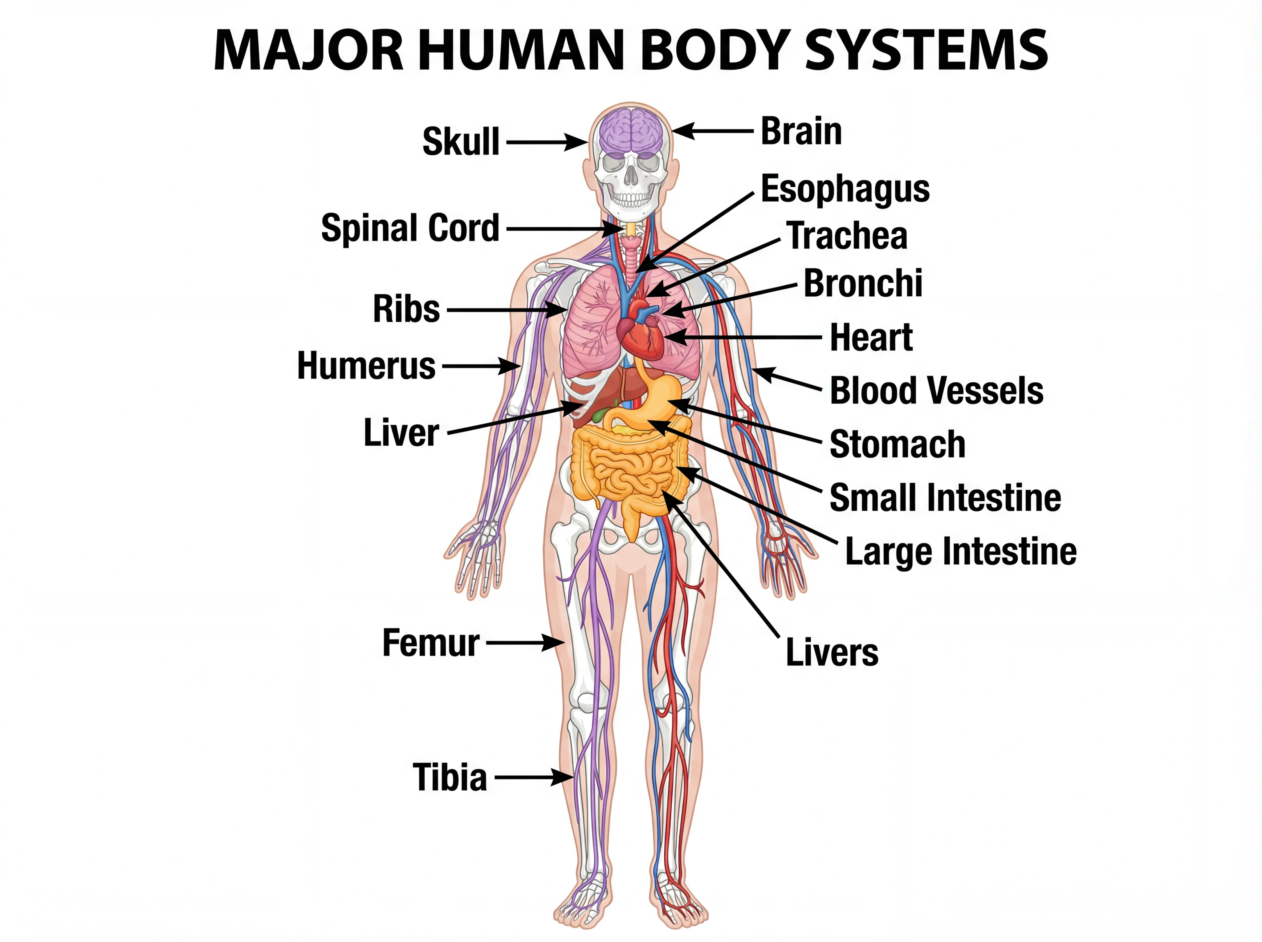
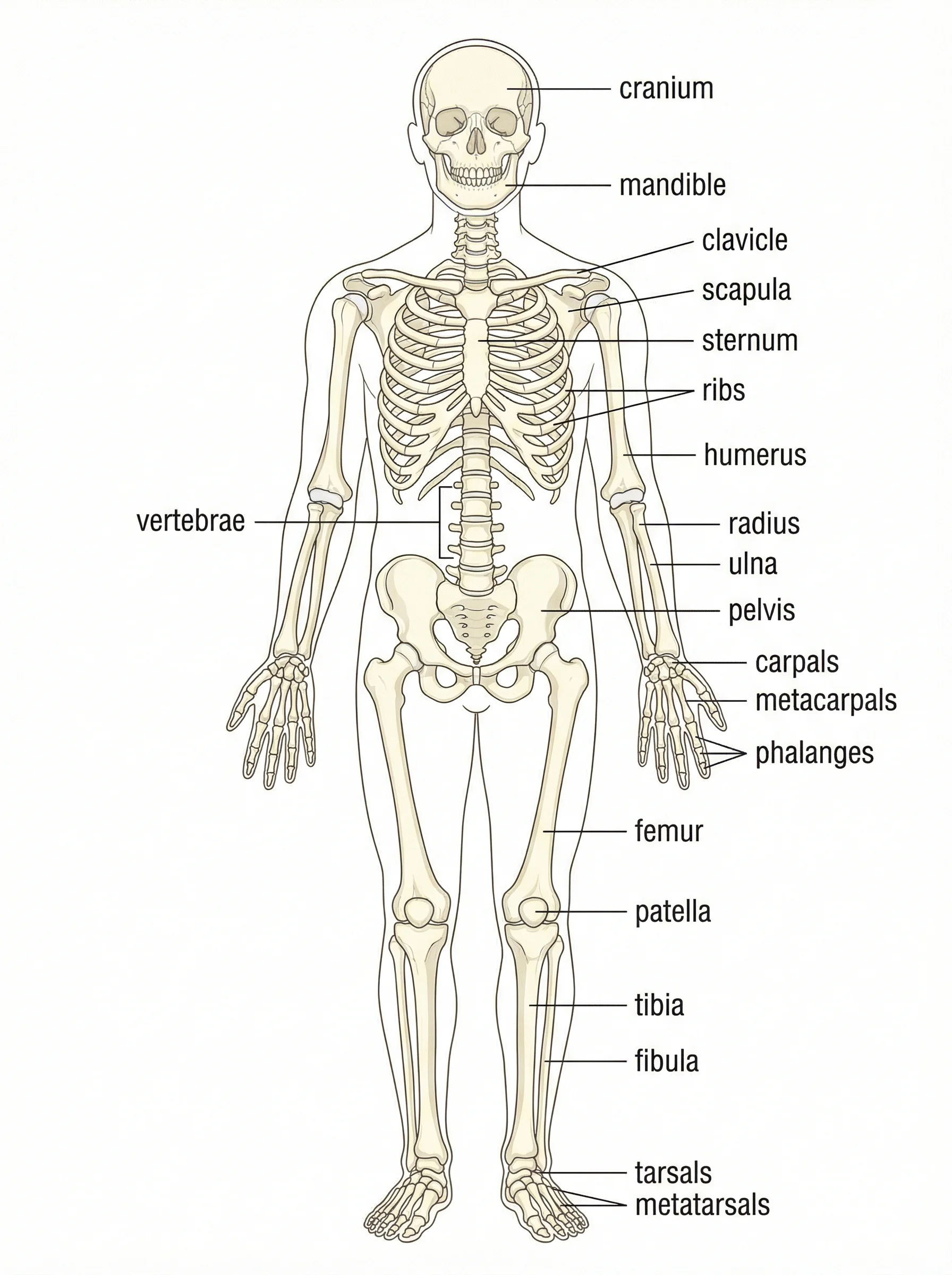
Human Body Systems Diagram Generator
Draw labeled diagrams of the body's organ systems, from skeletal to circulatory.
 Biology
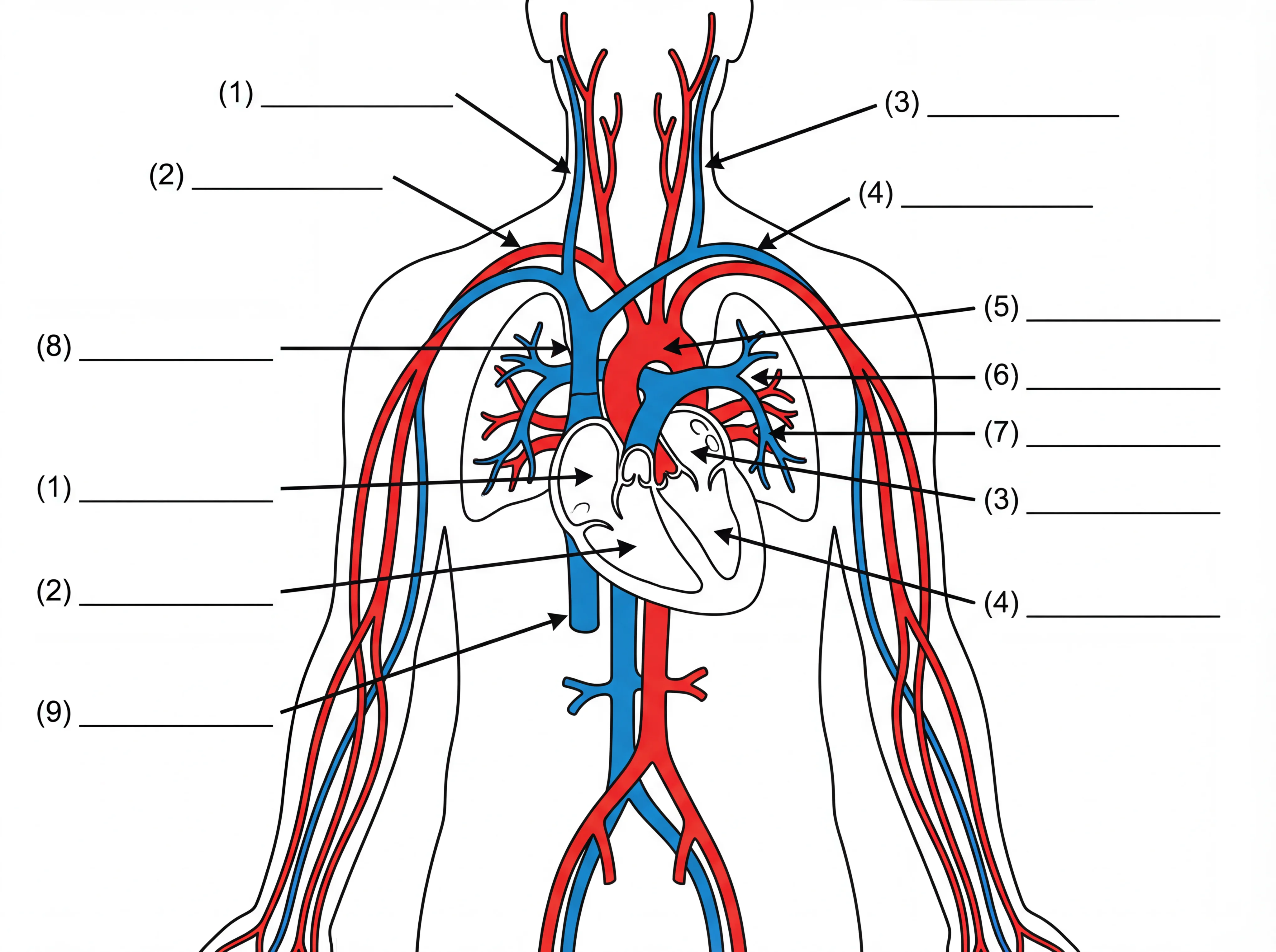
Heart Diagram Generator
Create labeled heart diagrams showing chambers, valves, and blood flow pathways.
 Biology
Digestive System Diagram Generator
Generate labeled diagrams of the digestive tract from mouth to large intestine.