Bacteria Diagram Generator for the Labeled Bacterial Cell
Make a clearly labeled bacteria diagram in seconds. Show the cell wall, plasma membrane, cytoplasm, nucleoid (circular DNA), plasmids, ribosomes, flagella, pili, and capsule on a prokaryotic cell — labeled or blank for worksheets — then download your diagram, free.
Bacteria Diagram Generator
Free to try ·
Your diagram will appear here
Describe what you need and click Generate
Bacteria Diagram Examples
Labeled bacterial cells, shapes, and microbiology diagrams
Labeled Bacterial Cell
A full prokaryotic cell with every part labeled — cell wall, membrane, nucleoid, plasmids, ribosomes, flagella, pili, and capsule.
Bacterial Shapes Chart
Cocci, bacilli, and spiral forms (vibrio, spirillum, spirochete) laid out in one labeled comparison chart.
Gram-Positive vs Gram-Negative
Compare the thick versus thin peptidoglycan wall and the Gram-negative outer membrane, side by side.
Binary Fission Stages
How bacteria reproduce — five labeled stages from one parent cell to two identical daughter cells.
E. coli Structure
The classic model organism, labeled — flagella, pili, and a cutaway view of the nucleoid, ribosomes, and plasmids.
Antibiotic Resistance
Four labeled resistance mechanisms — efflux pumps, enzyme degradation, target modification, and reduced permeability.
What is a bacterial cell?
A bacterium is a single-celled microorganism and the most common example of a prokaryote — a cell that has no nucleus and no membrane-bound organelles. Instead of keeping its DNA inside a nucleus, a bacterial cell holds a single loop of circular DNA in an open region of the cytoplasm called the nucleoid. Bacteria are tiny, usually a few micrometres across, yet they live almost everywhere on Earth and play roles in digestion, decomposition, fermentation, and disease. This generator draws a typical bacterial cell as a clearly labeled diagram so students can see how a prokaryotic cell is put together.
The labeled parts of a bacterial cell
- Cell wall: a rigid outer layer of peptidoglycan that gives the cell its shape and protects it from bursting.
- Plasma membrane: the thin layer just inside the wall that controls what enters and leaves the cell.
- Cytoplasm: the jelly-like interior where the cell’s reactions take place.
- Nucleoid: the region holding the single loop of circular DNA — the bacterium’s main chromosome.
- Plasmids: small extra rings of DNA, often carrying genes such as antibiotic resistance.
- Ribosomes: tiny structures that build proteins (smaller than those in eukaryotic cells).
- Flagella: long whip-like tails used to swim and move toward food or away from danger.
- Pili and fimbriae: short hair-like fibres used to stick to surfaces and to other cells.
- Capsule / slime layer: a sticky outer coat that helps the cell attach and resist drying out and host defences.
What each part of a bacterial cell does
Every structure in a bacterial cell has a job. The cell wall and plasma membrane work together as the outer boundary: the wall keeps the cell’s shape and stops it bursting, while the membrane is the gatekeeper that lets nutrients in and waste out. The cytoplasm is the workspace where chemical reactions happen, and floating in it are the ribosomes that read genetic instructions and build proteins. The nucleoid stores the main circular chromosome, while plasmids carry useful extra genes that bacteria can even pass to one another. On the outside, flagella drive movement, pili and fimbriae provide grip and a way to exchange DNA, and the capsule shields the cell. Because this tool labels each part, learners can connect a structure to its function at a glance.
Bacterial shapes: cocci, bacilli, spirilla, and vibrio
Bacteria are often classified by their shape, which is one of the first things you observe under a microscope. Cocci are spherical and can appear singly, in pairs (diplococci), in chains (streptococci), or in clusters (staphylococci). Bacilli are rod-shaped and may form chains (streptobacilli). Spiral forms include the comma-shaped vibrio, the rigid corkscrew spirillum, and the flexible spirochete. Knowing these shapes helps with identification and is a common exam topic, so the generator can draw a single shape or a full morphology chart that lays the main types out side by side.
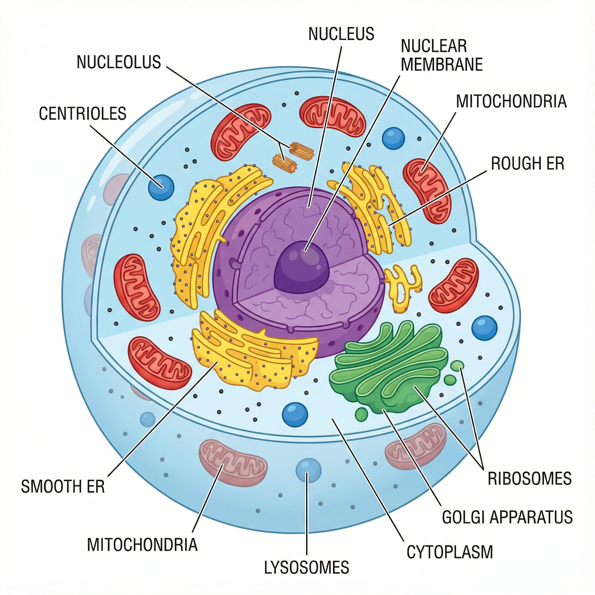
Prokaryote vs eukaryote: how bacteria differ
Bacteria are prokaryotes, and they differ from eukaryotic cells (such as animal and plant cells) in a few key ways. Prokaryotes have no nucleus — their DNA sits free in the nucleoid as a single circular chromosome — and they have no membrane-bound organelles like mitochondria or a Golgi apparatus. They are also smaller and have smaller ribosomes. Eukaryotic cells, by contrast, keep their DNA inside a nucleus, contain many specialised organelles, and are generally much larger. A side-by-side prokaryote-versus-eukaryote comparison is a popular way to make these differences stick, and you can generate one here from a short description.
Labeled vs blank diagrams for worksheets
For teaching, you often need two versions of the same diagram. A labeled diagram names every structure and is ideal for notes, slides, and revision. A blank or unlabeled diagram keeps the leader lines but removes the words, so students can fill them in as a quiz or worksheet. Because you describe what you want in plain English, you can ask for a fully labeled bacterial cell for the lesson and then a printer-friendly, black-and-white blank version of the same layout for assessment — no redrawing by hand.
Frequently Asked Questions
Related Biology Tools
 Biology
Animal Cell Diagram Generator
Create labeled animal cell diagrams with the nucleus, mitochondria, and other organelles — a eukaryote to compare with bacteria.
 Research
AI Scientific Image Generator
Generate clean, publication-style scientific illustrations from a plain-English description.
 Biology
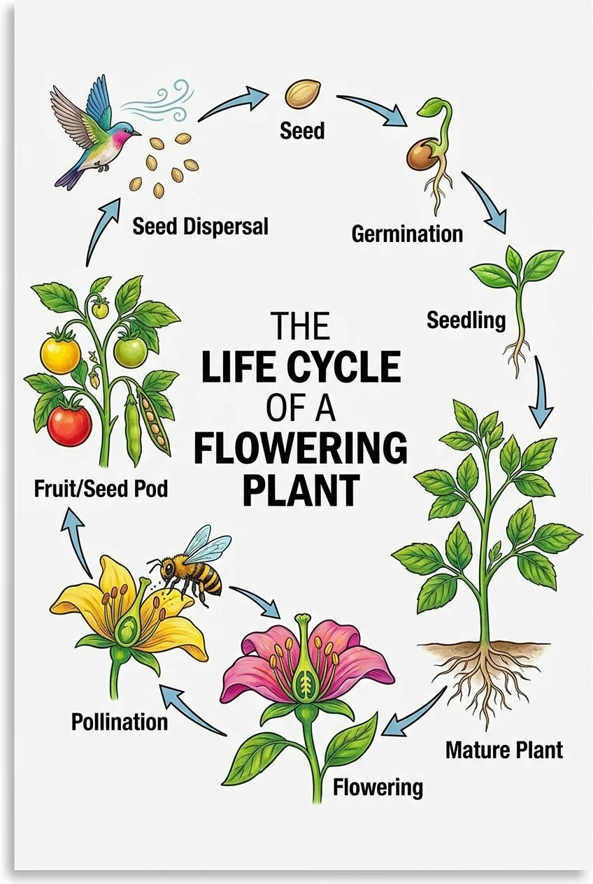
Life Cycle Diagram Generator
Make labeled life cycle diagrams showing each stage and the arrows between them.