Protein Structure Diagram Generator for the Four Levels of Structure
Create clear, labeled protein structure diagrams with AI. Show the four levels of protein structure — primary, secondary (alpha helix & beta sheet), tertiary, and quaternary — for biology class, notes, and presentations. Download as PNG.
AI Protein Structure Diagram Generator
免费试用 ·
Your protein structure diagram will appear here
Describe the level and features you want labeled
Protein Structure Examples
Labeled diagrams of the four levels of protein structure
Four Levels of Protein Structure
Primary → secondary → tertiary → quaternary, all in one labeled overview.
Primary Structure
The amino acid sequence — a linear chain joined by peptide bonds.
Secondary Structure
Alpha helices and beta-pleated sheets held together by hydrogen bonds.
Tertiary Structure
A single chain folded into its 3D shape by R-group interactions.
Quaternary Structure
Several folded subunits assembled into one functional protein.
Hemoglobin
A real example — hemoglobin’s four subunits, each with a heme group.
What is protein structure?
Protein structure is the three-dimensional arrangement of atoms in a protein, which determines its function. It is described at four levels: primary, secondary, tertiary, and quaternary. Each level builds on the previous one — from the simple sequence of amino acids up to a fully assembled, functional protein. This tool generates clear, labeled diagrams of any level for biology classes and study notes.
The four levels of protein structure
- Primary structure: the linear sequence of amino acids in a polypeptide chain, joined by peptide bonds.
- Secondary structure: local folding into regular patterns — alpha helices and beta-pleated sheets — stabilized by hydrogen bonds along the backbone.
- Tertiary structure: the overall 3D shape of a single polypeptide, held together by interactions between R-groups (hydrogen bonds, ionic bonds, disulfide bridges, and hydrophobic interactions).
- Quaternary structure: the assembly of two or more folded polypeptide subunits into one functional protein, such as hemoglobin.
Secondary structure: alpha helix vs beta sheet
The two main secondary structures are the alpha (α) helix, a right-handed coil stabilized by hydrogen bonds between every fourth amino acid, and the beta (β) pleated sheet, where segments of the chain line up side by side, connected by hydrogen bonds. Both arise from hydrogen bonding in the polypeptide backbone, not the R-groups, and they are the building blocks of the tertiary fold.
What determines a protein’s shape?
A protein’s final shape is determined by its amino acid sequence (primary structure) and the interactions between R-groups: hydrogen bonds, ionic (salt) bridges, disulfide bonds, and hydrophobic interactions that bury non-polar side chains away from water. When these interactions are disrupted by heat or pH (denaturation), the protein unfolds and usually loses its function.
Why protein structure matters
Structure dictates function: enzymes have active sites shaped to fit specific substrates, antibodies bind specific antigens, and structural proteins like collagen form strong fibers. A single change in the primary sequence can alter the fold and the function — as in sickle-cell hemoglobin. Visualizing the levels of structure makes these structure–function relationships clear.
Tips for a clear protein structure diagram
Describe the level you want (primary, secondary, tertiary, or quaternary) and any features to label — peptide bonds, hydrogen bonds, alpha helices, beta sheets, R-group interactions, or specific proteins like hemoglobin. Ask for a clean, textbook style with clear labels. Generate a few variations and pick the clearest, then download it for your worksheet or slides.
常见问题
Related Biology Tools
 Biology
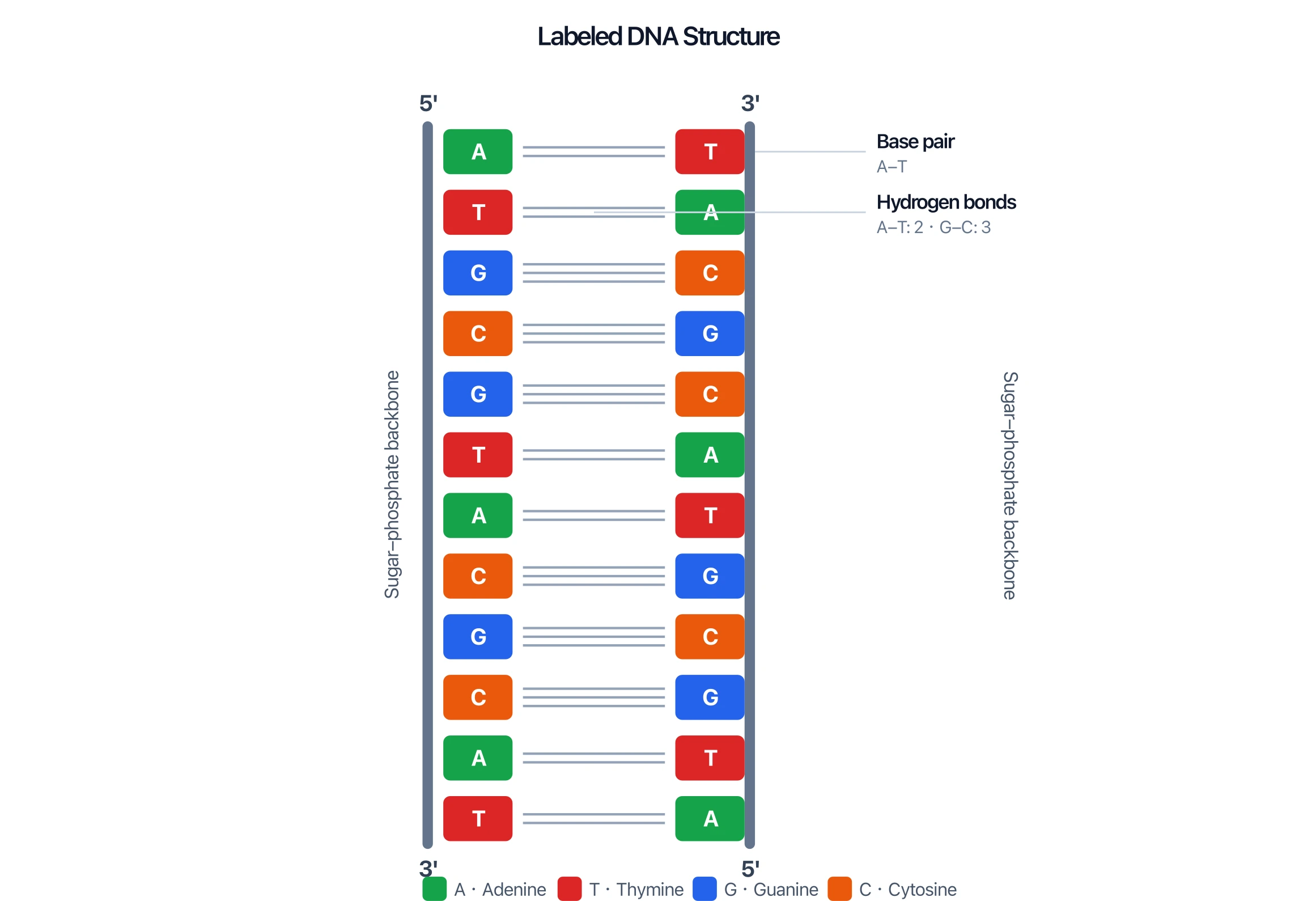
DNA Structure Diagram Generator
Make labeled DNA double-helix diagrams with base pairs, hydrogen bonds, and antiparallel strands.
 Biology
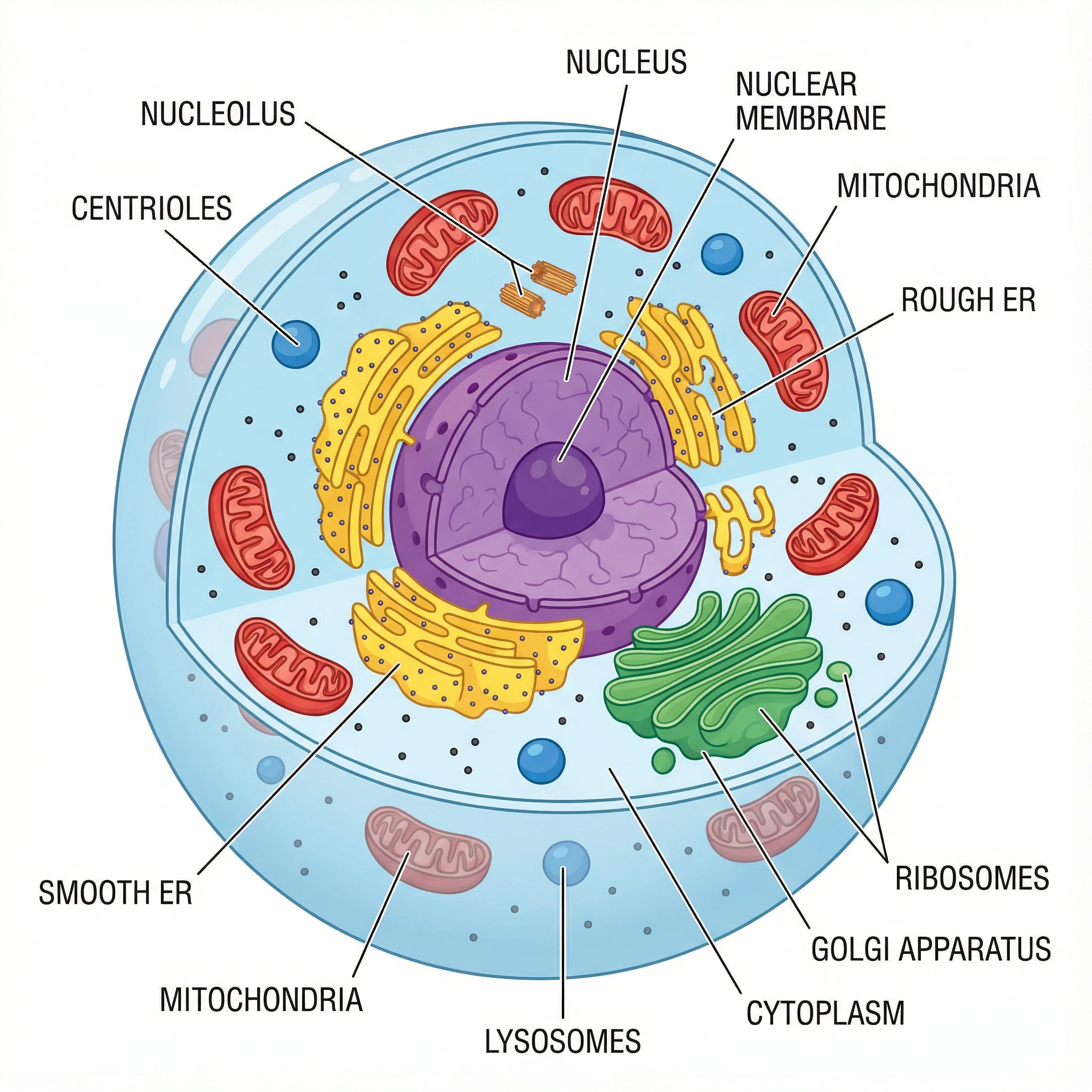
Animal Cell Diagram Generator
Create labeled animal cell diagrams with the nucleus, mitochondria, and other organelles.
 Chemistry
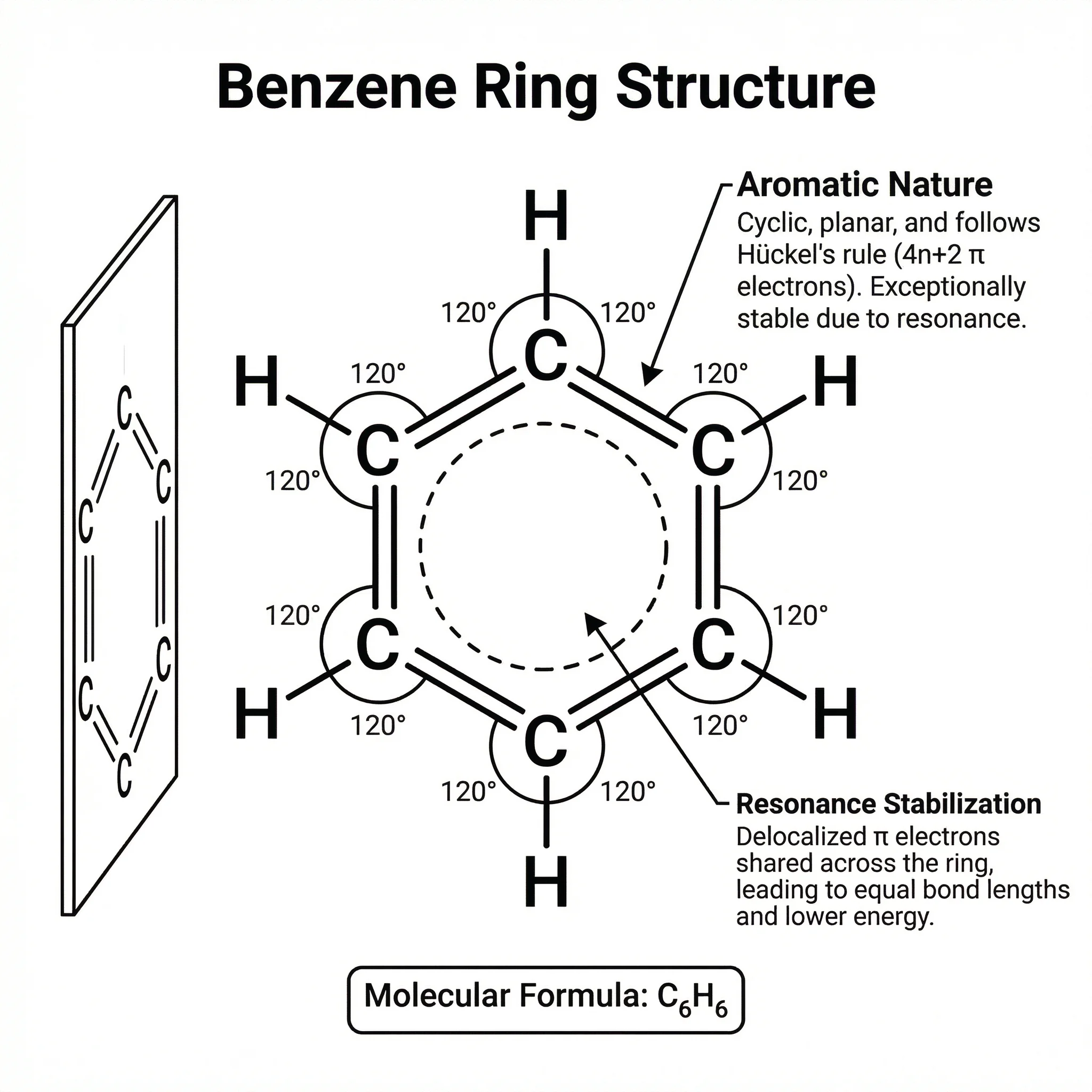
Chemistry Structure Generator
Draw molecules, mechanisms, and chemical structures with clean bonds and labels.