Golgi Apparatus Diagram Generator Labeled & Unlabeled
Create a clearly labeled Golgi apparatus diagram in seconds. Show the stacked cisternae, the cis face that receives proteins from the ER, the trans face that ships them, and the transport and secretory vesicles — or generate a blank, unlabeled version for worksheets.
Golgi Apparatus Diagram Generator
Free to try ·
Your diagram will appear here
Describe what you need and click Generate
Golgi Apparatus Diagram Examples
Labeled, unlabeled, and worksheet-ready diagrams of the Golgi body
Labeled Golgi Apparatus
Every major part labeled — cis face, trans face, cisternae stacks, and the transport and secretory vesicles.
Vesicle Trafficking
Vesicles arrive at the cis face from the ER and bud off the trans face — arrows show the direction of protein flow.
The Secretory Pathway
The Golgi in context: from the rough ER, through the cisternae, out to the plasma membrane and lysosomes.
Cis vs Trans Face
Compare the receiving cis face with the shipping trans face — the two ends of the Golgi stack.
Protein Processing
How proteins are modified — glycosylation and phosphorylation step through the cisternae from cis to trans.
Blank Worksheet
A blank, unlabeled outline — print it for labeling exercises, quizzes, or a coloring worksheet.
What is the Golgi apparatus?
The Golgi apparatus — also called the Golgi body, Golgi complex, or simply the Golgi — is the cell's processing and shipping center. It is a membrane-bound organelle found in nearly all eukaryotic cells, both animal and plant. Picture a stack of flattened, pancake-like sacs sitting near the nucleus and the endoplasmic reticulum (ER). Proteins and lipids made elsewhere in the cell arrive here to be modified, sorted, and packaged before they are sent to their final destinations. This generator draws that organelle with every structural part labeled, so students can see exactly what the Golgi apparatus is and how its pieces fit together.
The labeled parts of a Golgi apparatus diagram
- Cisternae: the stack of flattened membrane sacs at the core of the Golgi — usually four to eight per stack — where the actual processing happens.
- Cis face (receiving side): the end of the stack closest to the ER, where incoming transport vesicles fuse and deliver their cargo.
- Trans face (shipping side): the opposite end, where finished, sorted material is packaged into vesicles and sent on its way.
- Transport vesicles: small membrane bubbles that carry proteins and lipids from the ER to the cis face of the Golgi.
- Secretory vesicles: vesicles that bud off the trans face carrying the finished products to the cell membrane or other organelles.
What does the Golgi apparatus do?
The Golgi apparatus modifies, sorts, packages, and ships the proteins and lipids it receives from the endoplasmic reticulum. As cargo moves from the cis face toward the trans face, enzymes inside the cisternae chemically modify it — for example by adding sugar chains in a process called glycosylation. At the trans face, the finished molecules are sorted by destination and loaded into vesicles: some go to the plasma membrane to be secreted, some become lysosomes, and others are sent to different parts of the cell. A useful classroom analogy is a post office or a warehouse shipping department — receive, label, package, and dispatch.
Where is the Golgi apparatus in the cell?
The Golgi apparatus sits in the cytoplasm, usually near the nucleus and right next to the endoplasmic reticulum, because the two work together so closely. The ER and the Golgi are both part of the endomembrane system, the network of membranes that processes and transports material through the cell. Its position matters: the cis face is oriented toward the ER so it can receive transport vesicles, while the trans face points away, toward the cell membrane, so finished products have a clear path out. Seeing the Golgi in its place alongside the ER, nucleus, and other organelles helps students connect structure to function.
Labeled vs blank: diagrams for worksheets and study
- Labeled diagram: every structure named with clean leader lines — ideal for notes, slides, and explaining the organelle in class.
- Blank / unlabeled diagram: the same structure with empty label lines, perfect for quizzes and homework where students fill in the parts themselves.
- Coloring-page style: thick black outlines and no shading, ready to print as a low-cost, hands-on worksheet for younger students.
- To create any of these, just describe what you need in the box below — for example "unlabeled Golgi apparatus with blank label lines" — and the tool generates it. Pick the 4:3 aspect ratio for a tidy worksheet layout.
Using these diagrams in the classroom (and a note on accuracy)
These diagrams are made for biology students and teachers: drop a labeled Golgi into a slide deck, hand out a blank version as a labeling quiz, or print the coloring page for a hands-on activity. Because the diagrams are generated by AI from your description, always review the result for scientific accuracy before using it in graded material — check that the cis and trans faces are correct, that the cisternae are stacked the right way, and that the labels match what you are teaching. If something looks off, refine your prompt and generate again. Treat the output as a fast, editable starting point rather than a verified textbook figure.
Frequently Asked Questions
Related Biology Tools
 Biology
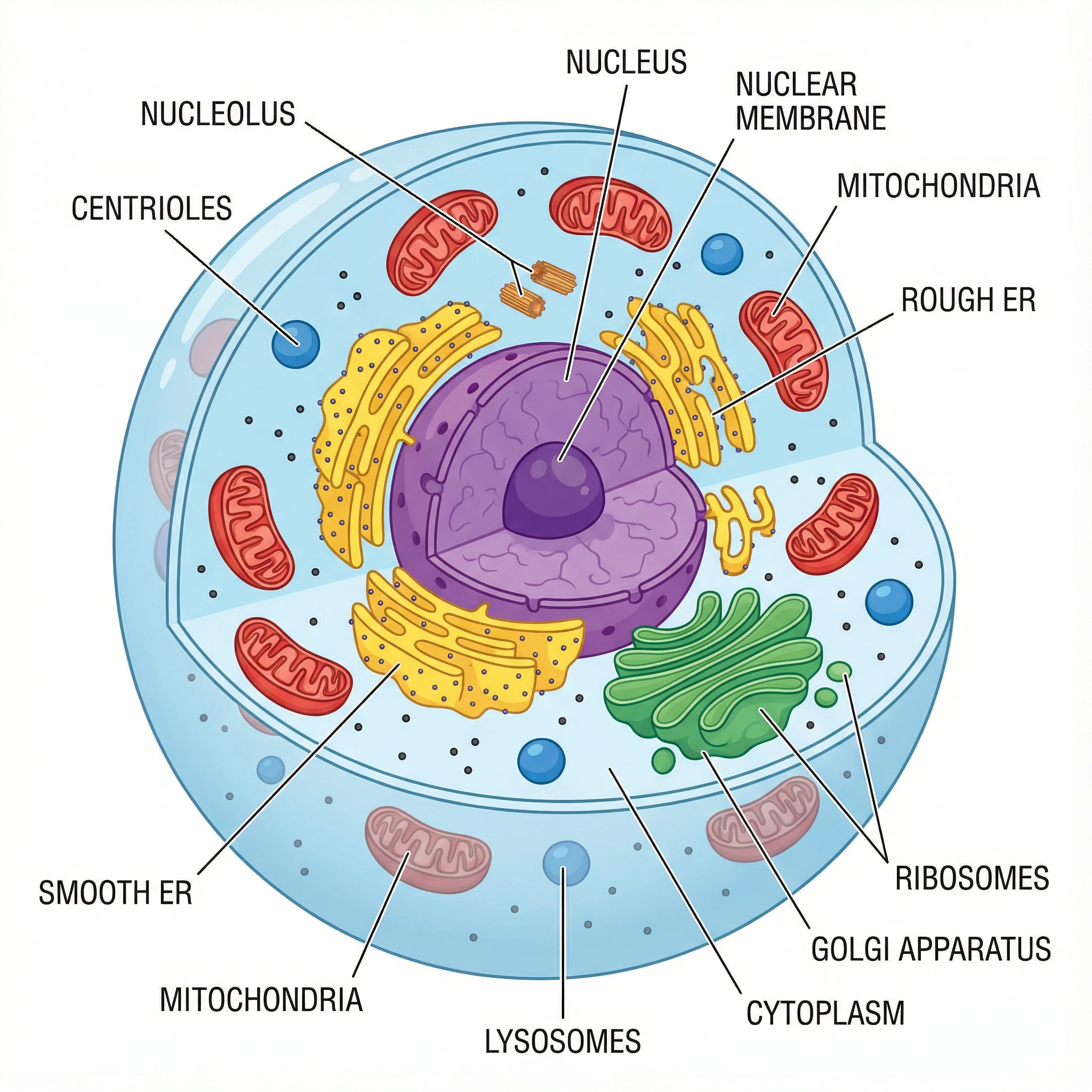
Animal Cell Diagram Generator
Create labeled animal cell diagrams with the nucleus, mitochondria, Golgi, and other organelles.
 Biology
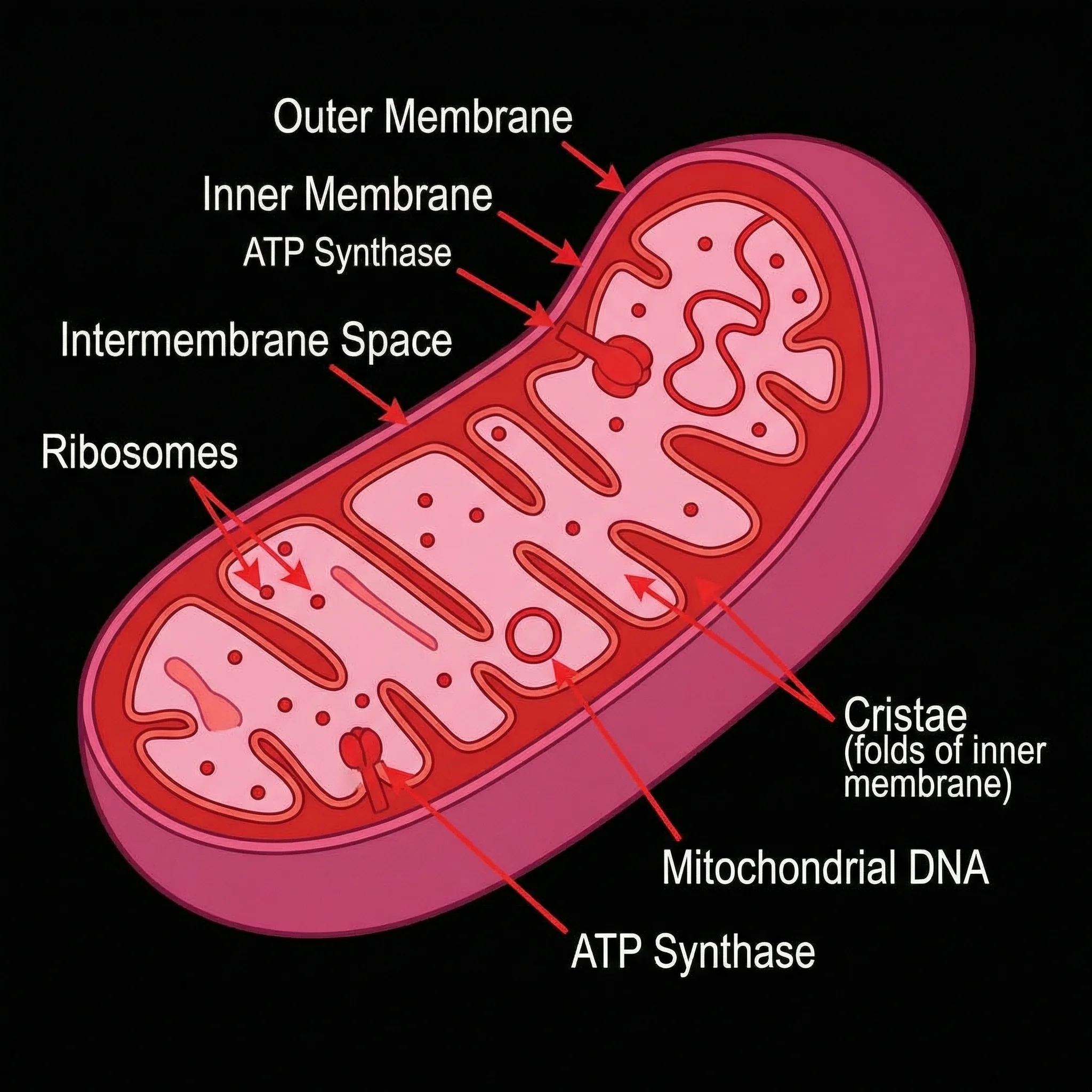
Mitochondria Diagram Generator
Draw labeled mitochondria diagrams showing the cristae, matrix, and inner and outer membranes.
 Biology
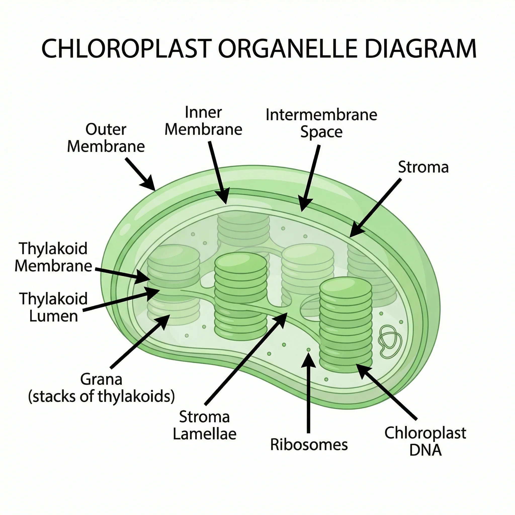
Chloroplast Diagram Generator
Make labeled chloroplast diagrams with the thylakoids, grana, stroma, and outer membrane.