
Eye Anatomy: The Parts of the Human Eye Explained
A clear guide to eye anatomy: the cornea, iris, pupil, lens, retina, and optic nerve, what each part does, how vision works, and how to read an eye diagram.
The human eye is a small organ that does an enormous job: it gathers light, focuses it, converts it into nerve signals, and sends those signals to the brain to build the image you see. To understand how that works, it helps to know the parts of the eye and what each one does.
This guide walks through eye anatomy from the outside in — the cornea, iris, pupil, lens, retina, macula, and optic nerve — explains how vision works, and shows how to read a labeled eye diagram.
Quick Answer: What Are the Main Parts of the Eye?
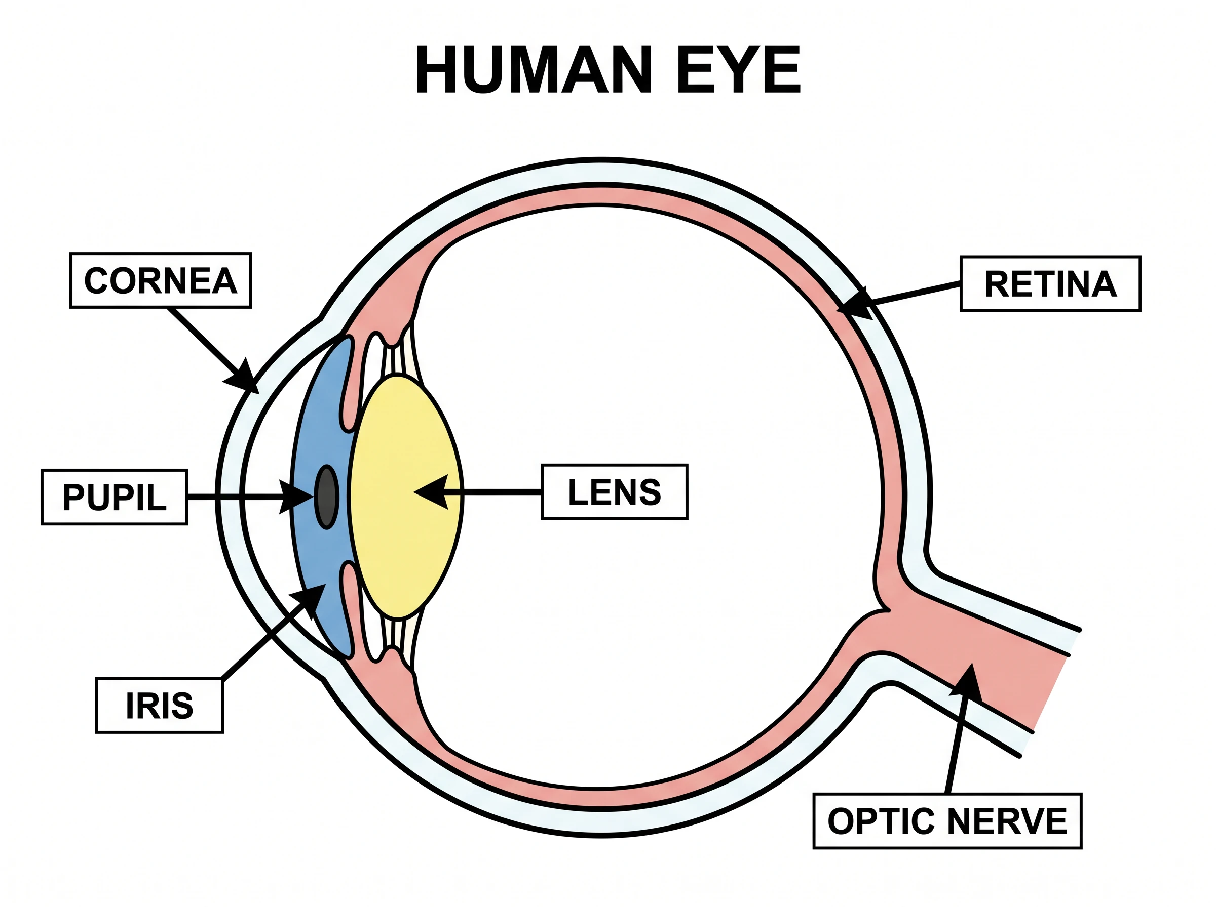
The eye is built from three layers and a clear interior. Light enters through the cornea, passes through the pupil (the opening in the colored iris), is focused by the lens, and lands on the retina at the back of the eye. The retina turns light into electrical signals, and the optic nerve carries them to the brain.
The Three Layers of the Eye
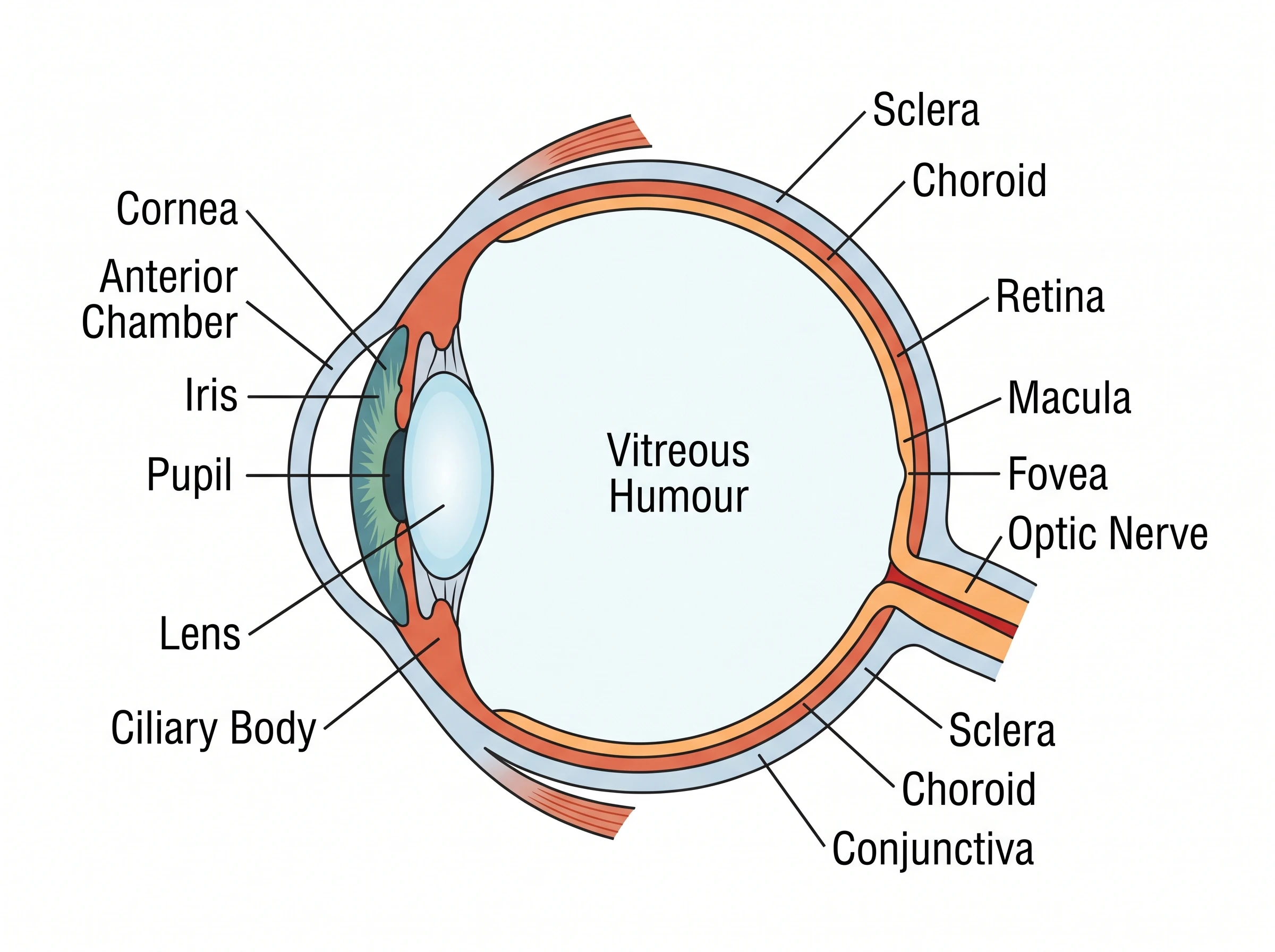
Anatomists group the eyeball into three concentric layers, or "tunics":
- Outer (fibrous) layer — the tough white sclera and the clear cornea at the front.
- Middle (vascular) layer, also called the uvea — the iris, ciliary body, and choroid.
- Inner (neural) layer — the retina, the light-sensitive lining at the back.
Knowing these layers makes a labeled diagram far easier to read, because every structure belongs to one of them.
Eye Anatomy Diagram Generator
Create clear, labeled diagrams of the human eye — cross-sections, front views, and how-vision-works illustrations — and download them free for study or class.
Make an eye diagram ->Outer Layer: Cornea and Sclera
The cornea is the clear, dome-shaped front window of the eye. It does most of the eye's focusing — bending incoming light before it ever reaches the lens. Because it has no blood vessels, it stays transparent and gets its nourishment from tears and the fluid behind it.
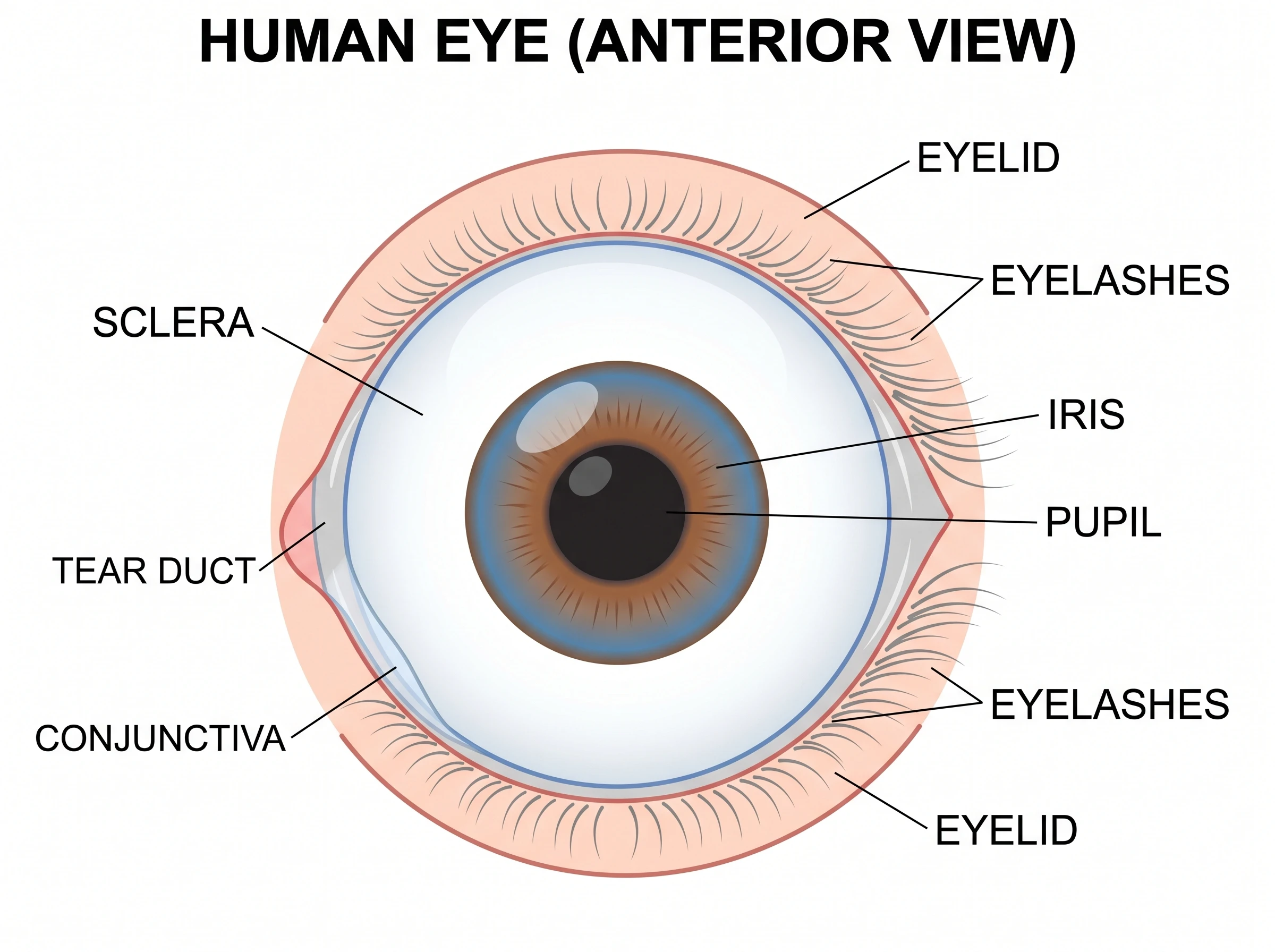
The sclera is the white of the eye. It is tough, opaque connective tissue that gives the eyeball its shape and protects the delicate structures inside.
Middle Layer: Iris, Pupil, Lens, and Ciliary Body
The iris is the colored ring you see in the mirror. It is a muscle that adjusts the size of the pupil — the dark opening in its center — to control how much light gets in. In bright light the pupil shrinks; in dim light it widens.
Just behind the pupil sits the lens, a clear, flexible disc that fine-tunes focus. The ciliary body is the ring of muscle that changes the lens shape: it thickens the lens for close objects and flattens it for distant ones, a process called accommodation. The ciliary body also produces the watery fluid at the front of the eye.
Inner Layer: Retina, Macula, and Optic Nerve
The retina lines the back of the eye and contains millions of photoreceptor cells — rods for dim light and motion, and cones for color and detail. The macula is a small, sensitive central zone of the retina, and at its center the fovea delivers the sharpest, most detailed vision. The optic nerve gathers the retina's signals and carries them to the brain; the point where it leaves the eye has no photoreceptors, creating the natural blind spot.
The Humors That Fill the Eye
Two clear fluids hold the eye's shape and keep it nourished:
- Aqueous humor — the thin, watery fluid in the front chamber between the cornea and lens. It feeds the cornea and lens and is constantly replaced.
- Vitreous humor — the thick, gel-like substance that fills the large space between the lens and the retina, helping the eyeball keep its round form.
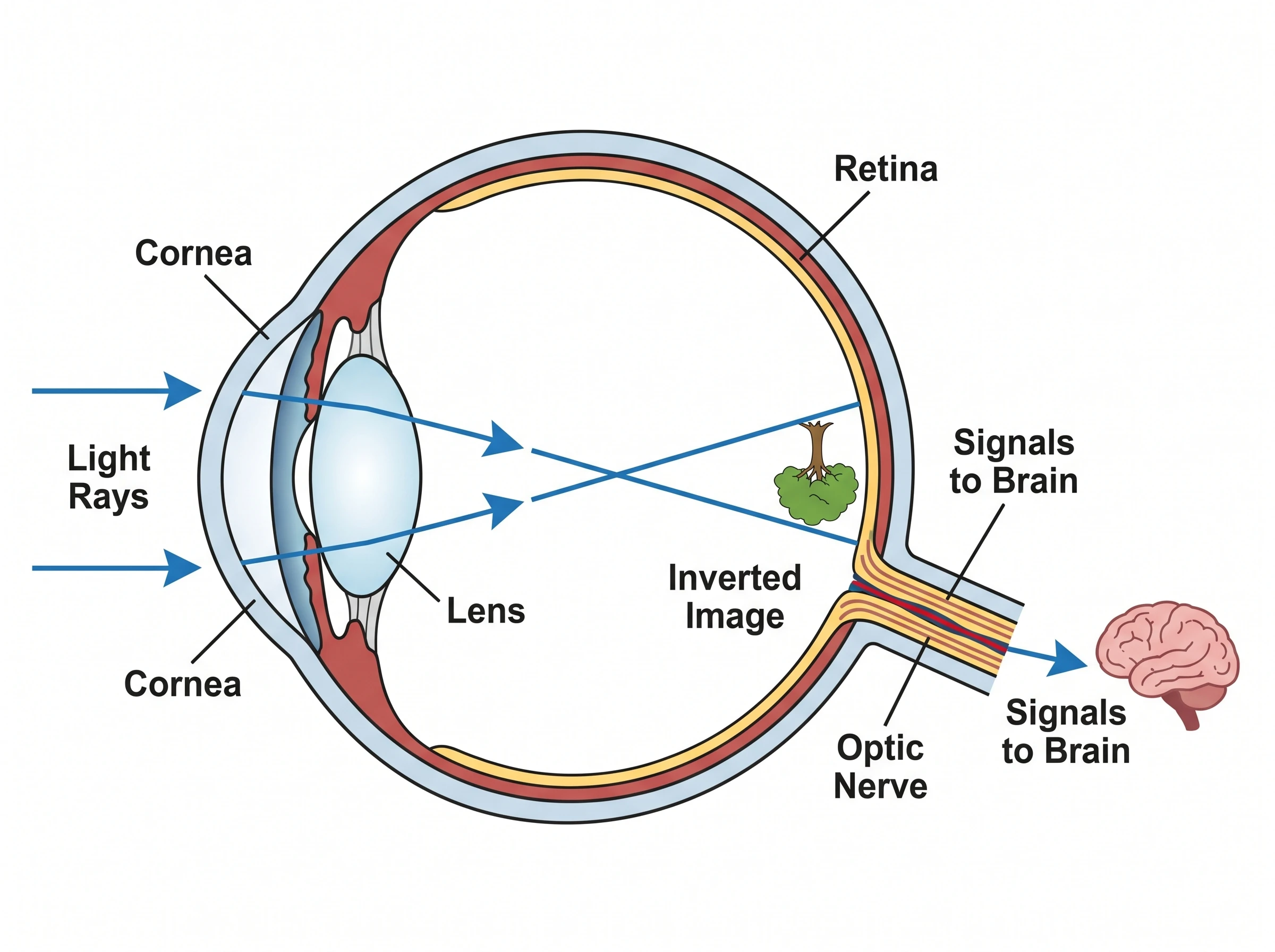
How Vision Works: From Light to Brain
Vision is a chain of steps, and a diagram makes the path easy to follow:
- Light enters through the cornea, which begins focusing it.
- The pupil adjusts to let in the right amount of light.
- The lens fine-tunes the focus, projecting a sharp — but upside-down — image onto the retina.
- The retina converts that light into electrical signals.
- The optic nerve carries the signals to the brain, which flips the image right-side up and interprets it.
How to Read a Labeled Eye Diagram
A good eye diagram is usually a side-on cross-section. To read one:
- Find the front of the eye — the cornea and pupil are on the side light enters.
- Trace the layers outward to inward: cornea/sclera, then iris/ciliary body/choroid, then retina.
- Locate the focusing parts — cornea then lens — along the light's path.
- Follow the optic nerve out the back to the brain.
- Match each label to its structure using the leader lines.
Common Mistakes
- Confusing the iris and the pupil. The iris is the colored muscle; the pupil is the opening it controls.
- Thinking the lens does all the focusing. The cornea does most of it; the lens fine-tunes.
- Mixing up rods and cones. Rods handle dim light and motion; cones handle color and detail.
- Forgetting the image is inverted. The retina receives an upside-down image; the brain flips it.
- Mislabeling the two humors. Aqueous (watery) is in front of the lens; vitreous (gel) is behind it.
Eye Anatomy Diagram Generator
Generate labeled human eye diagrams for biology study, class handouts, and presentations.
FAQ
What are the main parts of the human eye?
The main parts are the cornea, sclera, iris, pupil, lens, ciliary body, choroid, retina, macula, and optic nerve, plus the aqueous and vitreous humors that fill the eye.
What does the cornea do?
The cornea is the clear front surface of the eye. It protects the eye and does most of the focusing, bending incoming light before it reaches the lens.
What is the difference between the iris and the pupil?
The iris is the colored ring of muscle; the pupil is the dark opening in its center. The iris adjusts the pupil's size to control how much light enters the eye.
What does the retina do?
The retina is the light-sensitive layer at the back of the eye. Its rod and cone cells convert light into electrical signals that the optic nerve sends to the brain.
How does the eye work to create vision?
Light passes through the cornea, pupil, and lens, which focus it onto the retina. The retina turns the light into signals, and the optic nerve carries them to the brain, which interprets the image.
Further Reading
分类
更多文章

How to Make a Histogram in Google Sheets: Bins, Frequency Tables, and Chart Editor Tips
Learn how to make a histogram in Google Sheets, change bucket size, use frequency tables, fix common chart issues, and format a readable distribution chart.

How to Draw a Neural Network Architecture Diagram: A Complete Guide
Learn how to draw neural network architecture diagrams for CNNs, RNNs, Transformers, and more. Step-by-step guide with tools, examples, and best practices for researchers and engineers.

Scientific Illustration Guide 2026: Types, Tools & How to Get Started
What is scientific illustration? Learn the types (botanical, medical, technical), best tools (free & paid), and how to create effective scientific visuals. With career tips.