
DNA Structure: The Double Helix and Base Pairs Explained
A clear guide to DNA structure: the double helix, nucleotides, the sugar-phosphate backbone, A-T and G-C base pairing, antiparallel strands, and grooves.
DNA stores the instructions for building and running every living thing, and almost all of that information sits in one elegant shape: the double helix. Once you understand how the two strands twist together and how the bases pair up, the rest of molecular biology — replication, transcription, mutation — starts to make sense.
This guide explains DNA structure from the ground up: the double helix, the nucleotides and sugar-phosphate backbone, the four bases and how they pair (A with T, G with C), why the strands run in opposite directions, and how to read and label a DNA diagram.
Quick Answer: What Is the Structure of DNA?
DNA (deoxyribonucleic acid) is a double helix made of two long strands wound around each other like a twisted ladder. Each strand is a chain of nucleotides, and each nucleotide has three parts: a phosphate group, a deoxyribose sugar, and one of four nitrogenous bases — adenine (A), thymine (T), guanine (G), or cytosine (C).
The sugars and phosphates form the two outer backbones (the sides of the ladder), and the bases point inward and pair up across the middle: A always pairs with T, and G always pairs with C. These base pairs are the rungs of the ladder, and the two strands run in opposite (antiparallel) directions.
The Double Helix
The double helix is the iconic shape proposed by James Watson and Francis Crick in 1953, building on X-ray data from Rosalind Franklin and Maurice Wilkins. Picture a ladder, then grab the ends and twist: that spiral is the double helix.
Two key ideas define it:
- Two strands, not one. DNA is double-stranded. The two strands are held together across the middle by weak hydrogen bonds between the bases.
- A regular, repeating shape. The most common form (B-DNA) makes one complete turn about every 10 base pairs, giving the helix a consistent width of roughly 2 nanometers.
Because the bonds holding the two strands together are weak, the helix can be "unzipped" when the cell needs to read or copy the information — which is exactly what happens during replication and transcription.
Nucleotides and the Sugar-Phosphate Backbone
The building block of DNA is the nucleotide. Every nucleotide has the same three parts:
- A phosphate group
- A deoxyribose sugar (a five-carbon sugar)
- One nitrogenous base (A, T, G, or C)
Nucleotides link together when the phosphate of one joins the sugar of the next. This repeating sugar–phosphate–sugar–phosphate chain forms the backbone — the structural sides of the ladder. The backbone is identical all the way along; only the attached base changes from one rung to the next.
That design is important: the constant backbone keeps the molecule stable and uniform, while the changing sequence of bases carries the actual genetic message.
The Four Bases and Base Pairing
DNA's information is written with a four-letter alphabet:
- Adenine (A) and guanine (G) are purines (larger, double-ring bases).
- Thymine (T) and cytosine (C) are pyrimidines (smaller, single-ring bases).
The bases do not pair randomly. They follow strict complementary base pairing, also called Watson-Crick pairing:
- A pairs with T (held by 2 hydrogen bonds)
- G pairs with C (held by 3 hydrogen bonds)
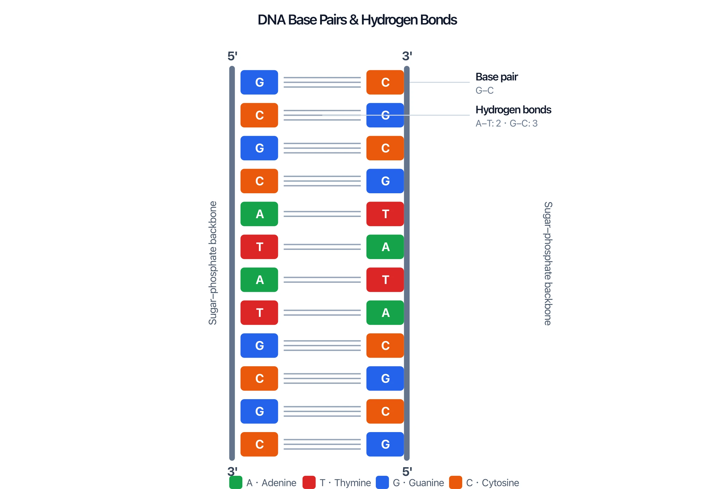
A purine always pairs with a pyrimidine, which keeps the width of the ladder constant. Because the pairing is fixed, the two strands are complementary: if you know the sequence of one strand, you automatically know the other. A strand reading A-T-G-C is paired with T-A-C-G.
This complementarity is the basis of accurate copying. When DNA replicates, each strand acts as a template for building a new partner strand.
Antiparallel Strands and the 5' and 3' Ends
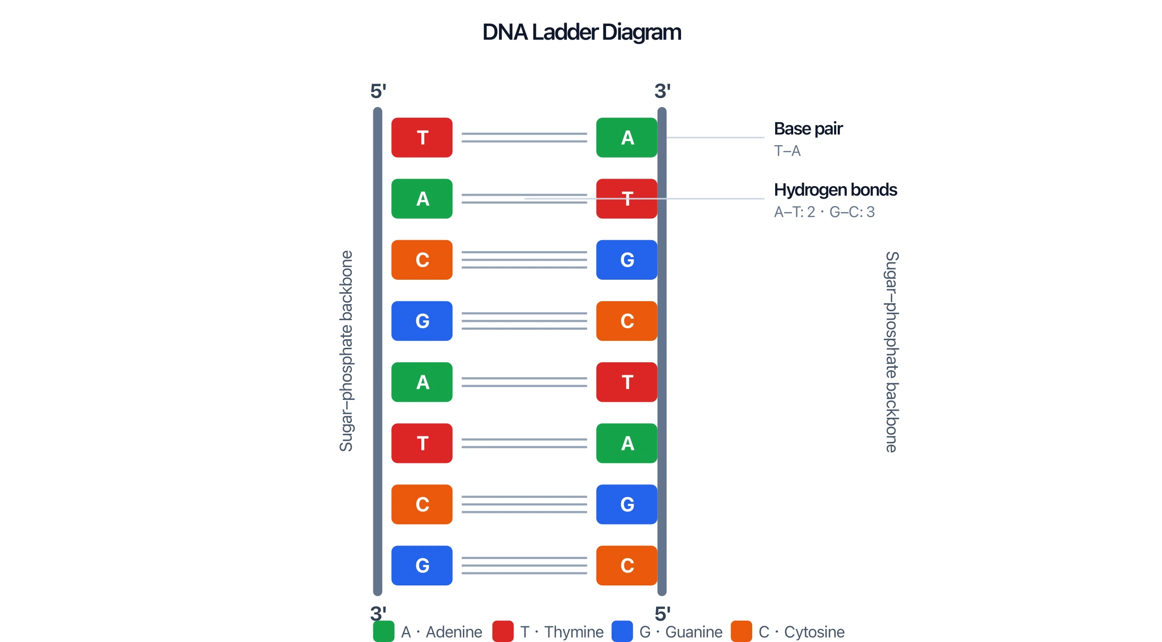
The two strands of DNA are antiparallel — they run in opposite directions. Each strand has a chemical orientation defined by its sugar carbons, labeled 5' (five prime) at one end and 3' (three prime) at the other.
In a DNA molecule, one strand runs 5' → 3' from top to bottom, while its partner runs 3' → 5' in the same view. On a diagram you will see one strand labeled 5' at the top and 3' at the bottom, and the opposite strand labeled the reverse. This antiparallel arrangement is required for the bases to line up correctly and for enzymes to read and copy the strands.
Dimensions: Grooves and the Twist
Twisting the ladder into a helix creates two uneven gaps that spiral along the outside:
- The major groove — the wider gap
- The minor groove — the narrower gap
These grooves matter because proteins such as transcription factors and repair enzymes dock into them to recognize specific sequences without unwinding the DNA. The major groove offers more chemical "fingerprints" for proteins to read, so it is the more important recognition site.
Why the Structure Matters
The double-helix structure is not just elegant — it directly enables DNA's two essential jobs:
- Information storage. The order of the four bases along a strand spells out genes. With a stable backbone protecting the message and four letters to work with, a single DNA molecule can encode enormous amounts of information.
- Faithful replication. Because A only pairs with T and G only with C, the molecule can unzip and each old strand can template a perfect new partner. This is why a cell can copy its entire genome before dividing.
In Watson and Crick's famous understatement, the specific pairing "immediately suggests a possible copying mechanism for the genetic material." The structure is the explanation for heredity.
How to Read and Label a DNA Diagram
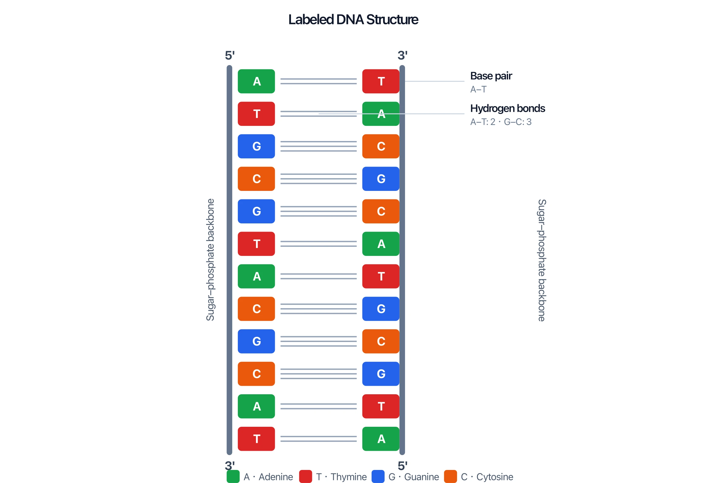
When you look at (or build) a DNA diagram, check for these labeled features:
- Two backbones running down the outside, labeled sugar-phosphate backbone.
- Base pairs as rungs in the middle, with A-T and G-C matched correctly.
- Hydrogen bonds between paired bases — usually 2 lines for A-T and 3 lines for G-C.
- 5' and 3' ends marked on both strands, running in opposite directions (antiparallel).
- Optionally, the major and minor grooves if the diagram shows the twisted helix.
A clear DNA diagram makes the relationships obvious: which base pairs with which, how many bonds hold them, and which way each strand runs.
DNA Structure Diagram Generator
Type any DNA sequence and get a labeled diagram with the correct A-T and G-C base pairs, hydrogen bonds, antiparallel strands, and sugar-phosphate backbone.
Make a DNA structure diagram ->Common Mistakes and Confusions
- Wrong base pairs. A pairs only with T, and G only with C. A diagram showing A-G or T-C is incorrect.
- Swapping bond counts. A-T has 2 hydrogen bonds; G-C has 3. Drawing 3 bonds on an A-T pair is a common error.
- Including uracil. Uracil (U) belongs to RNA, not DNA. DNA uses thymine (T).
- Parallel strands. The strands are antiparallel (opposite directions), not parallel. Both 5' ends should not sit on the same side.
- Forgetting the backbone is constant. The sugar-phosphate backbone never changes; only the bases vary along it.
FAQ
What are the four bases in DNA?
The four bases are adenine (A), thymine (T), guanine (G), and cytosine (C). A and G are purines; T and C are pyrimidines. The order of these bases along a strand encodes genetic information.
How do the bases pair in DNA?
Adenine pairs with thymine (A-T) using two hydrogen bonds, and guanine pairs with cytosine (G-C) using three hydrogen bonds. This fixed, complementary pairing is called Watson-Crick base pairing.
What does the sugar-phosphate backbone do?
The sugar-phosphate backbone forms the two structural sides of the DNA ladder. It is made of alternating deoxyribose sugars and phosphate groups, and it holds the bases in place while keeping the molecule stable.
Why are DNA strands antiparallel?
The two strands run in opposite directions — one 5' → 3' and the other 3' → 5'. This antiparallel orientation lets the complementary bases line up correctly across the helix and allows enzymes to read and copy each strand properly.
What is the difference between the major and minor groove?
As the double helix twists, it forms two unequal gaps: the wider major groove and the narrower minor groove. Proteins use these grooves — especially the major groove — to recognize specific DNA sequences without unwinding the helix.
DNA Structure Diagram Generator
Turn any DNA sequence into a labeled double-helix or ladder diagram with correct A-T and G-C base pairing.
Further Reading
分类
更多文章
")
8 Best Free ChemDraw Alternatives in 2026 (For Drawing Chemical Structures)
Compare the best free ChemDraw alternatives: ChemSketch, MarvinSketch, MolView, ChemDoodle & more. Draw chemical structures without expensive subscriptions.
")
Mapping Diagrams Explained: Complete Guide with Examples (2026)
Learn what mapping diagrams are, how to create them, and when to use them in mathematics and research. Complete guide with function mapping examples and free tools.
")
How to Create a Network Diagram: Types, Symbols & Step-by-Step Guide (2026)
Learn how to create network diagrams for IT infrastructure. Covers logical vs physical diagrams, standard symbols, topology types, and real-world examples.