Virus Diagram Generator for the Labeled Virus Structure
Generate a clearly labeled virus diagram in seconds. Show the capsid, capsomeres, genetic material (DNA or RNA), envelope, glycoprotein spikes, and matrix protein — or pick a specific virus shape such as icosahedral, helical, bacteriophage, or enveloped influenza/coronavirus — then download your diagram, free.
Virus Diagram Generator
Free to try ·
Your diagram will appear here
Describe what you need and click Generate
Virus Diagram Examples
Labeled virus structures, bacteriophages, and virology diagrams
Labeled Virus Structure
A complete enveloped virus with every layer labeled — envelope, glycoprotein spikes, matrix protein, capsid, capsomeres, and the nucleic acid core.
Bacteriophage Labeled
The classic T4 phage in full detail — head, collar, tail sheath, base plate, and tail fibers, every part labeled.
Enveloped Virus Diagram
Focus on the envelope: phospholipid bilayer, transmembrane spike proteins, matrix protein, and the nucleocapsid within.
Coronavirus Structure
Spike (S), envelope (E), membrane (M), and nucleocapsid (N) proteins all labeled on a coronavirus cross-section.
Virus vs Bacteria
Virus versus bacterium side by side — size scale bar, structural labels, and the key differences highlighted.
Blank Virus Worksheet
A printer-ready worksheet — leader lines, blank fill-in spaces, no labels — for classroom quizzes and self-testing.
What is a virus?
A virus is an ultramicroscopic infectious agent that can only replicate inside the living cells of a host organism. Unlike bacteria, viruses are not cells — they have no membrane, no cytoplasm, no ribosomes, and no metabolism of their own. A virus particle (virion) is essentially a protein shell wrapped around a small amount of genetic material, either DNA or RNA. Because viruses sit at the edge of what we define as "living," they are studied both in biology and in medicine. This generator draws a clearly labeled virus diagram so students can see the key structural components and how they compare across virus types.
The labeled parts of a virus
- Capsid: the protein coat that surrounds and protects the genetic material, made up of repeating subunits.
- Capsomeres: the individual protein subunits that assemble together to form the capsid.
- Genetic material: the nucleic acid core — either double-stranded DNA, single-stranded DNA, double-stranded RNA, or single-stranded RNA depending on the virus type.
- Envelope: a lipid bilayer membrane (derived from the host cell) that wraps around the capsid in enveloped viruses.
- Glycoprotein spikes: protein-sugar complexes that project from the envelope and are used to recognise and attach to host cell receptors.
- Matrix protein: a layer of protein between the envelope and the capsid that helps maintain the structure of enveloped viruses.
- Tail (bacteriophage only): the assembly that injects DNA into bacterial host cells — includes a collar, tail sheath, tail tube, base plate, and tail fibers.
Common virus shapes and types
Viruses come in three main shapes. Icosahedral viruses have a roughly spherical capsid built from 20 equilateral triangular faces — examples include adenovirus, poliovirus, and herpes simplex virus. Helical viruses wrap their capsid in a helix around the genome — the tobacco mosaic virus is the classic example, and many animal viruses (such as influenza before the envelope is added) are helical at the nucleocapsid level. Enveloped viruses such as influenza, HIV, and coronavirus surround their helical or icosahedral nucleocapsid with a lipid envelope studded with glycoprotein spikes derived from the host cell membrane. Bacteriophages are a special category of complex morphology: they have an icosahedral head containing DNA, a tail sheath, and tail fibers that inject DNA into bacteria.
Virus structure vs bacterial cell structure
A virus is not a cell and should not be confused with a bacterium. Bacteria are single-celled prokaryotes with a cell wall, plasma membrane, cytoplasm, ribosomes, and circular DNA; they can reproduce independently by binary fission. Viruses have none of those structures — just a capsid, genetic material, and in some cases an envelope and spikes. Viruses are also far smaller: most virions are 20–300 nm across, while a typical bacterium is 1–10 µm — about 10–100 times bigger. Because viruses depend entirely on host cell machinery to replicate, antibiotics that target bacterial structures (cell walls, ribosomes) have no effect on them. A side-by-side comparison diagram of a virus and a bacterium drawn to scale is one of the most useful illustrations for making these differences tangible.
How glycoprotein spikes work
Glycoprotein spikes are the recognition system of an enveloped virus. They are transmembrane proteins that project from the lipid envelope, and their outermost domain binds specifically to receptor molecules on the surface of host cells. In influenza, the hemagglutinin (HA) spike binds to sialic acid on respiratory epithelial cells; the neuraminidase (NA) spike helps new virions escape by cleaving sialic acid. In SARS-CoV-2, the spike (S) protein binds to ACE2 receptors. These spikes are also the primary target for neutralising antibodies produced during infection or vaccination, which is why they appear so prominently on virus diagrams used in immunology courses.
Labeled vs blank diagrams for worksheets
For teaching, you often need two versions of the same diagram. A labeled version names every structure and is ideal for lecture slides, revision notes, and textbook illustrations. A blank version keeps the leader lines but removes the text, so students can fill in the labels themselves as a quiz or a worksheet. Because you describe what you want in plain English, you can request a fully labeled enveloped virus cross-section for the lesson and then a printer-friendly, black-and-white blank version of the same layout for the accompanying test — no manual redrawing required.
Frequently Asked Questions
Related Biology Tools
 Biology
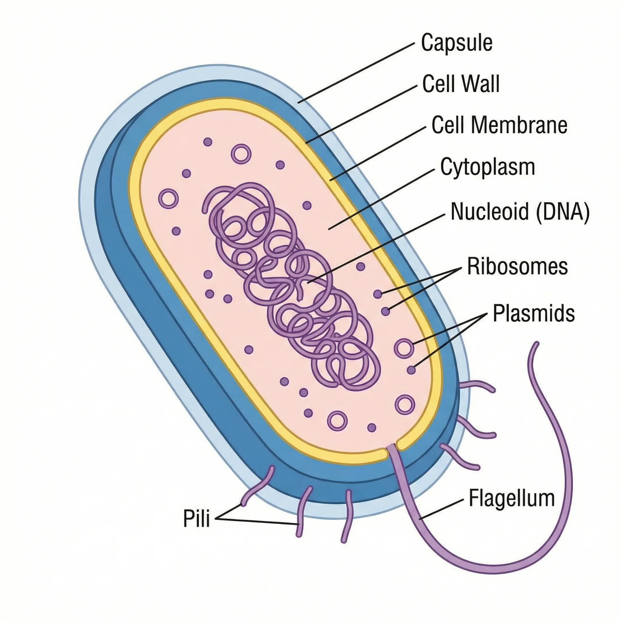
Bacteria Diagram Generator
Create labeled prokaryotic cell diagrams showing the cell wall, nucleoid, flagella, ribosomes, and more — the cellular counterpart to a virus.
 Biology
Plant Cell Diagram Generator
Generate labeled plant cell diagrams showing the chloroplast, cell wall, vacuole, nucleus, and other organelles.
 Biology
Animal Cell Diagram Generator
Create labeled animal cell diagrams with the nucleus, mitochondria, Golgi apparatus, and all major eukaryotic organelles.