Eye Anatomy Diagram Generator Labeled Human Eye Diagrams
Create labeled eye anatomy diagrams with AI. Show a cross-section of the human eye — cornea, iris, pupil, lens, retina, macula, and optic nerve — or how vision works, for biology and health class. Download as PNG.
AI Eye Anatomy Diagram Generator
By using ConceptViz, you agree not to generate or edit adult, sexual, explicit, unsafe, or policy-violating content. See Content Policy.
免費試用 ·
Your eye anatomy diagram will appear here
Describe the view (cross-section, front) and parts to label
Eye Anatomy Examples
Labeled diagrams of the human eye and how vision works
Labeled Eye Cross-Section
A full cross-section with every major structure labeled — the classic eye diagram.
Simple Eye Diagram
A clean, simplified version with just the main parts — great for younger students.
How Vision Works
The light path — cornea and lens focusing an image on the retina.
Retina Detail
A close-up of the retina — rods, cones, macula, fovea, and the optic disc.
Extraocular Muscles
The six muscles that move the eye, labeled — for physiology lessons.
Front View
The external eye — what you see from the front, labeled.
What does an eye anatomy diagram show?
An eye anatomy diagram shows the structures of the human eye and how they work together to produce vision. A typical labeled cross-section includes the cornea, iris, pupil, lens, retina, macula, fovea, optic nerve, sclera, and choroid. This generator creates clear, labeled diagrams of the eye for biology, anatomy, and health classes, study notes, and presentations.
The main parts of the eye
- Cornea: the clear front layer that bends (refracts) incoming light.
- Iris and pupil: the iris is the colored ring of muscle that controls the size of the pupil, the opening that lets light in.
- Lens: focuses light onto the retina, changing shape to focus on near or far objects (accommodation).
- Retina: the light-sensitive layer at the back, containing rod and cone photoreceptor cells; the macula and fovea give sharp central vision.
- Optic nerve: carries visual signals from the retina to the brain.
How the eye works (vision)
Light reflects off an object and enters the eye through the cornea, which does most of the focusing. It passes through the pupil and the lens, which fine-tunes the focus, and forms an inverted image on the retina. Photoreceptor cells convert the light into nerve signals, which travel along the optic nerve to the brain, where the image is interpreted (and flipped the right way up).
Rods, cones, and the retina
The retina contains two types of photoreceptor: rods, which are sensitive in dim light and detect black and white, and cones, which work in bright light and detect color. Cones are concentrated at the fovea, the spot of sharpest vision within the macula. Where the optic nerve leaves the eye there are no photoreceptors — the blind spot.
Tips for a clear eye diagram
Choose the view you need — a labeled cross-section for the internal structures, a front view for the external features, or a "how vision works" diagram for the light path. Mention the specific parts you want labeled and the level (simple for younger students, detailed for higher grades). Generate a few options and download the clearest for your worksheet or slides.
常見問題
Related Science Tools
 Physics
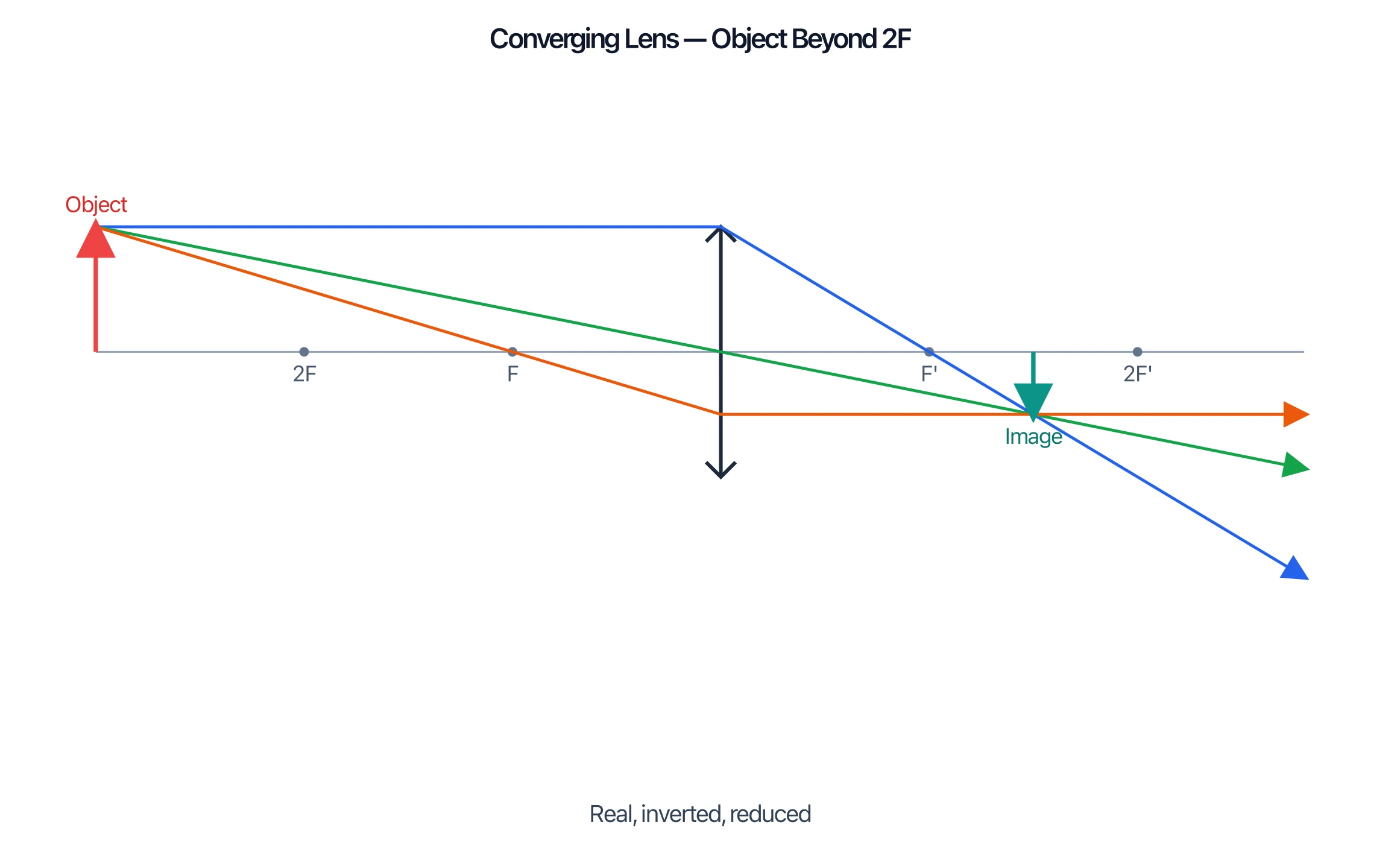
Ray Diagram Generator
Draw lens and mirror ray diagrams — perfect for showing how the eye focuses light.
 Biology
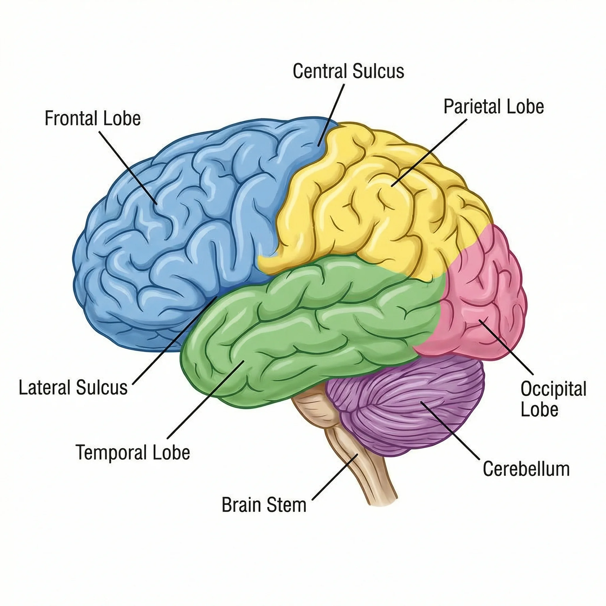
Brain Diagram Generator
Create labeled brain diagrams with lobes, regions, and structures.
 Medical
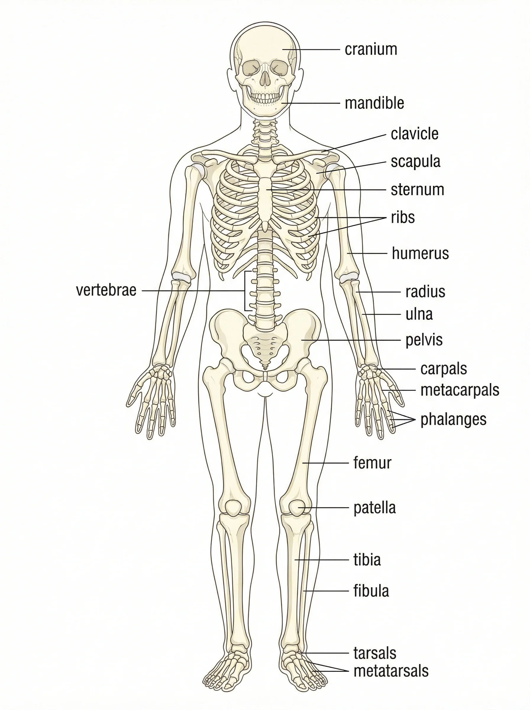
Anatomical Drawing Generator
Generate labeled anatomy illustrations of body systems and organs.