高基氏體圖生成器 標注版與詳細版
用 AI 製作專業高基氏體圖。生成標注版、空白版及路徑圖,適合細胞生物學及蛋白質處理教學。
高基氏體圖生成器
By using ConceptViz, you agree not to generate or edit adult, sexual, explicit, unsafe, or policy-violating content. See Content Policy.
免費試用 ·
圖表將顯示於此
描述所需內容後點擊生成
高基氏體圖範例
瀏覽我們的範例集,或在上方生成您的自訂圖表
高基氏體 - 標注版圖表
詳細標注高基氏體圖,呈現高中及大學生物所需的所有主要結構。
高基氏體 - 囊泡詳細圖
高基氏體囊泡出芽、融合及運輸路徑的詳細視圖。
分泌路徑中的高基氏體
完整分泌路徑圖,呈現高基氏體在蛋白質處理與分選中的角色。
順式與反式高基氏體網絡比較
比較圖,呈現順式與反式高基氏體網絡在蛋白質處理中的不同角色。
高基氏體 - 蛋白質處理圖
進階圖表,呈現高基氏體內的糖基化及分選等蛋白質修飾步驟。
高基氏體 - 著色學習單
適合學生學習單及標注練習的印表機友善高基氏體著色頁。
什麼是高基氏體?
高基氏體(又稱高爾基複合體或高爾基體)是存在於大多數真核細胞中的膜性胞器。1898 年由義大利醫師 Camillo Golgi 發現,是細胞的中央處理與運輸中心。高基氏體接收來自內質網的蛋白質與脂質,進行修飾與包裝後,再將其送往細胞內外的最終目的地。它在蛋白質的轉譯後修飾、分選及運輸中扮演關鍵角色。
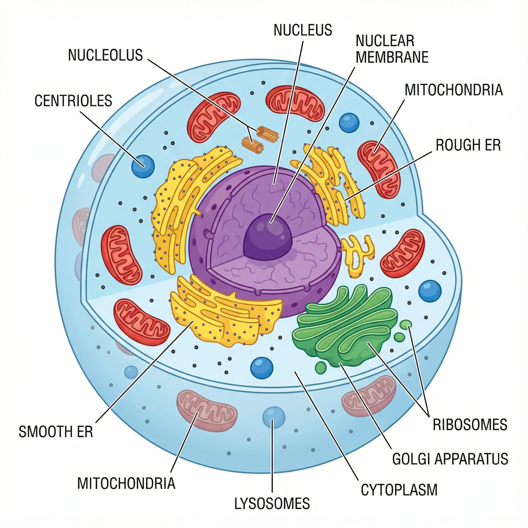
高基氏體的結構
- 扁平囊:構成高基氏體核心的扁平膜囊堆疊(通常每堆 4-8 個),每個扁平囊具有獨特的生化環境,負責蛋白質的序列性處理。
- 順式面(接收端):高基氏體朝向內質網的入口,來自內質網的運輸囊泡在此融合,輸送新合成的蛋白質。
- 反式面(輸出端):高基氏體朝向細胞膜的出口,修飾與分選後的蛋白質從此面以囊泡形式出芽,送往目的地。
- 順式高基氏體網絡(CGN):位於順式面的管狀與囊泡網絡,接收並分選來自內質網的入境貨物。
- 反式高基氏體網絡(TGN):位於反式面的分選站,將蛋白質包裝進不同囊泡,送往溶酶體、細胞膜或分泌路徑。
- 運輸囊泡:在高基氏體扁平囊之間穿梭蛋白質,並將蛋白質送往細胞各最終目的地的小型膜性載體。
- 分泌囊泡:從反式面出芽,攜帶已處理蛋白質運往細胞外分泌的囊泡。
高基氏體的功能
高基氏體執行細胞中多項重要功能。它透過糖基化(添加糖鏈)、磷酸化及蛋白質水解切割來修飾蛋白質;根據分子標籤對蛋白質進行分選,引導至正確的細胞目的地;將蛋白質與脂質包裝進囊泡,運往細胞膜、溶酶體或分泌路徑。高基氏體也合成部分多醣及糖脂,並透過以甘露糖-6-磷酸標記溶酶體酵素,在溶酶體形成中扮演關鍵角色。
分泌路徑中的高基氏體
高基氏體是內膜系統與分泌路徑的核心樞紐。在粗面內質網核糖體上合成的蛋白質,以 COPII 包被囊泡運輸至高基氏體。蛋白質依序通過順式、中間及反式扁平囊,進行逐步修飾。在反式高基氏體網絡,完全處理的蛋白質被分選至不同囊泡群:組成性分泌囊泡持續釋放內容物,調節性分泌囊泡在訊號觸發前儲存貨物,溶酶體囊泡將酵素送往溶酶體。這種有組織的運輸確保每種蛋白質到達正確的目的地。
在生物課教授高基氏體
我們的高基氏體圖專為各年級生物教育工作者設計。使用標注版圖表向學生介紹胞器結構與功能。空白版及著色頁版本適合作為測驗材料及標注練習。分泌路徑圖幫助學生理解高基氏體在更廣泛內膜系統中的連結。進階課程可使用蛋白質處理圖,說明每個扁平囊層發生的生化修飾。可搭配讓學生追蹤蛋白質從核糖體到最終目的地旅程的實作活動。