
Ear Anatomy: Parts of the Human Ear Explained
Learn human ear anatomy: the outer ear (pinna, ear canal), middle ear (eardrum, ossicles), and inner ear (cochlea, semicircular canals), plus how hearing works.
The human ear is far more than the flap you see on the side of your head. It is a precision instrument that catches sound waves from the air, amplifies them, and turns them into electrical signals your brain can understand — while also keeping you balanced.
This guide explains the anatomy of the ear part by part: the outer, middle, and inner ear, what each structure does, and how a sound travels all the way from the air to your brain.
Quick Answer: The Parts of the Ear
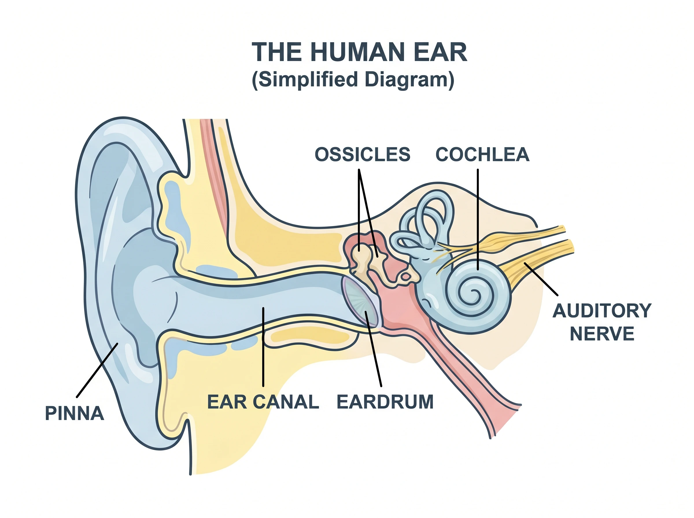
The ear is divided into three sections:
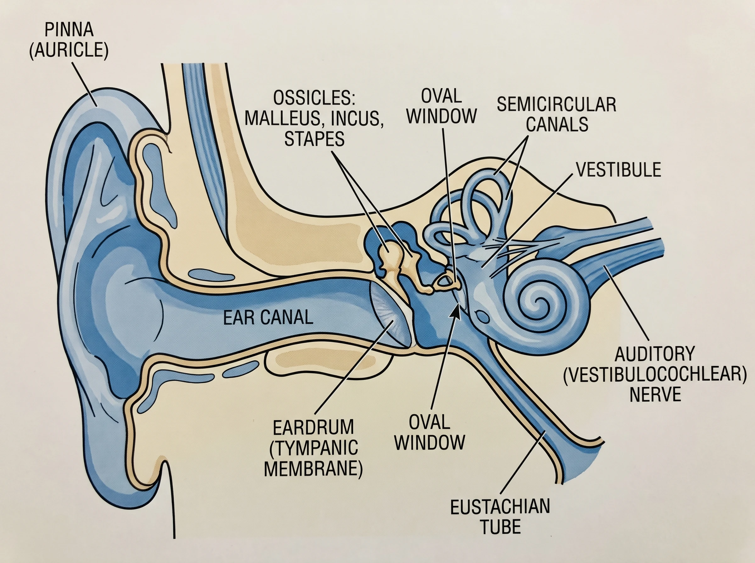
- Outer ear — the pinna (auricle) and the ear canal, which collect sound and funnel it inward.
- Middle ear — the eardrum (tympanic membrane) and three tiny bones, the ossicles (malleus, incus, stapes), which amplify the vibration.
- Inner ear — the cochlea (hearing) and the semicircular canals plus vestibule (balance), connected to the brain by the auditory nerve.
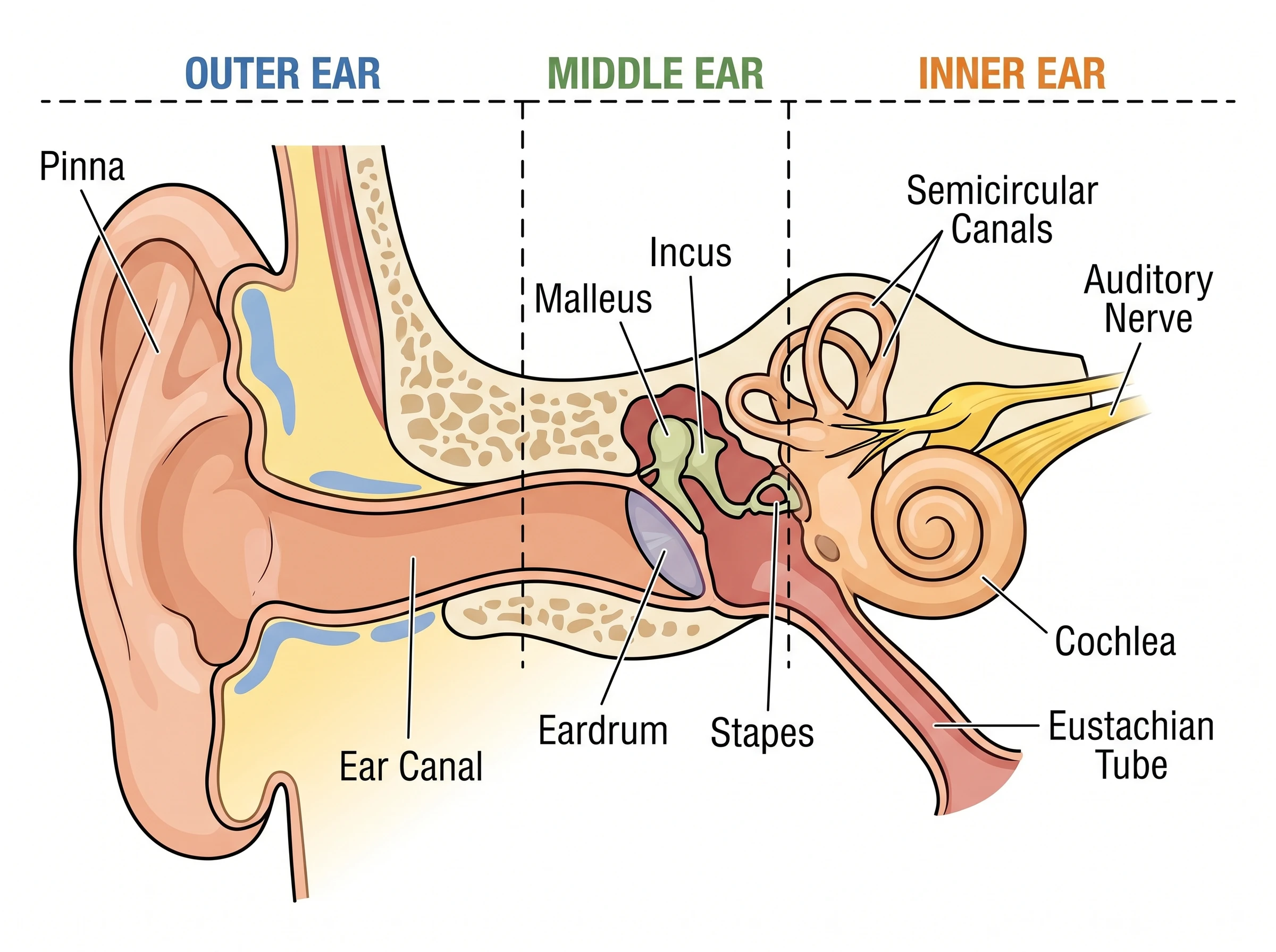
The Outer Ear
The outer ear is the only part you can see. The pinna (also called the auricle) is the curved, cartilage flap that acts like a satellite dish — its ridges gather sound waves and help you tell where a sound is coming from. Those waves travel down the ear canal (external auditory canal), a short tube lined with skin, fine hairs, and glands that produce earwax (cerumen) to trap dust and protect the canal. At the inner end of the canal sits the eardrum, which marks the boundary with the middle ear.
The Middle Ear
The middle ear is a small, air-filled cavity that turns sound waves into mechanical motion.
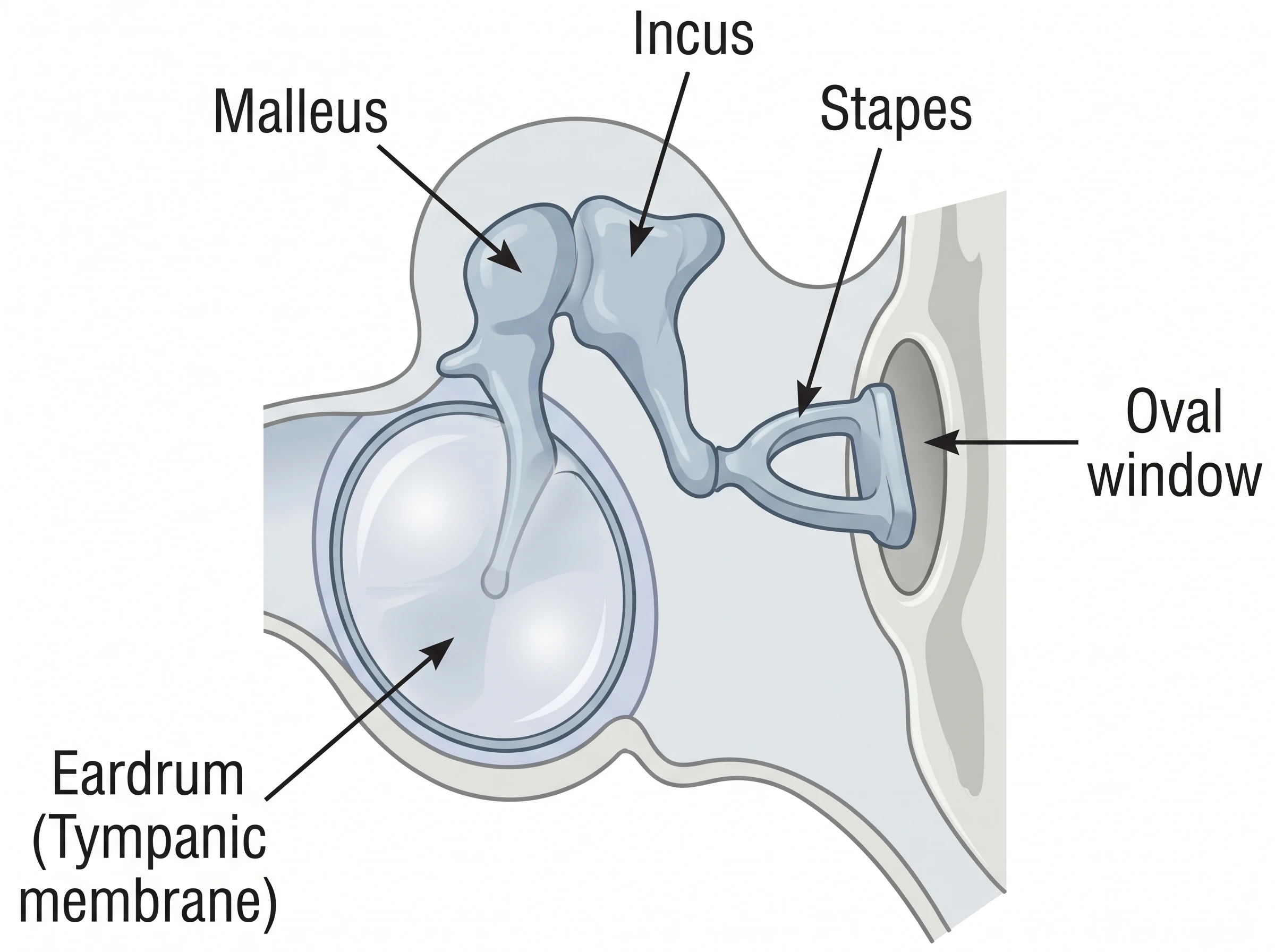
- Eardrum (tympanic membrane). A thin, tightly stretched membrane that vibrates when sound waves hit it.
- Ossicles. Three of the smallest bones in the body form a lever chain: the malleus (hammer) attaches to the eardrum, passes vibration to the incus (anvil), which passes it to the stapes (stirrup). The stapes pushes on the oval window, the doorway to the inner ear. This chain amplifies the force of the vibration so it can move the fluid inside the cochlea.
- Eustachian tube. A narrow passage connecting the middle ear to the back of the throat. It opens when you swallow or yawn to equalize air pressure — the reason your ears "pop" on a plane.
Ear Anatomy Diagram Generator
Create clean, labeled ear anatomy diagrams of the outer, middle, and inner ear, then download them free as PNG for study notes, slides, or worksheets.
Make an ear diagram ->The Inner Ear
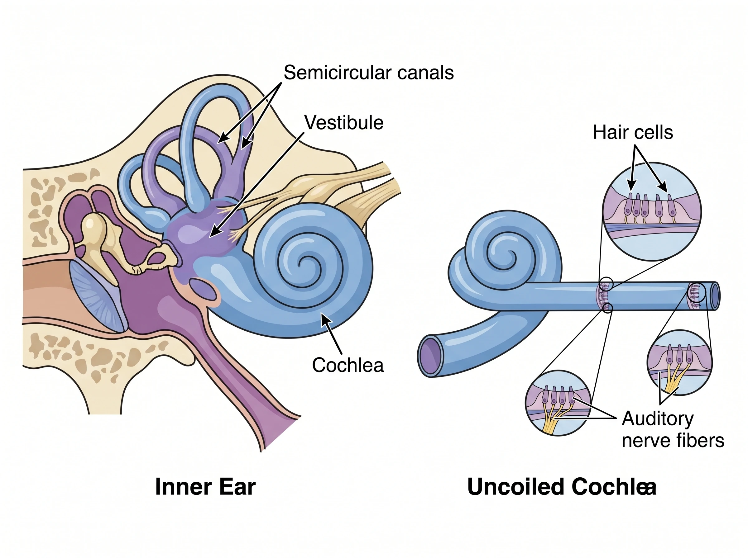
The inner ear is a maze of fluid-filled chambers carved into the temporal bone, sometimes called the labyrinth. It handles two jobs at once: hearing and balance.
- Cochlea. The snail-shaped hearing organ. It is filled with fluid and lined with thousands of tiny hair cells. When the stapes pushes the oval window, it sends waves through the cochlear fluid, bending the hair cells, which convert the motion into electrical signals.
- Semicircular canals. Three looping tubes set at right angles to one another. As your head turns, fluid inside them shifts and moves sensory hairs, telling your brain about rotation.
- Vestibule. The chamber between the cochlea and the canals; together with the canals it forms the vestibular system that keeps you balanced and oriented.
- Auditory (vestibulocochlear) nerve. Carries both the hearing signals and the balance signals from the inner ear to the brain.
How Hearing Works
Hearing is a relay that converts air pressure into brain signals in five steps:
- Collect. The pinna gathers sound waves and the ear canal funnels them inward.
- Vibrate. The waves strike the eardrum, making it vibrate.
- Amplify. The malleus, incus, and stapes pass and magnify the vibration, and the stapes pushes the oval window.
- Transduce. Ripples travel through the cochlear fluid, bending hair cells that turn motion into electrical signals.
- Interpret. The auditory nerve carries those signals to the brain, which decodes them as speech, music, or noise.
The whole journey takes a fraction of a millisecond.
Ear Anatomy Diagram Generator
Generate labeled diagrams of the outer, middle, and inner ear for notes, slides, and worksheets.
How to Read and Label an Ear Diagram
- Find the three zones first — outer, middle, inner — usually shown left to right in a cross-section.
- Trace the sound path from the pinna, down the ear canal, to the eardrum and ossicles, then to the cochlea.
- Label the ossicles in order: malleus, then incus, then stapes.
- Separate hearing from balance in the inner ear: the coiled cochlea is hearing; the looping semicircular canals are balance.
- Add the connectors — the Eustachian tube (middle ear to throat) and the auditory nerve (inner ear to brain).
Common Mistakes
- Mixing up the ossicle order. Sound goes eardrum -> malleus -> incus -> stapes, not the reverse.
- Confusing the cochlea with the semicircular canals. The spiral cochlea handles hearing; the loops handle balance.
- Forgetting the Eustachian tube. It is part of the middle ear and is what equalizes pressure.
- Calling the whole visible part the "ear." The pinna is only the outer ear; most of the organ is hidden inside the skull.
- Putting the eardrum in the inner ear. The eardrum is the start of the middle ear.
FAQ
What are the three parts of the ear?
The ear has three parts: the outer ear (pinna and ear canal), the middle ear (eardrum and the ossicles — malleus, incus, and stapes), and the inner ear (cochlea, semicircular canals, and vestibule).
What are the three bones in the ear called?
The three middle-ear bones, called the ossicles, are the malleus (hammer), incus (anvil), and stapes (stirrup). They are the smallest bones in the human body and amplify sound vibrations.
What does the cochlea do?
The cochlea is the spiral, fluid-filled hearing organ of the inner ear. Its tiny hair cells convert sound vibrations into electrical signals, which the auditory nerve carries to the brain.
Which part of the ear controls balance?
Balance is controlled by the vestibular system in the inner ear — the three semicircular canals and the vestibule. They sense head movement and orientation and send that information to the brain.
How does sound travel through the ear to the brain?
Sound is collected by the pinna and funneled down the ear canal, vibrates the eardrum, is amplified by the ossicles, ripples through cochlear fluid to bend hair cells, and is sent as electrical signals along the auditory nerve to the brain.
Related Guides
- Knee Anatomy: Bones, Ligaments & Diagram — another labeled human-anatomy guide, covering the bones, ligaments, and cartilage of the knee.
- Neuron Diagram Generator — generate a labeled diagram of the nerve cell that carries the signals your ear sends to the brain.
- Kidney Diagram Generator — generate a labeled diagram of another organ, showing the kidney's structure and the nephron.
Further Reading
分類
更多文章
")
How to Create Scatter Plots in Excel: Step-by-Step Guide (2026)
Learn how to make scatter plots in Excel with trend lines, labels, and formatting. Complete guide with screenshots and tips for research data visualization.

How to Present References in a Scientific Poster: Complete Citation Guide
Learn the best practices for formatting and presenting references on scientific posters. Includes citation styles, font sizes, placement tips, and real examples.

7 Free BioRender Alternatives 2026: Tested for Science & Research
7 free BioRender alternatives tested for scientific illustration — pricing, quality, ease of use compared. Best picks for researchers, teachers, and students.