Knee Anatomy Diagram Generator Labeled Knee Joint Diagrams
Create labeled knee anatomy diagrams with AI. Show the bones (femur, tibia, fibula, patella), ligaments (ACL, PCL, MCL, LCL), menisci, cartilage, and tendons of the knee joint, for anatomy, health, and sports science. Download as PNG.
AI Knee Anatomy Diagram Generator
By using ConceptViz, you agree not to generate or edit adult, sexual, explicit, unsafe, or policy-violating content. See Content Policy.
Gratuit à essayer ·
Your knee anatomy diagram will appear here
Describe the view and structures to label
Knee Anatomy Examples
Labeled diagrams of the bones, ligaments, menisci, and tendons of the knee
Labeled Knee Anatomy
A full front cutaway with every major structure of the knee labeled.
Knee Ligaments
The four stabilizing ligaments — ACL, PCL, MCL, and LCL — labeled.
Knee Bones
The bones that meet at the knee — femur, tibia, fibula, and patella.
Menisci
The C-shaped menisci that cushion the joint, viewed from above.
Side View
A lateral view showing how the patella and tendons sit over the joint.
Muscles & Tendons
The quadriceps and patellar tendons and hamstrings that move the knee.
What does a knee anatomy diagram show?
A knee anatomy diagram shows how the bones, ligaments, cartilage, and tendons of the knee joint fit together. A typical labeled diagram includes the femur, tibia, fibula, and patella; the four ligaments (ACL, PCL, MCL, LCL); the medial and lateral menisci; the articular cartilage; and the quadriceps and patellar tendons. This generator creates clear, labeled knee diagrams for anatomy, health, physiotherapy, and sports-science study.
The structures of the knee
- Bones: the femur (thigh bone) meets the tibia (shin bone), with the patella (kneecap) in front and the fibula beside the tibia.
- Ligaments: the ACL and PCL (cruciate ligaments) cross inside the joint and control forward/backward stability; the MCL and LCL (collateral ligaments) on the sides control side-to-side stability.
- Menisci: two C-shaped cartilage pads (medial and lateral) cushion and stabilize the joint between femur and tibia.
- Cartilage and tendons: articular cartilage covers the bone ends; the quadriceps tendon and patellar tendon connect the thigh muscle to the shin via the kneecap.
The knee ligaments: ACL, PCL, MCL, LCL
The knee has four main ligaments. The anterior cruciate ligament (ACL) and posterior cruciate ligament (PCL) cross each other inside the joint and stop the tibia sliding too far forward or backward. The medial collateral ligament (MCL) and lateral collateral ligament (LCL) run along the inner and outer sides and resist sideways forces. The ACL is the most commonly injured, especially in sports.
What do the menisci do?
The two menisci are crescent-shaped pads of tough cartilage that sit between the femur and tibia. They act as shock absorbers, spread load across the joint, and help the rounded femur sit stably on the flat top of the tibia. A torn meniscus is one of the most common knee injuries — which is why the menisci are a key feature to label.
Tips for a clear knee diagram
Pick the view that matches your lesson — a full front cutaway, a side view, a ligament-focused diagram, or a top-down view of the menisci. List the parts to label and the level of detail. Generate a few options and download the clearest for your worksheet, slides, or patient handout.
Questions Fréquentes
Related Anatomy Tools
 Medical
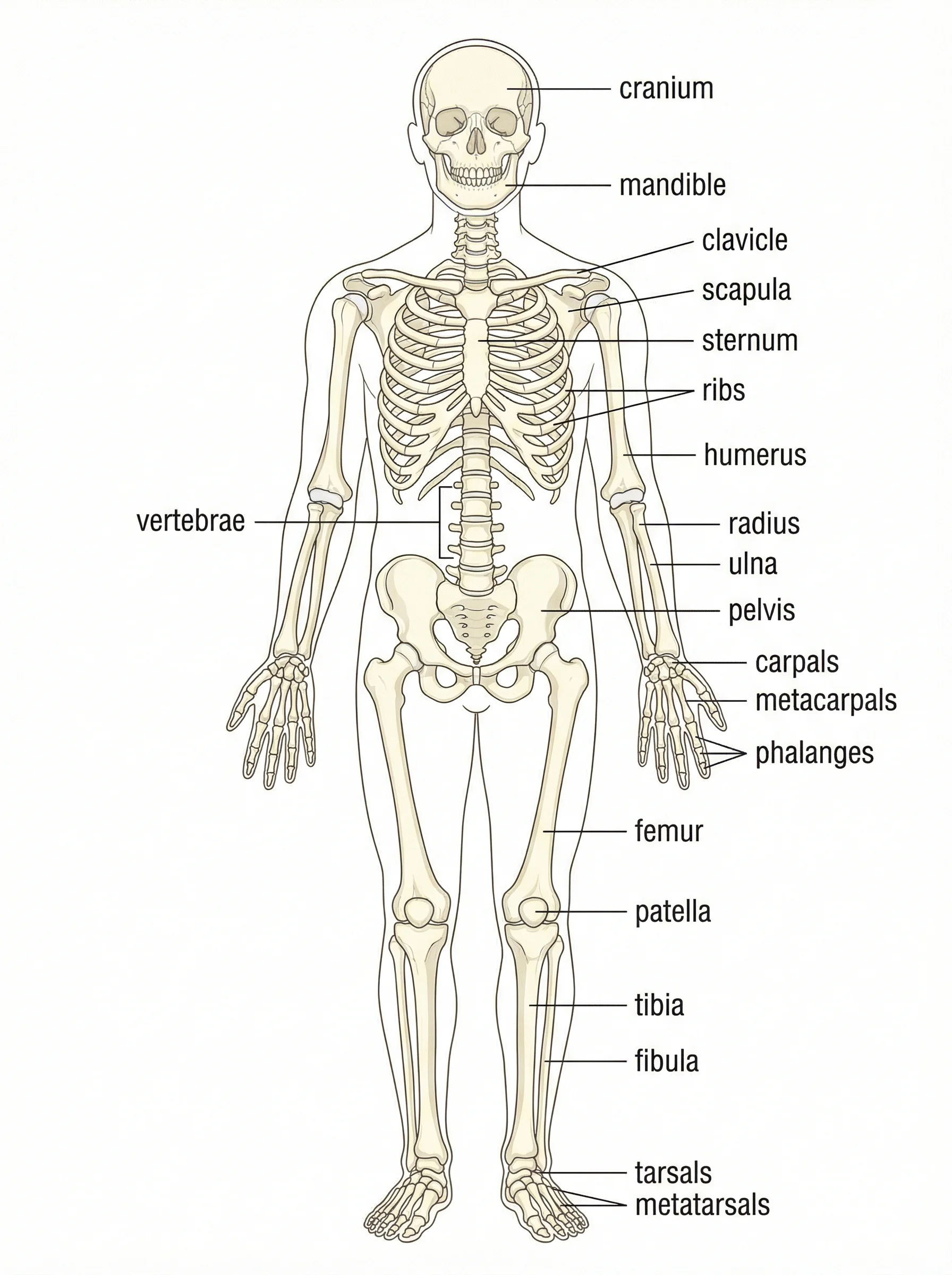
Anatomical Drawing Generator
Generate labeled anatomy illustrations of body systems, bones, and organs.
 Medical
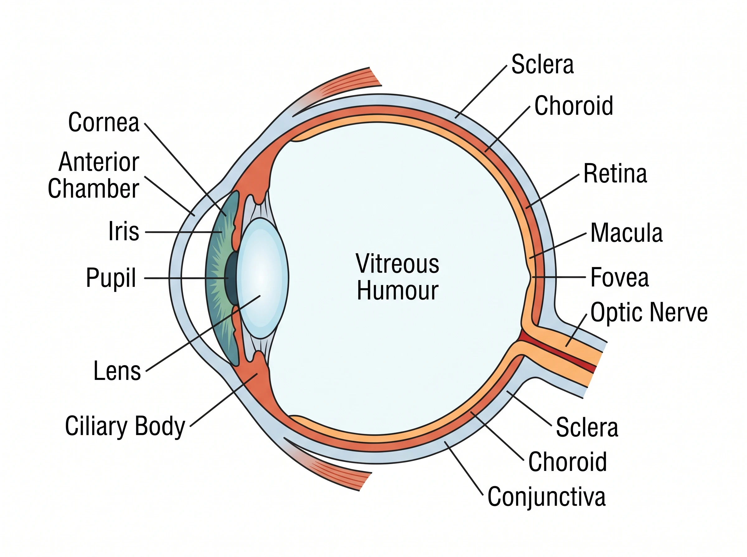
Eye Anatomy Diagram Generator
Create labeled eye diagrams — cornea, lens, retina, optic nerve — and how vision works.
 Medical
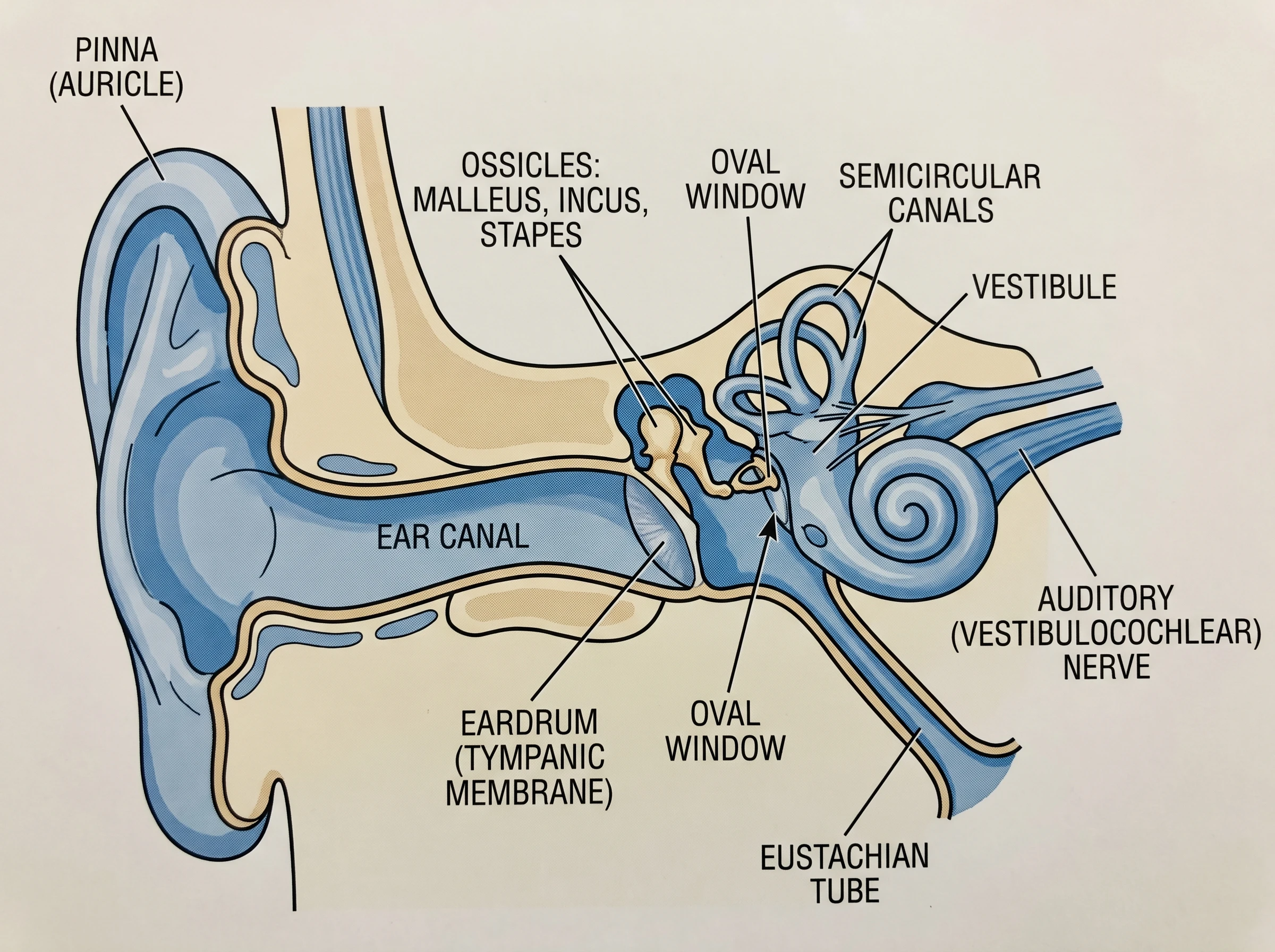
Ear Anatomy Diagram Generator
Create labeled ear diagrams of the outer, middle, and inner ear, and how hearing works.