
Knee Anatomy Explained: Bones, Ligaments & Diagram
A clear guide to knee anatomy: the bones, cartilage, four main ligaments and tendons, how the knee joint moves, and how to read and label a knee diagram.
The knee is the largest joint in the body and one of the most commonly injured. It carries your weight, bends and straightens thousands of times a day, and holds together under heavy load — all thanks to a precise arrangement of bones, cartilage, ligaments, and tendons.
This guide walks through knee anatomy one layer at a time: the bones that form the joint, the cartilage that cushions it, the four main ligaments that stabilize it, and the tendons that move it. You will also learn how the knee actually moves and how to read or label a knee diagram correctly.
Quick Answer: What Makes Up the Knee?
The knee joint is where three bones meet — the femur (thigh bone), the tibia (shin bone), and the patella (kneecap) — with the fibula sitting just to the side. Between them, two C-shaped menisci and a layer of articular cartilage absorb shock. Four ligaments (ACL, PCL, MCL, LCL) hold the bones together, and the quadriceps and patellar tendons let the muscles straighten the leg.
In short: bones for structure, cartilage for cushioning, ligaments for stability, and tendons for movement.
A Labeled Overview of the Knee
It helps to see all of the parts in one place before breaking them down. The diagram below shows a front view of a right knee with every major structure labeled.
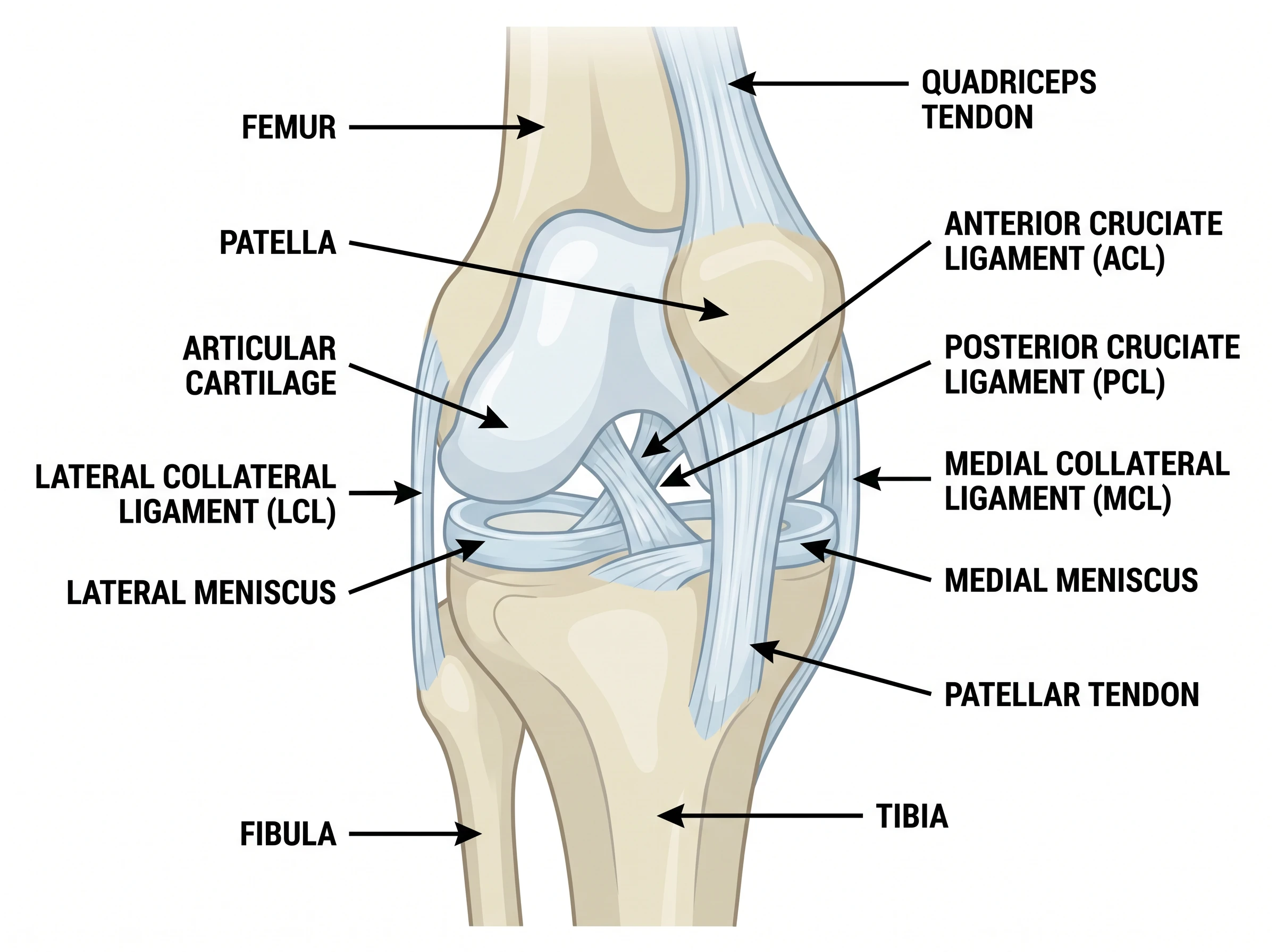
Notice how the patella sits in front of the joint, the ligaments cross and flank the space between femur and tibia, and the menisci rest on top of the tibia like two cushions. Keeping this layered picture in mind makes the individual parts much easier to remember.
The Bones of the Knee
Three bones form the knee joint, plus one that supports it:
- Femur — the thigh bone. Its rounded lower ends (the condyles) form the top of the joint.
- Tibia — the shin bone. Its flat top (the tibial plateau) forms the bottom of the joint and bears most of the body's weight.
- Patella — the kneecap. This small, triangular bone glides in a groove on the femur and protects the front of the joint.
- Fibula — the thinner bone beside the tibia. It does not bear much weight but anchors a key ligament on the outer side.
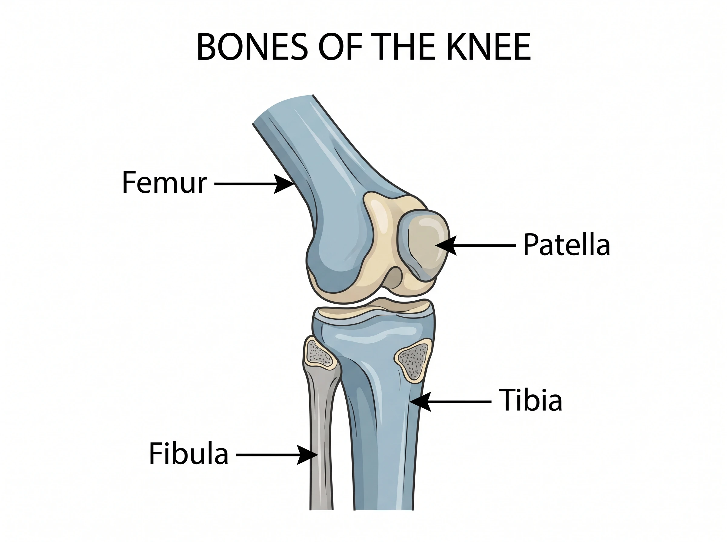
The femur and tibia meet to form the main hinge of the knee, while the patella rides over the front of the femur as the joint bends.
The Cartilage: Menisci and Articular Cartilage
Bone-on-bone contact would quickly wear the joint out, so two types of cartilage protect it:
- Articular cartilage is the smooth, glassy layer covering the ends of the femur, tibia, and back of the patella. It lets the bones glide over one another with almost no friction.
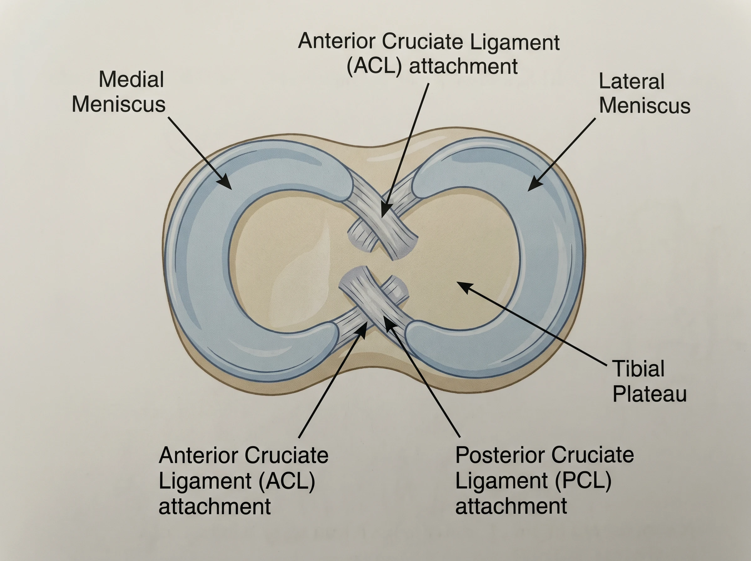
- The menisci are two C-shaped wedges of tougher fibrocartilage sitting between the femur and tibia — the medial meniscus on the inner side and the lateral meniscus on the outer side. They act as shock absorbers and help spread load evenly across the joint.
A torn meniscus is one of the most common knee injuries, which is why these two cushions get so much attention in sports medicine.
Knee Anatomy Diagram Generator
Describe the knee structures you need and get a clean, labeled anatomy diagram in seconds — perfect for study notes, slides, or worksheets.
Make a knee anatomy diagram ->The Four Main Ligaments
Ligaments are tough bands of tissue that connect bone to bone and keep the joint stable. The knee has four that matter most:
- Anterior cruciate ligament (ACL) — runs diagonally through the center of the knee and stops the tibia from sliding too far forward. It is the ligament most famously torn in sports.
- Posterior cruciate ligament (PCL) — crosses the ACL inside the joint and stops the tibia from sliding too far backward.
- Medial collateral ligament (MCL) — runs along the inner side of the knee and resists forces that push the knee inward.
- Lateral collateral ligament (LCL) — runs along the outer side and resists forces that push the knee outward.
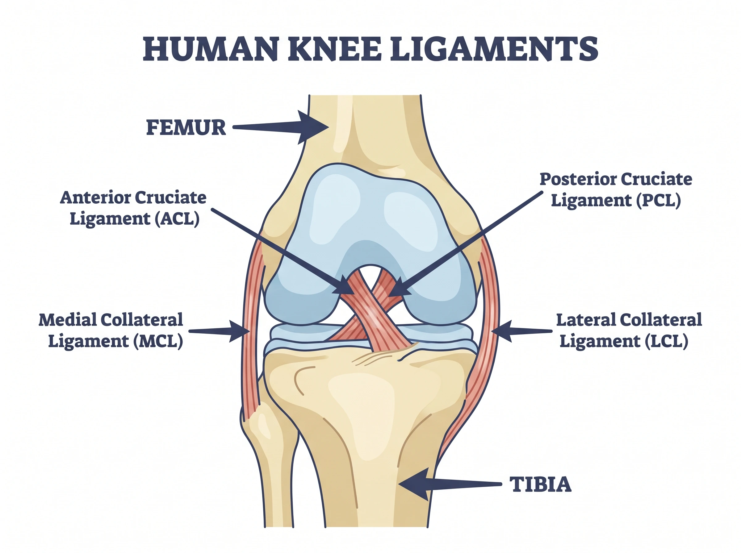
The ACL and PCL are called the cruciate ligaments because they cross like an X inside the joint. The MCL and LCL are the collateral ligaments because they sit on the sides (collateral) of the knee.
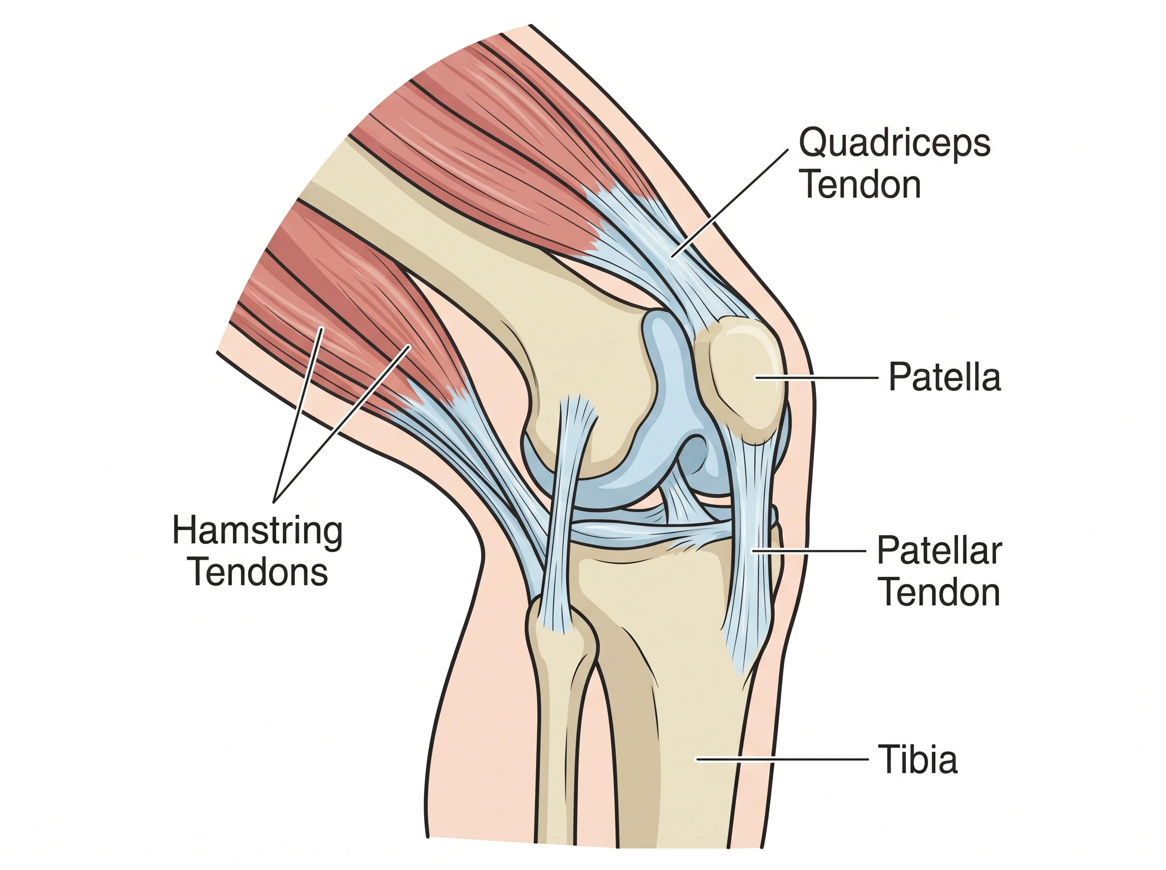
The Tendons That Move the Knee
Tendons connect muscle to bone, and two are central to the knee:
- The quadriceps tendon connects the large thigh muscles (the quadriceps) to the top of the patella.
- The patellar tendon continues from the bottom of the patella to the tibia.
Together they form a single pulley system. When the quadriceps contract, they pull on this tendon-patella-tendon chain to straighten the leg. The patella acts as a fulcrum that makes that pull more powerful.
How the Knee Moves
The knee is primarily a hinge joint — its main job is to bend (flexion) and straighten (extension). But it is not a simple door hinge. As the knee bends and straightens, the femur also rolls and glides on the tibia, and there is a small amount of rotation, especially when the knee is bent.
The four ligaments work together throughout this motion: the cruciates control front-to-back movement, and the collaterals control side-to-side stability. The menisci shift slightly to keep the contact surfaces cushioned, while the quadriceps and patellar tendons supply the force. When all of these parts cooperate, the knee feels effortless — which is exactly why an injury to any one of them is so noticeable.
How to Read or Label a Knee Diagram
When studying or drawing a knee diagram, work in the same order as this guide:
- Start with the bones — locate the femur on top, the tibia below, the patella in front, and the fibula to the side.
- Add the cartilage — place the menisci on the tibial plateau and shade the articular cartilage on the bone ends.
- Draw the ligaments — cross the ACL and PCL in the center, then add the MCL (inner) and LCL (outer).
- Finish with the tendons — the quadriceps tendon above the patella and the patellar tendon below it.
Labeling in layers like this keeps the diagram organized and prevents the most common mix-ups.
Knee Anatomy Diagram Generator
Turn a quick description into a labeled knee diagram showing bones, ligaments, menisci and tendons.
Common Mistakes and Confusions
- Swapping the ACL and PCL. The ACL controls forward slide of the tibia; the PCL controls backward slide. They cross, so it is easy to mislabel them.
- Confusing collateral and cruciate. Collateral ligaments (MCL, LCL) are on the sides; cruciate ligaments (ACL, PCL) are in the center and cross.
- Mixing up the menisci and articular cartilage. The menisci are the two C-shaped pads; articular cartilage is the thin coating on the bone surfaces.
- Calling the patellar tendon a ligament. It connects the patella (a bone) to the tibia, but it is the continuation of a muscle tendon, so it is usually named a tendon.
- Forgetting the fibula. It does not form the main joint, but it anchors the LCL and belongs on a complete diagram.
FAQ
What are the main parts of the knee?
The knee is made of three bones (femur, tibia, and patella, with the fibula alongside), two menisci and a layer of articular cartilage, four ligaments (ACL, PCL, MCL, LCL), and the quadriceps and patellar tendons.
What are the four ligaments of the knee?
The four main ligaments are the anterior cruciate ligament (ACL), the posterior cruciate ligament (PCL), the medial collateral ligament (MCL), and the lateral collateral ligament (LCL). The cruciates sit in the center and the collaterals sit on the sides.
What is the difference between the meniscus and articular cartilage?
The menisci are two C-shaped wedges of fibrocartilage that sit between the femur and tibia and act as shock absorbers. Articular cartilage is the thin, smooth layer covering the bone surfaces so they glide without friction.
What type of joint is the knee?
The knee is mainly a hinge joint that bends and straightens. It also allows the femur to roll and glide on the tibia and permits a small amount of rotation, especially when the knee is bent.
What do the quadriceps and patellar tendons do?
The quadriceps tendon connects the thigh muscles to the top of the patella, and the patellar tendon connects the bottom of the patella to the tibia. Together they let the quadriceps straighten the leg, using the patella as a fulcrum.
Further Reading
Catégories
Plus d'articles
")
Biology Drawing Guide: Create Scientific Diagrams (2026)
Learn how to create accurate biology drawings and scientific diagrams. Covers cell diagrams, organism drawings, labeling techniques, and AI-powered tools.

How to Create Science Animations for PowerPoint: Complete Guide for Researchers
Learn to create engaging scientific animations in PowerPoint with zero budget. Includes step-by-step tutorials, free tools comparison, and best practices for molecular biology, chemistry, and physics presentations.
: Tips, Slide Examples & Presentation Guide (2026)")
3 Minute Thesis (3MT): Tips, Slide Examples & Presentation Guide (2026)
3MT tips and winning slide examples. Learn how to design your 3 Minute Thesis slide, structure your presentation, and avoid common mistakes.