
Protein Structure: The 4 Levels Explained Simply
Protein structure made simple: the four levels (primary, secondary, tertiary, quaternary), the bonds that stabilize each, and why structure decides function.
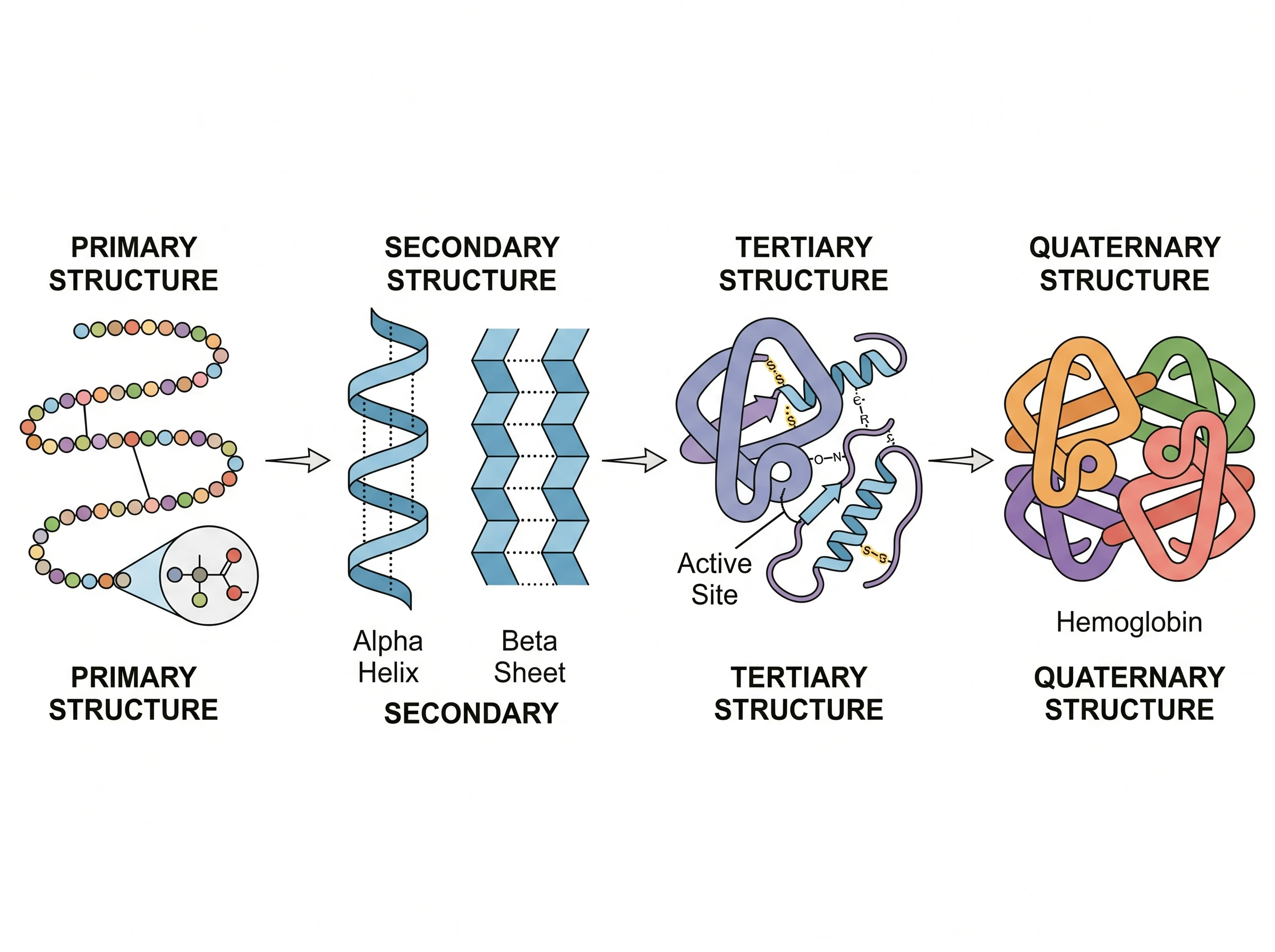
Proteins are the molecular machines of the cell, and almost everything they do depends on their shape. That shape is described at four levels of organization — primary, secondary, tertiary, and quaternary — each built on the one before it.
This guide explains protein structure the simple way: what each of the four levels is, which bonds hold it together, real examples, and why the final folded shape decides exactly what a protein can do.
Quick Answer: What Are the 4 Levels of Protein Structure?
Protein structure has four levels:
- Primary — the linear sequence of amino acids in the chain, joined by peptide bonds.
- Secondary — local folding into alpha helices and beta-pleated sheets, held by hydrogen bonds.
- Tertiary — the overall 3D fold of a single chain, driven by interactions between R-groups (side chains).
- Quaternary — the assembly of two or more folded chains (subunits) into one functional protein.
Each level is built from the level below it, and together they determine the protein's final shape and function.
What Determines a Protein's Structure?
The single most important factor is the amino acid sequence — the primary structure. The order of the 20 different amino acids decides how the chain will fold, because each amino acid's side chain (R-group) has its own chemistry: some are water-loving, some water-fearing, some positively or negatively charged.
As the chain is built, these side chains attract and repel one another, and the whole molecule settles into the lowest-energy, most stable shape it can find. In other words, sequence dictates folding, and folding dictates function.
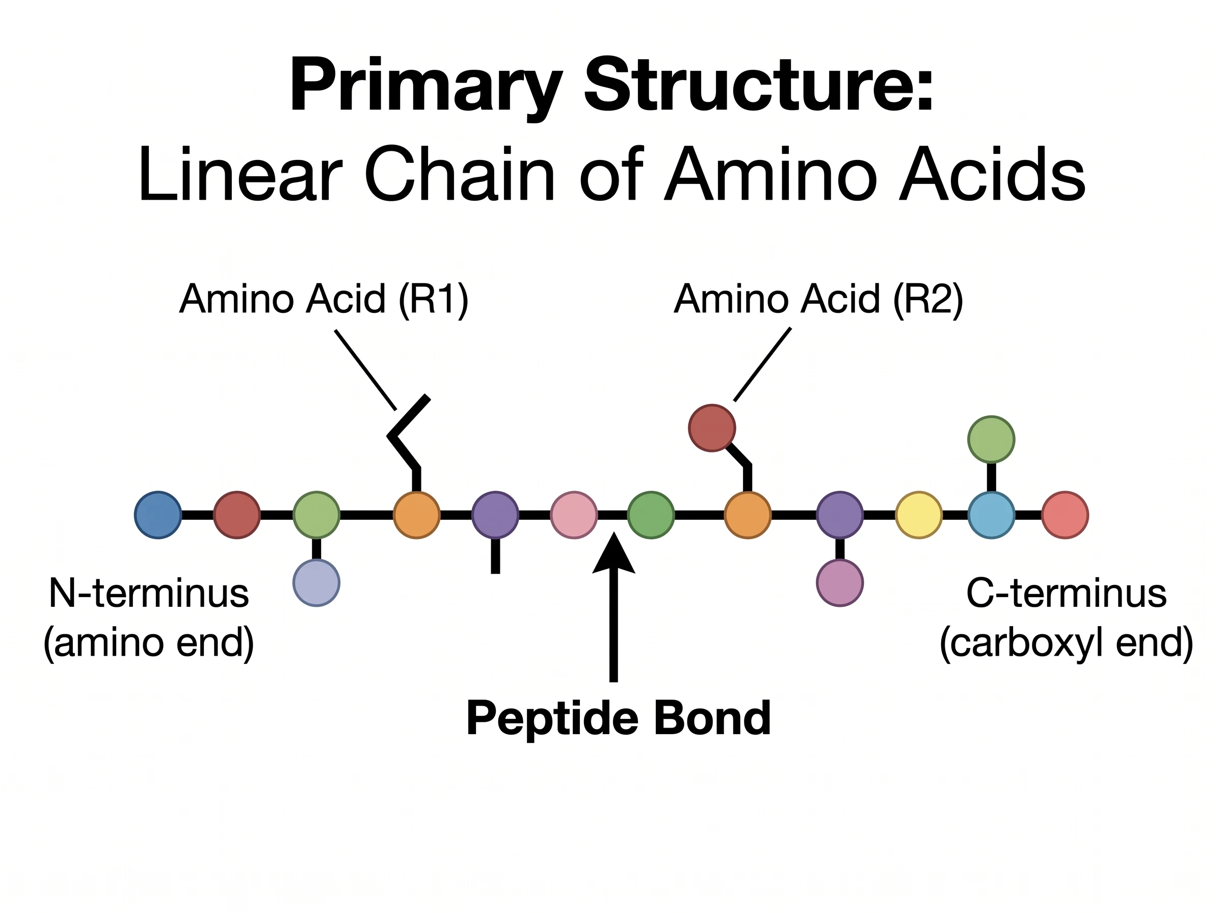
Primary Structure: The Amino Acid Sequence
The primary structure is simply the order of amino acids in the polypeptide chain, read from the N-terminus (amino end) to the C-terminus (carboxyl end). Neighboring amino acids are linked by peptide bonds — strong covalent bonds formed between the carboxyl group of one amino acid and the amine group of the next.
Even one wrong amino acid in this sequence can change everything. In sickle cell disease, a single swap (glutamic acid replaced by valine) in the hemoglobin chain is enough to make red blood cells deform — a vivid reminder that the primary structure is the blueprint for every level above it.
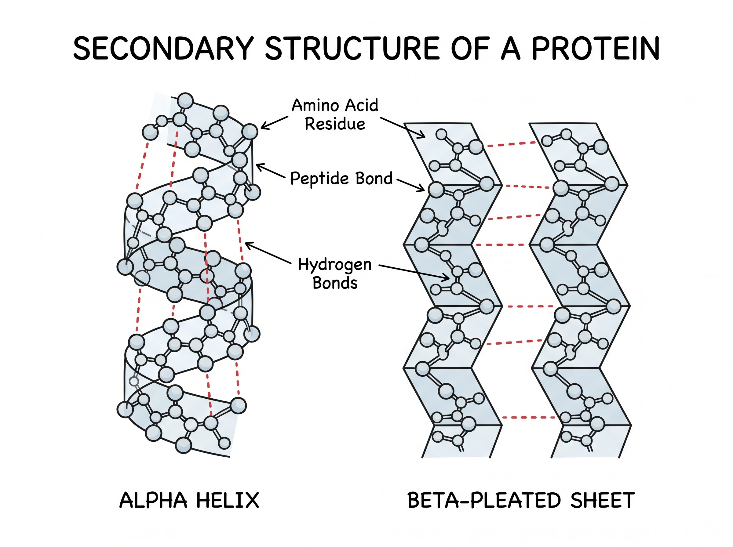
Secondary Structure: Alpha Helices and Beta Sheets
The secondary structure is local folding of the backbone into regular, repeating shapes. There are two main types:
- Alpha helix (α-helix) — the chain coils into a right-handed spiral, like a spring.
- Beta-pleated sheet (β-sheet) — stretches of the chain line up side by side into a flat, pleated arrangement.
Both shapes are held together by hydrogen bonds between the carbonyl (C=O) and amino (N–H) groups along the polypeptide backbone — not the side chains. Many proteins contain both helices and sheets connected by loops.
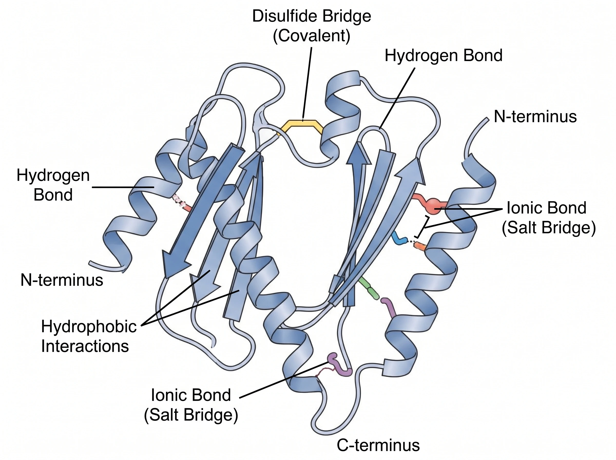
Tertiary Structure: The 3D Fold of One Chain
The tertiary structure is the complete three-dimensional shape of a single polypeptide chain — how all those helices, sheets, and loops pack together into one compact, functional unit.
This level is driven by interactions between the R-groups (side chains):
- Hydrophobic interactions — water-fearing side chains cluster in the protein's core, away from water.
- Hydrogen bonds — between polar side chains.
- Ionic bonds (salt bridges) — between oppositely charged side chains.
- Disulfide bridges — strong covalent bonds between two cysteine side chains.
Together these forces lock the chain into a precise shape, often forming pockets and clefts such as an enzyme's active site.
Protein Structure Diagram Generator
Create clear, labeled diagrams of the four levels of protein structure for study notes, slides, or reports — no drawing skills needed.
Make a protein structure diagram ->Quaternary Structure: Multiple Subunits Together
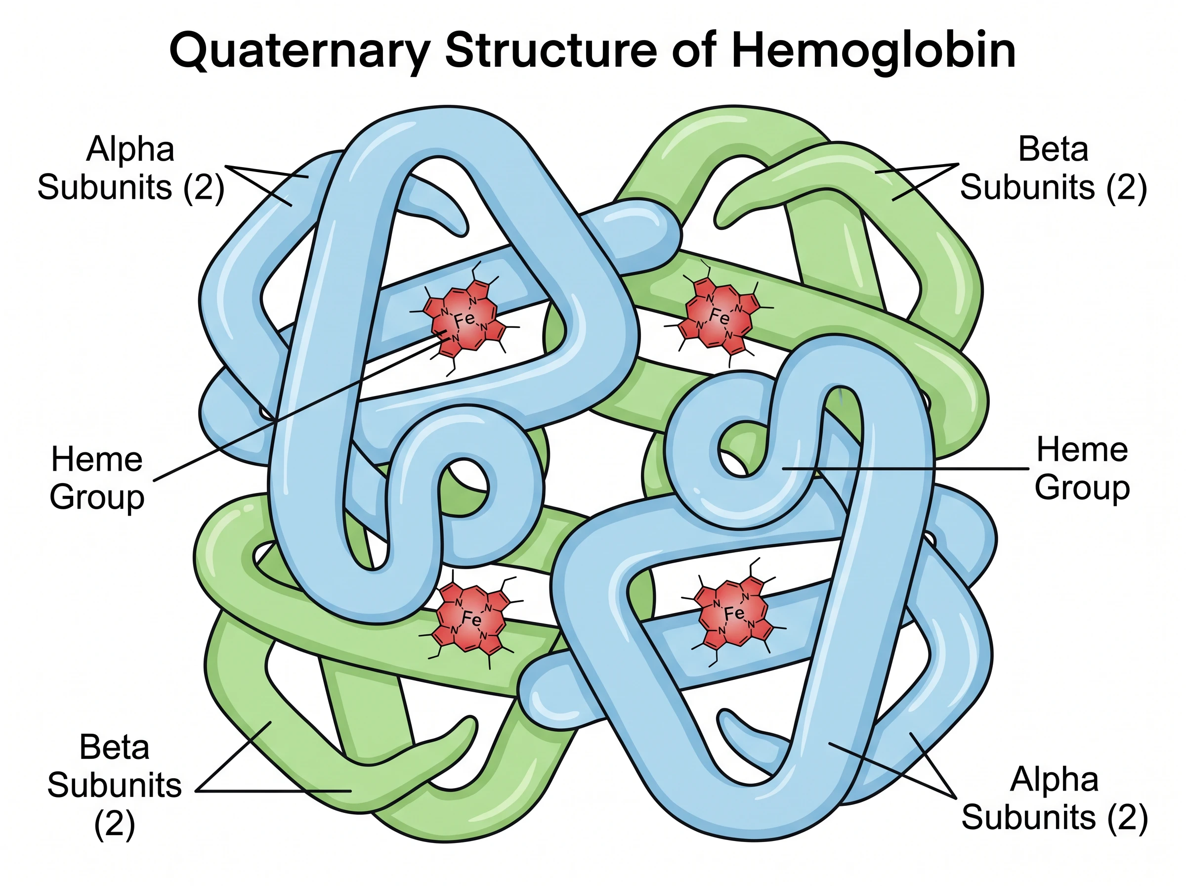
Not every protein has a quaternary structure — only those made of more than one polypeptide chain. The quaternary structure describes how these separate chains, called subunits, fit together into a single working complex.
The classic example is hemoglobin, the oxygen carrier in your blood. It has four subunits — two alpha and two beta chains — each cradling a heme group with an iron atom that binds oxygen. The four subunits cooperate, so binding oxygen at one site makes it easier for the others to bind too. The subunits are held together by the same forces that stabilize tertiary structure: hydrogen bonds, ionic bonds, hydrophobic interactions, and sometimes disulfide bridges.
Why Structure Determines Function (and Denaturation)
A protein's job depends on its exact 3D shape. An enzyme's active site has to fit its target molecule like a key in a lock; an antibody has to match its antigen. Change the shape, and the function is lost.
That loss of shape is called denaturation. Heat, extreme pH, or harsh chemicals can break the weak bonds (hydrogen, ionic, hydrophobic) that hold the higher levels together, causing the protein to unfold. The primary structure — the covalent peptide backbone — usually stays intact, but the protein still stops working. Cooking an egg is everyday denaturation: the clear, liquid proteins unfold and tangle into a solid white. Denaturation is sometimes reversible, but often it is permanent.
How to Read a Protein Structure Diagram
When you look at a protein structure diagram, use these cues to find your bearings:
- Beads on a string usually represent the primary structure — individual amino acids in sequence.
- Coils are alpha helices; flat arrows or ribbons are beta sheets (secondary structure).
- A single tangled, folded ribbon is one chain's tertiary structure.
- Several differently colored ribbons clustered together show quaternary structure — each color is a separate subunit.
- Dashed lines typically mark hydrogen bonds; solid yellow links often mark disulfide bridges.
Common Mistakes and Confusions
- Confusing secondary and tertiary bonds. Secondary structure is stabilized by backbone hydrogen bonds; tertiary structure depends on R-group (side-chain) interactions.
- Thinking every protein has four levels. Quaternary structure only exists in proteins with multiple subunits — many proteins stop at tertiary.
- Mixing up alpha helix and beta sheet. A helix is a coil; a sheet is flat and pleated.
- Believing denaturation breaks the primary structure. Denaturation unfolds the higher levels but leaves the peptide bonds (primary structure) intact.
- Forgetting that sequence drives shape. The amino acid order isn't just a list — it determines how the entire protein folds.
Protein Structure Diagram Generator
Turn any protein structure topic into a clean, labeled diagram for notes and slides.
FAQ
What are the four levels of protein structure?
The four levels are primary (the amino acid sequence), secondary (local alpha helices and beta-pleated sheets), tertiary (the full 3D fold of one chain), and quaternary (two or more chains assembled into one protein). Each level builds on the one before it.
What bonds hold each level of protein structure together?
Primary structure is held by covalent peptide bonds. Secondary structure is stabilized by hydrogen bonds along the backbone. Tertiary and quaternary structures rely on R-group interactions: hydrogen bonds, ionic bonds (salt bridges), hydrophobic interactions, and disulfide bridges.
What is the difference between secondary and tertiary structure?
Secondary structure is the local folding of the backbone into regular shapes (alpha helices and beta sheets) held by hydrogen bonds. Tertiary structure is the overall three-dimensional shape of the whole chain, driven by interactions between amino acid side chains.
Does every protein have a quaternary structure?
No. Quaternary structure only exists in proteins made of more than one polypeptide chain (subunits), such as hemoglobin. Proteins built from a single chain have only primary, secondary, and tertiary structure.
Why does protein structure determine function?
A protein's 3D shape creates the precise surfaces, pockets, and active sites it needs to do its job, such as binding a substrate or carrying oxygen. If the shape changes — for example, through denaturation — the protein usually loses its function.
Further Reading
分類
更多文章
")
How to Make a Bohr Model: Step-by-Step Guide for Students (2026)
Learn how to draw a Bohr model for any element. Covers electron shell rules, common elements, mistakes to avoid, and when Bohr models break down for heavier atoms.
")
7 Best Free Napkin AI Alternatives in 2026 (Text-to-Visual Tools)
Best free Napkin AI alternatives: Gamma, Canva, Piktochart, Venngage & more. Compare AI-powered text-to-visual tools for diagrams and infographics.
")
How to Make a Graphical Abstract: Free Maker & 7-Step Guide (2026)
Free graphical abstract maker + step-by-step guide. Create Elsevier & Cell journal-ready abstracts in minutes. Includes size specs (1328x531px, 1200x1200px), templates, and AI tools.